Fetal ECG Extraction Using ANFIS Trained
With Genetic Algorithm
A.Vigneswaran 1, N.S.Vijayalaksmi2, P.Esaiarasi3
Assistant Professor, Department of Electronics and Communication Engineering, SKP Engineering College
Tiruvannamalai, Tamilnadu, India1, 2, 3
ABSTRACT: Keeping the health condition of fetus in best conditions is one of the most important goals which be followed in pregnancy period. If physicians can be aware of the fetus’s health condition continuously, they can improve the level of their attentions and so they can take better decisions quickly in emergency situations. Here signal monitoring is one of the most important techniques which give physicians important information about fetal health condition. The fetal electrocardiogram (FECG) signal reflects the electrical activity of the fetal heart. The project proposes a new method for extracting the Fetal Electrocardiogram (FECG) signal form the two ECG signals recorded at the thoracic and abdominal areas of the mother’s skin. The thoracic ECG is assumed to be completely maternal ECG (MECG) while the abdominal ECG is assumed to be a combination of mother’s and fetus’s ECG signals and random noise. The maternal component in the abdominal ECG is a nonlinearly transformed version of the MECG. The method uses Adaptive Nero-Fuzzy Inference System (ANFIS) structure for identifying the nonlinear transformation. Using this transformation, we can cancel the maternal component in AECG and then we can estimate the FECG.
KEYWORDS: Artificial Intelligence, Neural Network, Fuzzy Systems, Genetic Algorithm (GA), Fetal Electrocardiogram (FECG) Signal.
I. INTRODUCTION
The fetal electrocardiogram (FECG) signal reflects the electrical activity of the fetal heart. It contains information on the health status of the fetus and, therefore, an early diagnosis of any cardiac defects before delivery increases the effectiveness of the appropriate treatment. There are several technical problems associated with the noninvasive extraction of FECG from ECG signals recorded at the abdominal surface. These problems are mainly due to the low power of the FECG signal which is contaminated by various sources of interference. These sources include the maternal ECG, the maternal electromyogram EMG, 50 Hz power line interference, baseline wander and random electronic noise . Assuming that we are using state of the art low noise electronic amplifiers with high common mode rejection ratio, the effect of the 50Hz interference and electronic random noise can be eliminated.The EMG noise can also be reduced but not necessarily eliminated with the use of classical low pass filtering techniques. Therefore, it is safe to say that if one is able to eliminate the maternal ECG component in the composite signal, a reasonable estimate of the FECG signal can be obtained. To further enhance this FECG estimate, especially its P and T waves, one needs to apply postfiltering techniques. These techniques include nonlinear filtering via wavelet denoising. Many signal-processing-based techniques for FECG extraction have been introduced with varying degrees of success. These techniques include adaptive filters, correlation techniques, singular-value decomposition (SVD), wavelet transform, neural networks, and blind source separation (BSS). BSS via independent
maternal component in the extracted FECG especially when the R wave of maternal and fetal QRS overlap. Recently, we have proposed a new FECG extraction technique based on polynomial networks which showed encouraging results on extracting FECG from two ECG recordings.
II. THEORY OF NON-INVASIVE FECG EXTRACTION
As presented before, the aim of non-invasive methods is to extract FECG signal from the signal recorded on mother’s abdomen (AECG). Also, it has been noted that AECG signal is a combination of FECG signal, a distorted version of MECG signal and interferences. Therefore, to obtain better results in extracting FECG component of AECG, we need to attenuate the power of maternal component and decrease the effects of interferences. There are so many ways to nullify interference as briefly mentioned in the literature. So, if we can find the maternal component of AECG, after subtracting it from AECG signal we can find a promising estimation for FECG component. The aim of this paper is to find the maternal component of AECG signal. In another word, the measured signal from the mother’s abdomen (AECG) is usually dominated by maternal heartbeat signal that propagates from the chest cavity to the abdomen. Based on this statement, we should find the transformation of this propagation of mother’s ECG signal from the chest to the abdomen. For better understanding of this signal propagation we have figured the problem as figure 1.
Figure1.Maternal signal propagation through the abdomen.
The method in this study uses two recorded signals, one is the signal recorded from the thoracic area (we called it m(n)) and the other recorded from mother’s abdomen which is a combined signal (we called it a(n)). In the method, it is assumed that the thoracic signal is just mother’s ECG and we use it as a reference signal. We can formulate the problem as follows:
a(n)= m^(n)+f(n)+n(n), m^(n)=T{m(n)}
Where m(n) and a(n) are thoracic and abdominal ECG signals respectively. n(n) represents random noise. m(n)is a deformed version of m(n) after applying T transformation. This deformation is due to the fact that the signal is recorded far away from its source, mother’s heart. All we should do is to find the transformation. If the transformation is linear, the problem could be a simple aligning of m(n) and a(n) via correlation; then a good estimation of m^(n) could be made by subtracting the aligned m(n) from a(n).But, as we know, transformation T is not linear. We have used ANFIS network trained by GA to approximate this non linear transformation. The transformation will operate on m(n) and results in signal m^(n). which is perfectly aligned with the deformed maternal component in a(n). We can use this alignment to remove the maternal component in a(n) and then we have the FECG component of a(n).
III. ADAPTIVE NEURO-FUZZY INFERENCE SYSTEM (ANFIS)
A. ANFIS STRUCTURE
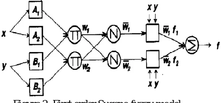
Compared with linear systems and neural networks there are many benefits to use ANFIS in pattern learning. These benefits are due to the fact that ANFIS collects the capabilities of both neural networks and fuzzy systems in learning nonlinearities. Fuzzy techniques incorporate base information sources into a fuzzy rule that represents the knowledge of the network structure so that structure learning techniques can easily be accomplished. Moreover, ANFIS architecture requirements and initializations are fewer and simpler in comparison with neural networks which require so many trails and errors for optimized updating of the weights. Based on these specifications, we have selected ANFIS as a reliable tool for determining the non linear transformation. In the algorithm, we have used first-order Sugeno fuzzy model as the structure.
Figure 2. First order Sugeno fuzzy model.
This ANFIS’s network composes of two parts. The first is the antecedent and the second one is the conclusion part; they are connected to each other by the rules in network frame. As figure 3 shows, the structure has five layers. The first layer executes the fuzzification process. All the nodes in this layer are adaptive nodes. The second layer executes
the fuzzy AND of the antecedent part of the fuzzy rules. The nodes in this 198 are fixed. The operator Π indicates that
they play as a simple multiplier. The third layer normalizes the membership function (MF). Nodes of the third layer are also fixed nodes and operator N represents the normalization role of these nodes. The fourth layer executes the consequent part of the fuzzy rules. The nodes in this layer are adaptive nodes.
The last layer computes the output of fuzzy system by summing up the outputs of the previous layer. The related equations of ANFIS model shown in figure 2 are as follows:
wi = µAi (x) * µBi (y)
ANFIS uses MFs for splitting each input dimension to many local regions to model complex nonlinear systems. The input space is covered by MFs with overlapping that means several local regions can be activated simultaneously by a single input. Bell-shaped with maximum of 1 and minimum of 0 is usually used as MFs.
B. ANFIS TRAINING
1. Model Optimization Using Genetic Algorithm
GA is currently one of the most popular stochastic optimization techniques. It is inspired by natural genetics and the biological evolutionary process, and can be characterized by the following features .
A scheme for encoding solutions to a problem in the form of a chromosome (chromosomal representation).
An evaluation function which indicates the fitness of each chromosome relative to the others in the current set of chromosomes (referred to as population).
An initialization procedure for the population of chromosomes.
Genetic operators which are used to manipulate the composition of the population
A set of parameters that provide the initial settings for the algorithm: The population size and probabilities employed by the genetic operators.
The GA uses three basic operators to manipulate the genetic composition of a population: reproduction, crossover and mutation Reproduction consists in copying chromosomes according to their objective function (strings with higher merit will have more chances to survive). The crossover operator mixes the genes of the two chromosomes selected in the phase of reproduction, in order to combine the features, especially their positive ones.
Mutation is occasional, producing with low probability an alteration of some of the gene values in a chromosome. To perform the GA, it is first very important to define the fitness function. This fitness function is constructed bearing in mind that the output signal must be fit to our FECG template. For this purpose, we must utilize a measure of the secondary structural element (SSE).
A. FITNESS FUNCTION
To minimize the error between the model and the template, the following fitness function is considered in the GA process:
Fitness=∑ ( _model(i)FECG_Template(i))2
By minimizing this function, one could expect to reach to an exact mathematical model of the FECG template. The available parameters for GA to minimize the above fitness are 10 unknown factors in equation (3).The parameters of the GA are: population size = 50, number of generation = 500, crossover probability = 0.8 and probability distribution of mutation is set to be Gaussian. After executing the GA, SSE value of less than 0.004 is derived. The convergence process is shown in figure 6. The resulted optimized model is in the following form
FECG_Model= -0.018278+∑ isinc( bit+di)
2. Training ANFIS Using Genetic Algorithm
ANFIS has two types of parameters which are needed to be updated, the antecedent and conclusion part parameters. There are three sets of parameters in antecedent part ai,bi,ci. Each one of the parameters has N genes; N represents the
number of MFs.
The conclusion part parameters, pi,qi,ri,. also trained during optimization algorithm. Each chromosome in conclusion
part has (1+1)*R genes where R is the number of 199 applied rules and I is the dimension of the input data. Parameters are initialized randomly in the beginning. In each iteration, one of the parameters is updated using GA. For example, in the first iteration ai s are updated and then in next iteration, bi s are updated and so on.
IV. ANFIS NETWORK TRAINED WITH GENETIC ALGORITHM FOR FECG EXTRACTION
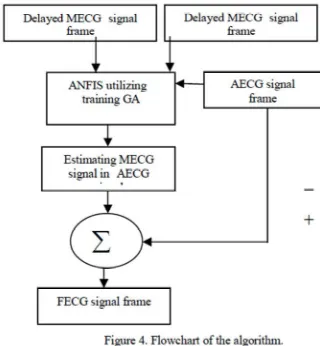
V. FLOWCHART OF THE ALGORITHM FOR EXTRACTING FECG
The algorithm uses thoracic, M(n), and abdominal, A(n), signals to extract FECG signal.
The basic assumption of this algorithm is the matter that the recorded signal from thoracic area consists only of mother’s ECG signal. Due to strong ECG signals of mother this assumption is a realistic one. The two recorded signals are segmented so that they are prepared to be trained by ANFIS. M(n) and A(n) signals are segmented in a way that each one is partitioned into N-sample segments. In this algorithm the overlapped segments are also considered; in other words, overlapping is N/2 samples. The ith segment of the signals is defined as follows. Accordingly, the training vector in ANFIS algorithm is obtained. The structure of ANFIS used in figure 3 has been repeated.
Figure 3. The structure of ANFIS used in the algorithm.
As illustrated in figure 4, ANFIS inputs are one of the vectors obtained from partitioning the thoracic signal and its delayed signal (the previous segment); the output of the network in training algorithm is the vector equivalent to the input vector in the set of the vectors obtained from abdominal signal segmentation. ANFIS parameters are separately adjusted for each pair of vector sets ( Mi s and Ai). After each training by one of paired vectors M vector is given as
ANFIS input. The obtained output is the same as transformed version of i M vector in abdominal area which we call i M. When all the i M s were obtained and the considered overlapping in segmentation process was taken into account, M signal (the transformed M signal in abdominal area) is created. Now we have fulfilled the predefined objective for we have been able to obtain mother ECG signal components after it passed thoracic area to reach abdominal area. At this time, we can subtract the obtained signal from abdominal signal in order to obtain an approximation of FECG signal which is the desired one. The flowchart of the algorithm is illustrated in figure 4.
VI. EVALUATION OF THE ALGORITHM
The method in this article uses two recorded signals to extract FECG signal components: the recorded signal at mother’s abdominal area and her thoracic area. MECG signal components in the recorded signal at the abdominal area are transformed versions of MECG signal. This transformation is because these components are recorded in some distance from their source (mother’s heart). Indeed, it should be noted that this is a nonlinear transformation. The method uses ANFIS and GA to model this transformation. The method uses GA to train ANFIS. By finding this transformation and applying it to MECG one can obtain the transformed version of MECG signal components in the combined signal.
In this study, in addition to visual criterion, two parameters have been also considered to compare the efficiency of algorithms. One is signal to noise ratio (SNR) parameter and the other is the number of signal channels necessary for implementing the algorithm. In this case, SNR parameter is defined as below. In using this parameter, it is assumed that the resulted signal from the algorithm only involves fetus signal components and uncorrelated noise. Given the above hypothesis, we divide the signal into some pulses first; then we take the pulses as columns of an imaginary matrix. Now, we obtain the eigenvalues of this matrix using Singular Value Decomposition (SVD) algorithm. We define SNR criterion as follows: In the, are eigenvalues corresponding to the matrix. SNRsvd parameter denotes the ratio of FECG signal components energy (first eigenvalue) to the energy of noise sources’ components (second eigenvalue onward) in the extracted signal. Now, if we apply normalization operation on matrix rows and if we define SNR parameter as a criterion to determine the level of correlation between two x(i) and x (j) pulses (columns of the imagined matrix), the above equation will be as follows:
We used the parameters defined in 10 and 11 equations, i.e. SNRcor and SNRsvd, to evaluate the efficiency of the parameters. By studying this table and comparing the illustrated signals in figure 5 we conclude that ANFIS based algorithms have done better than other algorithms. In fact, only the algorithm based on Independent Component Analysis has yielded a better result in SNRsvd criterion than the algorithm in this study; it owes this advantage to using several recorded signals in abdominal and thoracic areas instead of using just one recorded signal at abdominal area and another recorded signal in thoracic area which the algorithm uses; and, indeed, this is one of the negative aspects of ICA algorithm. B. The Results of Implementing the Algorithm on the Signals in PhysioBank Database As mentioned earlier, this database contains ECG signals recorded from thoracic and abdominal areas in different stages of gestation. We have used these signals to study the efficiency of the algorithm in different stages of pregnancy. Table 1 involves the values of SNRsvd and SNRcor parameters in different stages of pregnancy using the mentioned algorithms.
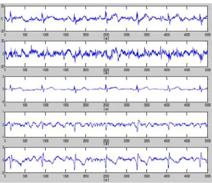
Figure 7. Comparing the efficiency of the algorithm with other algorithms in FECG signal extraction in 40th week of pregnancy; (a) the signal resulted from the algorithm, (b) the signal resulted from ANFIS based algorithm, (c) the signal resulted from ICA based algorithm, (d) the signal resulted from algorithm based on wavelet transform, (e) the signal resulted from SVD based algorithm.
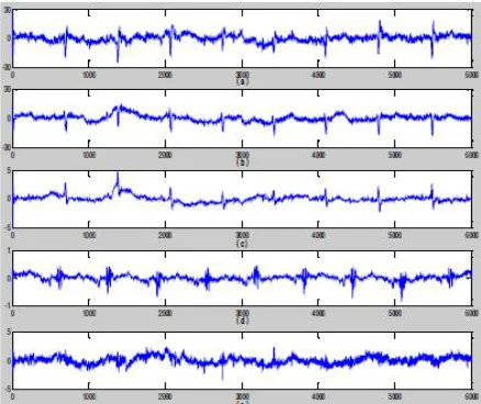
The illustrated signal in figure 6 is recorded in 21st week of pregnancy. One of the major problems in using different algorithm to extract FECG signal is the matter that some algorithms are not able appropriately isolate FECG signal in the early stages of fetus development. However, as we can see in figure 6, compared to other tested algorithms the extracted signal using the algorithm has been well able to extract FECG signal components.
VII. CONCLUSION
In this paper, we have applied ANFIS network trained by GA to extract the FECG signal using two ECG signals recorded at the thoracic and abdominal areas of the mother. Using only two recorded signals is one of the most important advantages of the ANFIS based method. In this algorithm, ANFIS has been used to determine the non linear transformation between the
REFERENCES
[1]. S. Sargolzaei, K. Faez, A. Sargolzaei, Signal Processing Based Techniques for Fetal Electrocardiogram Extraction, Proc. IEEE International Conf. on Biomedical Engineering and Informatics, Hanian, China, 2011, 492-496.
[2]. Ferrara, E. R., & Widrow, B.Fetal Electrocardiogram enhancement by time-sequenced adaptive filtering. IEEE, Trans. on Biomed. Eng. [3].L. Lathauwer, Database for the identification of systems: FECG data EAST/SISTA K.U. Leuven, Belgium[Online].Available:http://www.esat.kuleuven.ac.be/sista/daisy/
.[4]. A. Sargolzaei, K. Faez, S. Sargolzaei, “A New Robust Wavelet BasedAlgorithm for Baseline Wandering cancellation in ECG signals”, Internationalconference on Signal and Image Processing Applications (ICSIPA 2009), pp.33-38, Kuala lumpur, Malaysia.
[5]. D.A. Tong; K.A. Bartels; K.S. Honeyager, Adaptive reduction of motion artifact in the electrocardiogram.
[6] V. Vigneron; A. Paraschiv-Ionescu; A. Azancot; O. Sibony; C. Jutten,Fetal electrocardiogram extraction based on non-stationary ICA and wavelet denoising
[7] Xing Jiang; Liqing Zhang; Qibin Zhao; Sahin Albayrak,ECG, Arrhythmias Recognition System Based on Independent Component Analysis Feature Extraction
[8] Reza Sameni; Christian Jutten; Mohammad B. Shamsollahi,What ICA Provides for ECG Processing: Application to Noninvasive Fetal ECG Extraction
[9] X.Y. Li; T. Wang; P. Zhou; H.Q. Feng,ST-T,Complex automatic analysis of the electrocardiogram signals based on wavelet transform