Acknowledgements
I wish to thank my thesis supervisor Dr. Rüdiger Klein who gave me the opportunity to
do my PhD work in his lab and directed my scientific work all these years.
Special thanks to Dr. Tomoko Iwata for offering me a fruitful collaboration and for
supervising the first part of my PhD studies.
I thank all my collaborators, especially Guido Panté and Lu Qun for providing tools to do
my studies and interesting discussion. Special thanks also to lab members Joaquim Egea
and Jenny Köhler who very kindly provided DNA constructs and antibodies.
I thank Angel Nebreda at EMBL as member of my thesis committee for his precious
suggestions.
Special Thanks to Sidney Cambridge for critically reading and proofreading this thesis
and for his friendship.
I acknowledge all members of the Klein lab for providing a great environment both in the
lab and in the Biergarten. Especially I am grateful to Svetla Dimitrova, Joaquim Egea,
Jenny Köhler, Archana Mishra, Laura Knott, Elsa Martinez, Taija Mäkkinen, Stefan
Weinges and Manuel Zimmer, for their help, critical discussions and friendship.
I would like to thank all the people at the Max-Planck Institute of Neurobiology who
have supported and encouraged my work, especially the members of the labs of Amparo
A DNA-Microarray screening:
G
-Catenin, a new
mediator of Eph-ephrin signaling
Dissertation
Der Fakultät für Biologie der
Ludwig-Maximilians Universität München
Eingereicht am 20.5.2005 von
First “Gutachter“: Dr. Rüdiger Klein
Second “Gutachter“: Dr. Angelika Böttger
The work presented in this thesis was performed from March 2001 to August 2001 in the
laboratory of Dr. Rüdiger Klein at the European Molecular Biology Laboratories (EMBL)
Heidelberg, Germany; and from September 2001 to April 2005 at the Max-Planck
Erklärung
Ich versichere, daȕ ich meine Dissertation selbstständig, ohne unerlaubte Hilfe angefertigt, und mich dabei keiner anderen als der von mir ausdrücklich bezeichneten
Hilfen und Quellen bedient habe.
Die Dissertation wurde in der jetzigen oder ähnlichen Form bei keiner anderen
Hochschule eingereicht und hat noch keinen sonstigen Prüfungszwecken gedient.
______________________ ______________________
1. TABLE OF CONTENTS
1. TABLE OF CONTENTS……….6
2. ABBREVIATIONS………..……….9
3. PREFACE……...………...…………...………....14
4. SUMMARY………...………...…………16
5. INTRODUCTION………..……….17
5.1 Introduction outline……….18
5.2 The generation of the dendritic tree……….19
5.2.1 Dendrite outgrowth………...20
5.2.2 Dendrite guidance……….21
5.2.3 Dendrite branching………21
5.2.3.1 Role of Rho family GTPases in dendrite branching………..23
5.2.3.2 Downstream cellular effectors of Rho family GTPases ………...25
5.2.3.3 Extracellular cues that trigger Rho family GTPases signalling cascade…………28
5.2.3.4Spine formation………..29
5.3 Eph receptors and their ephrin ligands……….33
5.3.1 The Eph class of receptor tyrosine kinases………...33
5.3.2 Ephrin ligands………...34
5.3.3 Signaling mechanisms by Eph receptors and ephrin ligands………36
5.3.3.1 Mechanisms of Eph receptor forward signaling………46
5.3.3.2 Mechanisms of ephrin ligand reverse signalling………...40
5.3.4 Effects of Eph-ephrin signaling………43
5.3.4.1 Forward and reverse signaling during axon guidance………...43
5.3.4.2 Eph/ephrins and synaptic plasticity………...45
5.4 cDNA Microarrays………..47
5.5New candidate molecules for dendritic development:GCatenin………..………….50
5.5.1 Molecular structure………...50
5.5.2 Expression pattern……….52
5.5.3 The biological functions of GCatenin……….56
5.5.3.1GCatenin in cell junctions………56
5.5.3.3GCatenin and actin cytoskeleton dynamics………..60
6. RESULTS………...………..64
6.1 Identification of novel Eph-ephrin downstream effectors using a cDNA microarray screening:GCatenin……….65
6.2GCatenin and Eph-ephrin signaling………...76
6.2.1GCatenin is phosphorylated by Eph-forward signaling but not by ephrin-reverse.76 6.2.2GCatenin and EphA4 interact physically………79
6.3 Eph receptor forward signaling, but not ephrin reverse signaling, leads to the formation of G-Catenin aggregates ………87
6.4 GCatenin and Eph-ephrin signaling are required for the establishment of proper dendritic morphology……….92
6.4.1G-Catenin and the dynamics of filopodia formation………...…………....109
6G-Catenin and HeLa cell migration………...116
7. DISCUSSION………...………..120
7.1 Outlook………..134
8. MATERIALS AND METHODS……….….135
8. 1 Buffers and solutions………....135
8.2Media and antibiotics for bacterial culture ………...136
8.3 Bacterial strains………..137
8.4Media and supplements for tissue culture………..137
8.5 Media and supplements for primary culture of neurons………138
8.6 Cell lines………138
8.7Solutions for cell Transfection………...139
8.8 Solutions for Biochemistry………139
8.9 Plasmids……….140
8.9.1 Expression constructs……….140
8.10 Antibodies………141
8.11 Chemicals and commercial kits………...143
8.12Molecular biology………144
8.12.2 Enzymatic treatment of DNA………...144
8.12.3 Separation of DNA on agarose gels………..145
8.12.4 Purification of DNA………..145
8.12.5 Transformation of competent E. coli by electroporation………..145
8.13 Semi-quantitative real-time PCR……….146
8.13.1 cDNA preparation……….146
8.13.2 PCR primers and templates………...146
8.14 Primary culture of cortical neurons for biochemistry ……….147
8.14.1 Stimulation of cells………...147
8.15 Primary culture of neurons for immunocytochemistry, cell imaging and time lapse imaging………148
8.15.1 Transfection of cell lines and primary neurons……….148
8.15.2 Time-lapse imaging………..149
8.15.3Immunocytochemistry and cell imaging………...149
8.16Microarray………150
8.16.1 Microarray production………..150
8.16.2 RNA extraction and preparation of fluorescent probes………151
8.16.3 Microarray hybridization………..151
8.16.4 Microarray analysis………...151
8.16.5 Insert cloning and generation of probes for various uses……….152
8.17 Boyden chamber assay……….152
8.18 Sholl analysis………...153
8.19 Biochemistry………153
8.19.1 Cell lysis………...153
8.19.2 Immunoblotting and immunoprecipitation………...154
8.20 Histology………..154
8.20.1 Vibratome sections for in situ hybridization……….154
8.20.2 In situ hybridization………..155
9. BIBLIOGRAPHY………...….157
2. ABBREVIATIONS
Abi-1Abl interacting protein-1
AblAbelson kinase
ACpAnterior Commissure, posterior
AMPAdenosin-5’-monophosphate
AMPAD-2-amino-3-hydroxy-5-methyl-4-isoxazole-propionic acid
APAdaptor protein
APSAmmonium-persulfate
ArgAbelson related gene
ARP2/3Actin related protein 2/3
ATPAdenosin-5’-triphosphate
BBSBES buffered saline
BDNFBrain-derived neurotrophic factor
BESN,N-bis[2-hydroxyethyl]-2-aminoethanesulfonic acid
BoraxSodium tetraborate
bFGFBasic fibroblast growth factor
BSABovine serum albumine
CAMCell adhesion molecule
CamKIICa2+/Calmodulin-dependent kinase II
cAMPCyclic AMP
CAPCbl associated protein
Cdc42Cell division cycle 42
cDNAComplementary DNA
CNSCentral nervous system
CREBCa2+/cAMP-response element binding protein
CskC-terminal Src kinase
CXCRCXC motif receptor
DIVDaysin vitro
DMEMDulbecco’s modified Eagles medium
DNADeoxyribonucleic acid
D-PBSDulbecco’s phosphate buffered saline
EEmbryonic day
EDTAEthylenediamine-tetra acetic acid
EGFPEnhanced green fluorescent protein
EphErythropoietin-producing hepatocellular
EphexinEph-interacting exchange protein
EphrinEph family receptor interacting protein
ERKExtracellular regulated kinase
EYFPEnhanced yellow fluorescent protein
FAKFocal adhesion kinase
FBSFetal bovine serum
FNIIIFibronectin type III
FynFgr/Yes-related Novel gene
G Protein GTP binding protein
GDPGuanosine diphosphate
GEFGuanine nucleotide exchange factor
GlobGlobular (domain)
GluRGlutamate receptor subunit
GPIGlycosylphosphatidylinositol
GRIPGlutamate receptor interacting protein
HBSSHank’s balanced salt solution
Hepes(Hydroxyethyl)-piperanzine-ethane sulfonic acid
HGFHepatocyte growth factor
IFImmunofluorescence
IgImmunoglobulin
IHImmunohistochemistry
IPImmunoprecipitation
KDKinase dead
LBLuria-Bertani
LTDLong-term depression
LTPLong-term potentiation
MAPKMitogen-activated protein kinase
NckNoncatalytic region of tyrosine kinase
NGFNerve growth factor
NikNck interacting kinase
NMDAN-methyl-D-aspartate
N-WASPNeural Wiskott-Aldrich syndrome protein
PAKP21-activated kinase
PARGPTPL1-associated RhoGAP
PCRPolymerase chain reaction
PDBPDZ binding (domain)
PDGFPlatelet-derived growth factor
PDZPSD95/Discs large/ZO-1
PFAParaformaldehyde
PI3KPhosphatidylinositol 3-kinase
PICKProtein interacting with C kinase
PKAProtein kinase A
PKCProtein kinase C
PRK2PKC related kinase 2
PSDPostsynaptic density
PSD-95Postsynaptic density protein 95
PTP-BLPhosphotyrosine phosphatase-basophile like
PYPhospho-tyrosine
RacRas-related C3 botulinum toxin substrate
RapRas related protein
RasRat sarcoma
RGCRetinal ganglion cell
RGSRegulator of G-protein signaling
RhoRas homologous member
RNARibonucleic acid
RoboRoundabout
ROCKRho-associated, coiled-coil containing protein kinase
RTRoom temperature
RT-PCRReal time PCR
RTKReceptor tyrosine kinase
SAMSterile-D-motif
SCSuperior colliculus
SDF-1Stromal cell-derived factor 1
SDSSodium dodecyl sulfate
SFKSrc family kinase
SHSrc homology
SrcSarcoma virus transforming gene product
TEMEDN,N,N’,N’-Tetra-methylethylenediamine
VASPVasodilator-stimulated phosphoprotein
WBWestern blot
WntWingless-type mmtv integration site family
3. PREFACE
During the development of multicellular organisms cells divide, migrate, differentiate and
die in a spatially and temporally coordinated manner. One impressive example of
morphogenesis is the development of the brain. Billions of neuronal cells begin to form a
precise network of nerves and synaptic connections. Neurons are highly polarized cells,
with an axon that transmits information from the cell to the environment and a dendritic
tree that receives and decodes the signals arriving to the cell from the environment.
During development, the establishment of neuronal polarity is a crucial event and it
normally follows precise steps. Axonogenesis, or axon generation, is the first event of
neuronal polarization. An axon arises from young, undifferentiated neurons, and then
extends away from the cell to cover specific territories, often enduring particularly long
journeys in order to establish the right connection and finally take up to delivering a
specific electric message. The process of “dendritogenesis” or dendrite generation,
immediately follows, and it consists of the outgrowth of tiny single processes that
elongate, branch and further increase their complexity overtime resulting in the formation
of an extensive and intricate dendritic “tree”. Mature neurons come in contact with axons
of a number of other neurons through the complex dendritic network, receiving and
integrating electric input messages, and then further transmitting the packages of
information to the rest of the neuron and further.
The complex processes of axonogenesis, dendritogenesis and the establishment of the
proper neuronal networks require that cells communicate with other cells and with their
environment, the correct wiring of the nervous system relying on the ability of neurons to
find and to recognize their appropriate synaptic partners during development. A growing
axon with its highly dynamic growth cone encounters, for example, several
environmental cues on its way, each bearing a message that directs the growing process
in a defined direction. Extracellular information is received for example, via surface
in order to evoke the proper responses, which may translate in a “stop” message or a “go”.
Similar mechanisms govern the generation, correct extension and branching of the
dendritic tree. The molecular mechanisms and the environmental cues driving
axonogenesis and axonal guidance have been partially uncovered in the last years
whereas less is known about the processes leading to dendritogenesis. The identification
of new molecular players in dendritogenesis, both at a cellular level and as extracellular
4. SUMMARY
Signaling between ephrin ligands and their Eph receptors is crucial for several
developmental processes. During the development of the nervous system some growth
factors like the neurotrophins have been shown to be key regulators of dendritogenesis
and dendritic architecture. Eph-ephrin signalling instead, has been mostly shown to be
crucial for axon guidance and local events like the promotion of spine formation and
maintenance, and synaptic plasticity, at later stages of neuronal development. Very little
evidence is available that shows involvement of Eph-ephrin signalling in earlier stages of
neuronal morphogenesis and their role as modulators of dendritic development in a
broader structural way.
In the first part of the project, a microarray screening was carried out in order to identify
new players in the Eph-ephrin signalling pathway. GCatenin, a developmental protein was found to be strongly induced by the activation of the Eph-ephrin system.GCatenin is a member of the Catenin family, proteins that are mainly involved in the formation and
reinforcement of adherens junctions. Some members of this family, like ECatenin and the nervous system-specificGCatenin, have recently been found to be crucial players in dendritic morphogenesis.
The second part of the project was aimed at elucidating the activity of GCatenin in dendritogenesis, characterising its potential role as a new effector in Eph-ephrin
5.1 Introduction outline
The first part of this introduction will cover specific aspects of dendritogenesis, its
chronology, what it is known about the extracellular cues directing it and the molecular
mechanisms that lie downstream.
The second part will cover more specifically the nature of Eph-ephrin signalling, some of
the neuronal developmental processes influenced by it and the molecular machinery
downstream.
5.2 The generation of the dendritic tree
Although the processes of the generation of a dendritic tree are complex and diverse, they
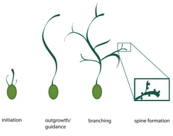
can be separated broadly into a few essential steps. First, dendrites grow out from
morphologically immature and unpolarized young neurons and attain characteristic
growth rates, lengths, diameters and molecular compositions, different from those
neurites which will later be defined as axons. Second, dendrites extend in a defined
direction and increase in diameter. Third, they start forming branches at defined intervals.
Fourth, as dendrites elaborate, many also generate filopodia and small specialized
protrusions called spines that are the major synaptic sites in the mammalian brain (see
figure 5.1)
5.2.1 Dendrite outgrowth
To form connections with the correct presynaptic axon, dendrites must sprout, and extend
away from the neuronal cell body into their target field. The outgrowth is initiated by a
local event of subcortical actin polymerization in an organized manner, similar to the
formation of lamellipodia. Bundles of actin filaments push the cellular membrane
forming a protrusion that is almost simultaneously invaded by microtubules,
polymerizing in an oriented manner, and contributing to the elongation and stabilization
of the process.
Several extracellular factors have been shown to contribute to dendritic sprouting and
elongation. One example is a GPI-linked candidate plasticity gene 15 (CPG 15) that
promotes tectal projection neurons to extend their dendrites when over-expressed in
Xenopus tectal cells (Nedivi et al., 1998). Another is osteogenic protein 1 (OP-1), a member of the transforming growth factor-E (TGF-E) superfamily, which has been demonstrated to stimulate dendrite growth from sympathetic neurons in culture with
nerve growth factor (NGF) as a cofactor and to promote primary dendrite elongation in
E18 mouse cortical neurons (Lein et al., 1995; Le Roux et al., 1999).
NGF, together with brain derived neurotrophic factor (BDNF), neurotrophin 3 (NT3) and
NT4/5, belongs to the family of neurotrophins; they are dimeric secretory proteins that
signal through receptor tyrosine kinases, known as TrkA, TrkB, TrkC and the co-receptor
p75NTR.
Neurotrophins have also been shown to increase dendritic length and complexity in
several studies in vivo andin vitro, in cultures and in brain slices. (McAlister et al., 1995; McAlister et al., 1996). Time lapse imaging of live rat hippocampal neurons in slice
The dynamics of dendritic sprouting and elongation and especially the cellular pathways
through which extracellular signals exert their effects have not yet been fully understood
and it is not clear whether these factors act directly by locally regulating the cytoskeleton
or if they turn on a specific genetic program for dendritic sprouting from undifferentiated
neurons. Rho family GTPases whose roles are much better characterized in later dendrite
development, could also be involved in dendritic sprouting.
5.2.2 Dendrite guidance
Similar to axonal processes, a growing dendrite has to steer toward its target and cross
territories in order to establish the right connections. Not so much is known about
dendritic guidance, whereas axonal guidance has been more extensively studied. In this
process diffusible proteins like semaphorins, for example, are important. These molecules
have been shown to be crucial for axonal growth cone guidance but they also seem to be
involved in dendritic growth cone guidance.
Semaphorin (Sema) 3A normally causes repulsion or collapse of axonal growth cones.
Interestingly, knock-out mice for Sema 3A also display defects in dendrite guidance of
cortical pyramidal neurons (Polleux et al., 1998; Polleux et al., 2000) together with the
expected axonal phenotypes. Sema 3A appears to serve as an attractant for the apical
dendrites of pyramidal neurons in cortical slices (Polleux et al., 2000) because cortical
pyramidal neurons send dendrites towards the source of endogenous Sema 3A (Giger et
al., 1996). This asymmetric mechanism could be dependent on different concentrations,
in dendrites and axons, of Guanylate cyclase (GCS), the enzyme that synthesizes cyclic
nucleotide guanosine 3’/5’-monophosphate (cGMP) ( Luo et al., 1993; Messersmith et al.,
1995). cGMP seems to be necessary for Sema 3A-mediated dendritic attraction and a
and dendritic growth cones may therefore share similar guidance mechanisms and
transduction machinery even if they are differentially regulated.
5.2.3 Dendrite branching
To cover the correct target fields and reach the final neuronal “complexity”, most
dendrites need to form an extensive set of dendritic branches. Two major mechanisms of
dendrite branch formation have so far been described: one is the ‘splitting’ of growth
cones which was first observed predominantly in in vitro studies (Bray et al., 1973), and second, the so called ‘interstitial’ branching, a process in which new branches arise along
the sides of established dendritic shafts. This latter mechanism appears to be predominant
in physiological conditions.
Time lapse studies of live pyramidal neurons from rat hippocampus slices have
demonstrated that each branch initially appears in the form of a single filopodium and
then it elongates assuming its defined morphology (Dailey and Smith, 1996).
Most filopodia emerge from and quickly retract into the dendritic shaft; some develop
further into growth cone-like structures which then mature into branches (Daley et al.,
1996). Stabilized branches extend and become substrate for further branch addition and
stabilization, so that the final complexity is reached by iterative cycles of branch addition,
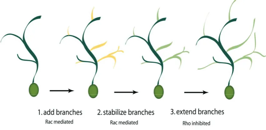
Figure 5.2. Rho GTPase-dependent stages of dendritic branching. For the correct development of dendrites, three distinct processes are required. Branches must be first added to the arbor and Rac plays a key role. Newly formed branches must then be stabilized and the process is also Rac-mediated. Finally, stabilized branches extend and these branches then support further branch additions in an iterative process. Branch extension is mediated by a decrease in RhoA activity. High levels of RhoA repress branch stabilization (modified from Cline and Van Aelst, 2004).
Newly generated filopodia arise mainly as bundles of actin filaments and F-actin seems
to be the main component until the filopodium is committed to form a new branch and is
therefore stabilized by the invasion of microtubules. The elongation is then elicited by a
dual process involving dynamic F-actin structures at the leading edge of the growth cone
and tubulin to stabilize and reinforce the newly formed process.
Similar to dendritic branch formation, the emergence of dendritic spines involves lateral
protrusions of membrane and cytoskeleton: some of the newly formed filopodia are not
stabilized in branches but mature into spines suggesting that the initial steps of the
formation of both spines and branches could be similarly regulated but then differ later
according to extracellular cues or neuronal activity (Altman et al., 1972; O’Leary et al.,
5.2.3.1 Role of Rho family GTPases in dendrite branching
Because of the crucial role played by actin in the early stages of dendritic branching, the
regulators of the actin cytoskeleton have been the most studied molecules, and
specifically the Rho family of small GTPases have emerged as key integrators of
environmental cues regulating actin dynamics in the dendritic cytoskeleton (see figure 5.2). The Rho family of small GTPases are low molecular weight guanine nucleotide binding proteins. They alternate between an ‘active’ GTP-bound state and an ‘inactive’
GDP-bound state. Only in their GTP bound state can those GTPases interact with
downstream molecules that mediate their effect. The ratio of the two forms is regulated
by a set of other molecules, for example the GTPase activating proteins (GAPs) which
increase the hydrolysis rate of bound GTP and thereby inactivate Rho. Other regulating
molecules are the guanine nucleotide exchange factors (GEFs) that instead favor the
exchange of hydrolyzed GDP for GTP and thereby activate Rho. The level of activity of
Figure 5.3. Molecular switch of Rho family GTPases. Inactive Rho bound to GDP is transformed into its active GTP bound state by a guanosine nucleotide exchange factor (GEF). Rho bound to GTP activates effector molecules. GTPase activity is promoted by a GTPase activating protein (GAP) that turns back Rho into its inactive GDP bound state.
The best studied members of the Rho family GTPases are cell division cycle 42 (Cdc42),
Ras-related C3 botulinum toxin substrate 1 (Rac1) and Ras homologous member A (Rho
A). Of the three molecules, Rac 1 and to some extent Cdc42, are key regulators of
dendritic branching and dendritic remodelling. Rho A instead, appears to play a key role
in regulating overall dendritic and branch lengths (see figure 5.2).
It has been shown in Xenopus retinal ganglion cells that the expression of dominant negative mutants of Cdc42 and Rac1 reduces dendritic complexity whereas RhoA
mutants has no noticeable effect; conversely a dominant active form of Rac1 but not of
Cdc42 favours dendritic branching (Ruchhoelt et al., 1999). Other studies with Xenopus
neurons showed that Rac1 promotes branch addition whereas Rho A mostly seems to
affect branch extension (Li et al., 2000) and interestingly, in chick retinal ganglion cells,
Differently to Cdc42 and Rac 1, RhoA activity appears to be mostly inhibitory, as active
RhoA functions as a repressor of dendritic growth when bound to GTP while allowing
dendritic growth when inactive. Under physiological conditions the Rho A pathway is
normally repressed and is locally activated when dendritic growth needs to be limited.
Expression of a constitutively active Rho A mutant in several neuronal systems generally
results in a decrease in dendritic growth. Conversely, dominant negative versions of Rho
A or RhoA selective inhibitors result in the opposite phenotype, i.e. a marked increase in
total dendritic length. (Nakayama et al., 2000; Ruchhoelt et al., 1999; Wong et al., 2000;
Lee et al., 2000).
Not surprisingly, Rho family GTPases are also important for dendritic spine formation; in
particular, dominant active forms of Rac1 promote the formation and maturation of
dendritic spines in Purkinje cells of transgenic mice and in rat hippocampal and cortical
neurons ( Luo et al., 1996; Nakayama et al., 2000; Tashiro et al., 2000). In contrast, Rho
A activity seems to block spine formation, maintenance and elongation.
Experimental evidence thus suggests that a differential engagement of GTPases controls
different structural changes to attain the final dendritic architecture and plasticity. In fact
the situation may even be more complex, given recent evidence that there could be a
cross talk between Rho GTPases and their upstream and downstream effectors, in the
form of feedback loops which ultimately determine the final architectural outcome.
Interestingly, recent studies have shown that intracellular distributions of RhoA, Cdc42
and Rac1 differ according to the neuronal developmental stage in rat hippocampal
cultures. During the early stages of process formation, that is during axon and dendritic
sprouting, the three GTPases are evenly distributed throughout the cell, suggesting a
possible interplay of the three different modulators, in a joint manner.
In fully developed neurons though, RhoA enriches in dendrites, Rac1 in axons, and
Cdc42 is equally abundant in both domains, so that at later stages, a polarized segregation
of the actin regulatory machinery might play an important role in axonal and dendritic
5.2.3.2 Downstream cellular effectors of Rho family GTPases
Members of the Rho family GTPase exert their function through the activation of a
cascade of factors all of which tend to affect the dynamics of the actin and/or microtubule
cytoskeleton.
Well-known effectors that can be activated by both Rac and Cdc42 belong to the
p21-activated kinase (PAK) family of serine/threonine kinases. PAK proteins have been
ascribed roles in regulating actin cytoskeleton dynamics and gene expression (Jaffer and
Chernoff 2002; Bokoch et al., 2003). These kinases exist in an inactive state in the
cytoplasm as a result of their N-terminal autoinhibitory region. Upon binding to Rac-GTP
or Cdc42-GTP, the inhibition is relieved, resulting in PAK activation and its
auto-phosphorylation. One mechanism by which PAKs affect the actin cytoskeleton involves
phosphorylation and activation of the Lin-11, Isl-1, and Mec-3 (LIM) domain-containing
kinases (Yang et al. 1998; Edwards et al. 1999; Dan et al. 2001). Once active, these
kinases phosphorylate and inhibit cofilin, an actin filament depolymerising factor, with
the result of stabilizing actin filaments and promote actin polymerization (Bamburg 1999;
Stanyon and Bernard 1999). The regulation of myosins is likely to be another component
of PAK-mediated cytoskeletal signalling. There is evidence that PAK1 can interfere with
myosin light chain (MLC) function via direct phosphorylation and inhibition of myosin
light chain kinase (MLCK) (Sanders et al. 1999; Bokoch 2003). This action of PAK may
assist in the disassembly of actin stress fibers triggered by PAK.
Another key mechanism by which Rac and Cdc42 relay signals to the actin cytoskeleton
involves the Aldrich-syndrome family of scaffolding proteins. The
Wiskott-Aldrich-syndrome protein (WASP) and its closest relative neuronal WASP (N-WASP)
are regulated by Cdc42 (Rohatgi et al. 1999). Three other members of this family,
WAVE1–3 (also known as Scar proteins) mediate actin-based processes triggered by Rac
(Miki et al. 1998; Machesky et al. 1999; Suetsugu et al.1999; Yamazaki et al. 2003; Yan
et al. 2003). Both the WASP and WAVE family members are linked to the actin
cytoskeleton through their interaction with the Arp2/3 complex. In the case of
WASP/N-WASP, these proteins have been shown to directly bind the activated form of Cdc42.
allowing it to activate the Arp2/3 complex to nucleate the formation of new actin
filaments in vitro (Machesky et al. 1999; Rohatgi et al.1999, 2000; Kim et al. 2000). The
WAVE proteins also mediate actin cytoskeletal changes downstream of Rac but without
directly binding to it. They, in addition to directly activating the Arp2/3 complex, also
influence the activity of profilin which binds a number of actin monomers, preventing
spontaneous nucleation and the addition of actin monomer to the pointed end of the
filament, but not to the barbed end, and thus directly controlling actin polymerization
dynamics (Pollard et al. 2000).
The major downstream effectors of RhoA that mediate GTPase’s effects on the
cytoskeleton are members of the Rho-kinase (also called ROK/ROCK) family (Leung et
al. 1995; Matsui et al., 1996; Nakagawa et al. 1996; Leung et al. 1996).
Rho-kinases are serine/threonine kinases that play several roles in RhoA-induced actin
reorganization. They control actin filament bundling by directly phosphorylating
and activating MLC, or by phosphorylating and inactivating MLC phosphatase, thereby
indirectly increasing MLC phosphorylation and activation (Amano et al. 1996; Kimura et
al. 1996). Furthermore, Rho-kinases may promote F-actin accumulation by
phosphorylating and activating LIM-K, which in turn phosphorylates and inactivates the
actin depolymerization factor (ADF) cofilin (Maekawa et al. 1999; Sumi et al. 1999,
Figure 5.4. The actin cycle: signalling downstream of Rho GTPases (see text for details). Rho, Rac and Cdc42 are positively regulated by GEFs and negatively regulated by GAPs. Other GTPases can
also influence the state of phosphorylation of Rho GTPases. Activation of Rho triggers a signalling cascade mediated by Rho kinase (ROCK) which phosphorylates and activates myosin also through the inhibition of myosin light chain phosphatase (MLC-Ptase). Rac and Cdc42 mediate part of their effects through p21 activated kinase (PAK). PAK can also phosphorylate myosin light chain (MLC) but its main activity is the activation of Lin-11, Isl-1, and Mec-3 kinase (LIM-K); which in turn negatively regulate cofilin contributing to the shift of the equilibrium between G-actin and F-actin towards the filamentous form. PAK may also induce WASP family Verprolin-homologous protein (scar/WAVE) activity positively regulating profilin and activating the complex Arp2/3 and therefore actin polymerization. Activation of Cdc42 positively regulates neuronal Wiskott-Aldrich-Syndrome protein (N-WASP) which in turn activates profilin and Arp2/3 complex promoting actin polymerization.
5.2.3.3 Extracellular cues that trigger Rho family GTPases signalling cascade
Several studies have shown that synaptic activity is able to regulate dendritic arbour
growth and branch dynamics. In vivo experiments with Xenopus tectal neurons showed that visual stimulation favours dendritic arbour growth and the rate of branch addition
and that this effect is abolished by the treatment with blockers of N-methyl-D-aspartate (NMDA) and Damino-3-hydroxy-5-methyl-4-isoxazole propionic acid (AMPA) glutamate receptors suggesting that glutamatergic synaptic transmission is required for
the dendritic growth rates (Jan et al., 2003).
Visual stimulation was also reported to promote endogenous Rac activity and to decrease
Rho A activity in the optic tectum. Similarly, glutamate receptor activity increases Rac
and Cdc42 activity (Li et al., 2002; Sin et al., 2002). Finally, it was shown that the use of
dominant negative forms of Rac and Cdc42 and of dominant active Rho A completely
blocks arbor development following visual stimulation (Li et al., 2002).
The other main upstream cues that lead to the activation of Rho GTPase signalling and
contribute to dendrite complexity, are extracellular signalling molecules whose activity is
in some cases, also connected to neuronal activity. These cues, among others, are
neurotrophins, semaphorins, slit and the ephrins. Most of the studies have been carried
out in the context of axonal growth and guidance and still little is known about the
mechanisms through which the above mentioned factors exert their effect on dendritic
development and spine formation.
Several studies show the involvement of neurotrophins in dendritic development through
Rho GTPase signalling in combination with neuronal transmission, in particular through
the NMDA receptors. (Engert et al., 1999; McAllister et al., 1997; McAllister et al., 1995;
Schumann, 1999).
Other studies trying to connect neurotrophins and Rho GTPases have suggested that in
cortical neurons BDNF is likely to increase arbor complexity by increasing the rate of
branch addition and stabilization in a very similar way to that induced by Rac
The already mentioned study by Yu and Malenka (Yu et al., 2003) has shown a
connection between dendritic development and the soluble factor Wnt (Wingless), via
ECatenin and through Rho GTPases. Over-expression of E-Catenin increases dendritic arbor growth by a mechanism that requires the interaction with its binding partner
N-Cadherin and neuronal activity. Wnt signalling starts from the interaction of the soluble
ligand with its membrane receptor, Frizzled, and this leads to the dissociation of
ECatenin from N-cadherin and to the subsequent enhancement of dendritic arbor growth. Neuronal activity induces a release of soluble Wnt and this positively regulates dendritic
growth.
5.2.3.4 Spine formation
(also see chapter in Eph-ephrin signalling)
Once a neuron has extended its dendritic tree and covered its target field it begins to
establish connections with the axonal projections of other neurons in its environment.
The major path of communication between dendrites and axons are the so
aforementioned “spines”, typical of excitatory synapses. Immature dendritic protrusions,
classified as filopodia, tend to be long and thin, while mature protrusions, or spines, tend
to have a well-defined head and neck structure. The cytoskeleton of spines and filopodia
is mainly actin based. Because of the importance of actin for spine shape and function,
the Rho GTPases play a predictable crucial role in spine formation, maintenance, and
physiology (Bonhoeffer and Yuste 2002; Lisman 2003). On the other hand, little is
known about the external cues that influence Rho GTPase activity in the context of spine
morphogenesis. In general, the effect of Rho A, Rac1 and Cdc42 on spine formation can
be generalized such that: activation of Rho causes spine loss whereas activation of Rac
and Cdc42 signalling promotes dendritic spine formation (RhoGTPase signalling and
Most of the evidence comes from the characteristic spine phenotypes produced by the
overexpression of Rho GTPase mutants in neurons or by the treatment with specific
inhibitors. Constitutively active Rho A decreases spine density and length in pyramidal
neurons in rat and mouse hippocampal slices and cultured rat hippocampal neurons. The
use of Rho A inhibitors though, has somewhat contradictory effects depending on the
system used: they cause elongation of spine necks, reduce spine density, and increases
filopodia density in cultured rat hippocampal neurons but they also increases the density
and length of spines in mouse cortical and hippocampal pyramidal neurons in
organotypic slices, although dominant negative Rho A does not affect pyramidal spine
density in rat hippocampal slices neurons (Nakayama et al. 2000; Tashiro et al. 2000;
Pilpel and Segal 2004). The effects of constitutively active Rho A on hippocampal spines
has been shown to be mediated by the Rho effector Rho kinase (Nakayama et al. 2000).
Rac1 has been shown to play a role in both, the formation and maintenance of dendritic
spines. Neurons expressing constitutively active Rac1 tend to form overlapping
protrusions which are composed of numerous “mini” spines. Rac activation decreases
spine size and increases their density in mouse and rat cortical and hippocampal
pyramidal neurons in slices and in cultured rat hippocampal neurons (Tashiro et al., 2000;
Nakayama et al., 2000; Pilpel and Segal 2004). In contrast, dominant negative Rac1
causes a reduction in spine density in rat and mouse pyramidal neurons in hippocampal
slices (Nakayama et al., 2000; Tashiro and Yuste 2004). Regulators of Rac in the context
of dendritic spine morphogenesis include Kalirin and E-PIX. Kalirin-7 is one of the most prevalent RhoA/Rac1 GEF isoforms in the adult rat brain. It contains only the Rac1 GEF
domain and it has been shown to activate Rac1 (Penzes et al. 2000, 2001b). In primary
cortical neurons, ectopically expressed Kalirin-7 is targeted to spines and increases the
number and size of spine-like structures. Conversely, reduced expression of Kalirin in
CA1 neurons in hippocampal slices or dissociated rat hippocampal neurons results in
reduced spine density (Ma et al.2003). Kalirin-7 has also been implicated in a signalling
cascade whereby ephrin-B1 treatment of cultured neurons induces phosphorylation and
activation of Rac1 and its effector PAK, leading to an increase in the number and size of
dendritic protrusions with different morphologies. Interestingly, dominant negative forms
of EphB receptor, catalytically inactive Kalirin, dominant negative Rac1 or inhibition of
PAK interfere with ephrin-B1-induced spine development (Penzes et al. 2003).
Another important player in Rac-mediated spine formation is E-PIX.E-PIX is a Rac GEF implicated in dendritic spine morphogenesis involving G protein-coupled receptor kinase
interacting protein (GIT)1. In cultured hippocampal neurons, a dominant negative GIT1
mutant results in a significant decrease in the number of synapses and normal
mushroom-shaped spines, with a concomitant increase in the number of long, thin dendritic
protrusions. This phenotype results from disruption of the synaptic localization of GIT1
and mislocalization of its binding partner PIX and Rac (Zhang et al. 2003).
Beta-PIX and GIT1 have also been found in a complex with PAK and Shank, a post-synaptic
scaffolding protein shown to interact with glutamate receptors and actin cytoskeletal
proteins (Ehlers 1999; Tu et al. 1999; Boeckers et al. 2002 ; Park et al. 2003).
The role of Cdc42 in spine morphogenesis is less defined compared to RhoA and Rac1.
Neither constitutively active nor dominant negative Cdc42 appear to have any significant
effect on spine density or length in mouse cortical and hippocampal pyramidal cells in
slices (Tashiro et al. 2000). Regulators and effectors of Cdc42 implicated in dendritic
spine regulation include intersectin-1, N-WASP, and insulin receptor tyrosine kinase
substrate (IRSp53). Activated EphB2 receptor physically associates with Cdc42 GEF
intersectin-1 and activates its GEF activity in cooperation with neural N-WASP, a
regulator of Arp2/3-mediated actin nucleation. This is turn activates Cdc42 and spine
morphogenesis in dissociated mouse hippocampal neurons. Interestingly, mutants of
intersectin-1, N-WASP, and Cdc42 are all capable of inhibiting spine formation in this
system, leading to an increase in protrusion length and a decrease in width, and
consequently a loss of mature spines and an increase in filopodia (Irie and Yamaguchi
5.3 Eph receptors and their ephrin ligands
The Eph receptors constitute the largest class of receptor tyrosine kinases in the human
genome. They are conserved among vertebrates, insects, nematodes and even sponges.
They function in a broad variety of developmental and adult processes ranging from cell
migration, to synapse plasticity. Ephs and ephrins signal in a bidirectional way, i.e. Eph
receptors as well as their ligands transduce signals into the cells expressing them, thus,
ephrin ligands also act as receptor-like molecules. Signalling downstream of the receptors
is termed as ‘forward’ and downstream of the ligands as ‘reverse’ signalling.
5.3.1 The Eph class of receptor tyrosine kinases
The human genome encodes 13 different Eph receptors. Based on sequence similarity
and ligand binding characteristics they are subdivided into 8 EphA receptors
(EphA1-EphA8) and 5 EphB receptors (EphB1-EphB4, EphB6). Chicken encode an additional
EphB receptor, EphB5 (Wilkinson, 2001). The genomes of the nematodeCaenorhabditis elegans and the fruitfly Drosophila melanogaster encode one Eph receptor each (Vab-1 and Dek respectively) (George et al., 1998; Scully et al., 1999). Ephs are type I
transmembrane receptors. Their extracellular domain consists of an N-terminal globular
ligand-binding domain, followed by a cysteine rich region and two fibronectin type III
repeats. The single transmembrane region is followed by a juxtamembrane region, a
tyrosine kinase domain, a sterile-D-motif (SAM)-domain and a C-terminal PDZ-binding domain. Ligand binding specificity is encoded within the N-terminal globular domain
Figure 5.6. Main structural features of Eph receptors and their ephrin ligands (see text for explanation)
5.3.2 Ephrin ligands
Similarly to the Eph receptors, the vertebrate ephrin ligands are also subdivided into two
classes. While all ephrins share a homologous N-terminal ephrin-domain, only the
ephrinB ligands have a single transmembrane domain and a cytoplasmic domain
ligands are linked to the membrane by a glycosyl-phosophatidyl-inositol (GPI)-anchor.
Vertebrates have 5 ephrinA ligands (ephrinA1-ephrinA5) and 3 ephrinB ligands
(ephrinB1-ephrinB3).
EphA receptors bind ephrinA ligands, whereas EphB receptors bind ephrinB ligands.
Cross-binding among subclasses is not very common with the exception of EphA4
(Wilkinson, 2000). Binding specificity within a subclass is low. All EphA receptors bind
to all ephrinA ligands. EphB1, EphB2, and EphB3 receptors bind equally well to both
ephrinB1 and ephrinB2. There is some specificity within subclass B: EphB4 appears to
only bind ephrinB2 (Wang et al., 1998; Adams et al., 1999; Gerety et al., 1999). In C. elegans there are 4 GPI-anchored ephrin ligands (EFN1-EFN4). The sequence of their N-terminal domains shares similarities with vertebrate A- and B-type ephrins (Chin-Sang et
al., 1999). In Drosophila, D-ephrin is the only known ligand for Dek. The D-ephrin coding sequence predicts 3 transmembrane domains, an extracellular ephrin domain after
the first transmembrane domain and a cytoplasmic domain with some sequence similarity
Figure 5.7. Eph receptor classes and their ligands. Eph receptors can be subdivided according to their binding specificity to their ephrin ligands. The B-subclass (in mammals EphB1-B4, EphB6) binds to ephrinB1-B3, whereas EphA receptors bind to ephrinA1-A5. Exceptions are EphA4 which can bind to both ephrinAs and ephribB2 and -B3; and EphB2 which also interacts with ephrinA5.
5.3.3 Signaling mechanisms by Eph receptors and ephrin ligands
5.3.3.1 Mechanisms of Eph receptor forward signaling
Most RTKs activate signalling pathways that target transcription in the nucleus leading to
proliferative and/or differentiation responses. By contrast, Eph receptors regulate cell
migration, repulsion, and attachment to the extracellular matrix. The signalling cascades
that they activate, therefore, ultimately converge on the cytoskeleton and on adhesion
The first step in initiation of Eph signalling is the recognition and binding of receptors
with ligands on opposing cell surfaces. Several studies lead to a model of a two step
mechanism: initially a high affinity heterodimer is formed between one N-terminal
globular domain of the Eph receptor and the N-terminal domain of an ephrin molecule.
Recognition proceeds via an induced fit mechanism where a loop of the ligand induces
the folding of a hydrophobic binding pocket on the receptor. This heterodimeric 1:1
complex then favors tetramerization to a 2:2 complex. Low affinity binding sites between
tetramers may then lead to further oligomerization (Himanen et al., 2001). It has been
shown that Eph-receptors require higher order oligomerized ligands for full activation
when presented as soluble recombinant Fc-fusion proteins (ephrin-Fc) (Stein et al., 1998).
It is thought that this oligomerization mimics the membrane anchorage of the ligand. (see
Figure 5.8. Mechanisms of Eph receptor activation. (A) In the unbound state, the cytoplasmic domain of Eph receptors is in a close and inactive conformation held by a fold in the juxtamembrane region. A tyrosine residue in the A-loop of the kinase domain keeps the enzyme inactive. (B) The binding of ligand induces a conformational change in the globular domain. Ligand-receptor interactions promote the formation of a heterotetramer followed by higher order heteromultimers (not shown). Kinase domains come close together and basal activity leads to trans-autophosphorylation. Phosphorylation of the A-loop tyrosine activates the kinase. Phosphorylation at the juxtamembrane tyrosines opens up the close conformation leading to full kinase activity.
There is no evidence that the ligand induced change in the secondary structure of the
receptor is transfered outside of the globular domain. Thus, the mechanisms of kinase
activation are rather similar to those of other RTKs. Ligand binding brings together two
catalytically autoinhibited receptors into an orientation that favors
trans-autophosphorylation on a tyrosine residue located on the so called activation-loop
(A-loop) within the kinase domain. In its unphosphorylated form, the A-loop folds into the
catalytic pocket of the kinase domain, thereby inhibiting its activity. Steric forces liberate
the phosphorylated loop outside the pocket, leading to kinase activation. Another
domain and the juxtamembrane region. Upon phosphorylation of two conserved tyrosines
within this region, this interaction folds up permitting the full activation of the receptor.
The activated receptors then interact with adaptor molecules that transmit signals into the
cell. Adaptor molecules contain functional protein-protein interaction domains such as
the src-homology-2 (SH2) and SH3 domains. SH2 domains bind to phosphorylated
tyrosine motifs of the receptor thereby connecting upstream and downstream signalling
events. The interaction of a variety of such adaptors with Eph receptors has been shown.
The SH2 domains of the non receptor tyrosine kinases Abl and Arg bind to the
phosphorylated juxtamembrane tyrosine residues. Abl and Arg are known regulators of
the actin cytoskeleton and activation of EphB1 receptor leads to a decrease in Abl activity.
Eph receptors also interact with the related non receptor tyrosine kinases of the Src
family (SFK) (Ellis et al., 1996; Zisch et al., 1998). SFKs have been implicated in
regulation of cytoskeletal dynamics and cell-substrate adhesion via integrins (Thomas
and Brugge, 1997).
The Rho GEF Ephexin (Eph interacting exchange protein) provides another link between
Eph receptors and Rho family GTPases. Ephexin binds to the kinase domain of EphA4
and has differential effects such that RhoA is activated and Rac as well as Cdc42 are
inhibited. The net result is a shift of actin dynamics towards contraction and reduced
extension. Dominant negative forms of Ephexin abolished Eph receptor dependent
growth cone collapse (Wahl et al., 2000; Shamah et al., 2001).
Eph receptors are regulators of substrate adhesion via integrins. Their action can lead to
enhanced or reduced adhesion. Activated EphB2 receptor in NIH3T3 fibroblasts leads to
phosphorylation of the GTPase R-Ras, a known regulator of integrin mediated adhesion.
Phosphorylation of R-Ras led to decreased adhesion (Zou et al., 1999). Focal adhesion
kinase (FAK) is an important regulator of integrins. EphA2 receptor was shown to
dephosphorylate FAK, suppressing the adhesion of prostate carcinoma cells in culture
(Miao et al., 2000). Other studies reported that Eph receptor signalling can lead to
enhanced adhesion by integrins. In human kidney cells EphB1 can promote integrin
interacting kinase (NIK) (Becker et al., 2000). In NIH3T3 and HEK293 cells activation
of EphA8 receptor caused the plasma membrane recruitment of the p110J subunit of phosphatidylinositol 3-kinase (PI3KJ), promoting integrin mediated adhesion and cell migration (Gu and Park, 2001; Gu and Park, 2003). The signalling events described are
summarized in figure 5.9.
As mentioned before, Eph receptors also regulate synaptic spine morphology (see section
about spine formation in the dendritic development chapter). In this case the neuronal cell
surface proteoglycan syndecan-2, a cell adhesion molecule, plays an important role in the
process of spine maturation (Ethell and Yamaguchi, 1999). EphB2 activation leads to
syndecan-2 clustering in cultured hippocampal neurons resulting in the formation of new
spines. This effect depends on the PDZ-domain protein syntenin, a known interactor of
syndecan-2 and EphB2, as well as tyrosine phosphorylation of syndecan-2 by EphB2
(Ethell et al., 2001). As mentioned before, filamentous actin is an important structural
feature of spines.
Stimulation of neurons with soluble ephrinB1-Fc activates the RhoGEF kalirin. A
pathway involving the Rho family GTPase Rac1 and its downstream effector p21
activated kinase (PAK) and links EphB receptors to actin dynamics in spines (Penzes et
al., 2003). EphB2 also interacts with the RhoGEF intersectin, an activator of Cdc42,
providing another link between EphB receptors and actin assembly in spines (Irie and
Yamaguchi, 2002). EphB2 receptor can directly modulate the activity of NMDA
receptors. In young immature neurons activated EphB2 caused the co-clustering of
NMDARs. EphB2 activates the cytoplasmic tyrosine kinase Src, which can
phosphorylate cytoplasmic tyrosine residues in NMDAR. Phosphorylation enhances the
ability of NMDAR to flux calcium ions, an important step in synaptic plasticity (Dalva et
al., 2000; Takasu et al., 2002).
5.3.3.2 Mechanisms of ephrin ligand reverse signalling
Much less is known about the signalling mechanisms that act downstream of ephrinB
ligands. The conservation of the five tyrosine residues in all ephrinBs suggests a role in
EphrinB immunoprecipitates from embryonic nervous tissue are tyrosine phosphorylated
(Brückner et al., 1997). Phosphorylation occurs mainly at tyrosine residues 23, 18 and
-4 (C-terminal residue counted as -1) (Kalo et al., 2001). Stimulation of cells
heterologously expressing ephrinB ligands with soluble Fc fusions of EphB receptors
(EphB-Fc) results in their subsequent phosphorylation on tyrosines (Holland et al., 1996;
Brückner et al., 1997). Ephrin phosphorylation was also induced in these cells by contact
with EphB expressing cells (Holland et al., 1996). Thus, trans-interactions of ephrinBs with their cognate receptors, activates signalling pathways, involving tyrosine kinases, in
the ligand expressing cells. Concomitant overexpression of ephrinB with Src leads to
ephrinB phosphorylation (Holland et al., 1996). Tyrosine phosphorylation of ephrinBs
can also be achieved in cis by activation of platelet derived growth factor (PDGF)-receptors or fibroblast growth factor (FGF)-(PDGF)-receptors if they are expressed in the same
cells (Brückner et al., 1997; Chong et al., 2000). Therefore it is likely that ephrinBs can
serve as components of the signalling pathways downstream of these receptors. So far
one adaptor molecule has been identified to bind to ephrinB ligands in a phosphotyrosine
dependent way: The SH2- and SH3 domain containing
Nck homologue Grb4 (NckE). Prolonged stimulation of cells heterologously expressing ephrinB molecules with EphB-Fc results in a reduction of F-actin stress fibers and the
disassembly of focal adhesions, leading to the detachment of the cells from the
substratum. This effect is accompanied by FAK phosphorylation and delivery of the focal
adhesion protein paxillin from the plasma membrane, suggesting a the regulated
disassembly of focal adhesion sites. The rearrangements in the actin cytoskeleton are
abolished by co-overexpression of a dominant negative form of Grb4. Grb4 binds to a
variety of other signalling molecules including Abl interacting protein-1 (Abi-1), axin, a
scaffold protein in the Wnt signalling pathway and the c-Cbl associated protein CAP
(Cowan and Henkemeyer, 2001). EhrinB ligands interact with several PDZ domain
proteins (Torres et al., 1998; Brückne et al., 1999; Lin et al., 1999; Lu et al., 2001). Some
of them like GRIP1, GRIP2 and syntenin are adaptor proteins containing only
PDZ-domains. Others are linked to functional domains like the protein interacting with
C-kinase (PICK1) or the tyrosine phosphatase PTP-BL. Interestingly the PDZ binding motif
studies revealed that phosphorylation might influence PDZ binding (Lin et al., 1999). Not
so much is known about ephrinB-PDZ interaction. Both, B- and A-type ephrins localize
to lipid rafts. Rafts are small subdomains in cell membranes rich in cholesterol and
sphingolipids. Rafts serve as signalling platforms harboring a variety of membrane
anchored signalling molecules (Simons and Toomre, 2000). EphrinB ligands recruit the
PDZ domain containing proteins GRIP1 and -2 to lipid rafts (Brückner et al., 1999). The
GPI anchor targets ephrinA ligands to rafts. EphrinA molecules engaged with their
cognate EphA receptor activate the Src family kinase (SFK) Fyn, which is also targeted
to rafts via its myristoyl moiety. Associations via rafts are thought to provide a
mechanism as to how ephrinA ligands are able to transduce signals despite the fact that
they lack a cytoplasmic domain. EphrinA reverse signalling regulates integrin mediated
adhesion (Davy et al., 1999; Davy and Robbins, 2000; Huai and Drescher, 2001). These
data together with recent findings in our lab (Palmer A. and Zimmer M.) lead to the
proposal of a ‘switch model’ for ephrinB reverse signaling. EphrinB engagement with its
EphB receptors induces the rapid co-clustering of ephrinB and SFKs, causing SFK
activation and ephrinB phosphorylation. Both active SFKs and phosphorylated ephrinB
activate signaling pathways, either independently or in concert with each other, involving
phosphotyrosine/SH2 interactions. With delayed kinetics, ephrinB clusters recruit
PTP-BL, which dephosphorylates
both Src and ephrinB, effectively turning off signaling by ephrinB and Src via
phosphotyrosine. The recruitment of PTP-BL to ephrinB may not terminate ephrinB
signaling completely, but rather shifts signaling from phosphotyrosine-dependent to
PDZ-domain-dependent signaling.
Figure 5.10. Ephrin reverse signaling. EphrinB ligands signal via PDZ-RGS to inhibit SDF-1/CXCR4 mediated cell migration. Upon receptor binding ephrinB ligands become tyrosine phosphorylated on their cytoplasmic domain. This provides docking sites for the SH2 adaptor Grb4. Grb4 signals via CAP to the cytoskeleton. The focal adhesion proteins FAK and Paxillin become tyrosine phosphorylated downstream of ephrinB ligands. Integrin adhesion can be modulated by both ephrinB and ephrinA reverse signaling. EphA receptor binding to ephrinA ligands leads to activation of Fyn kinase. The mechanism of Fyn activation may rely on interactions within rafts.
5.3.4 Effects of Eph-ephrin signaling
As previously mentioned, Eph-ephrin signaling is at the basis of several crucial
developmental and adult processes, axon pathfinding and topographic mapping, synapse
morphology and synapse plasticity, but also cell migration, segmental patterning,
angiogenesis and tumorigenesis. The influence of Eph-ephrin activation on cell behaviour
cellular processes, such neuronal growth cones. In a few cases Eph-ephrin activation
leads to increased adhesion/attraction. In molecular terms many of the signalling
pathways downstream of Eph and ephrins converge to regulate the cytoskeleton.
Eph-ephrin mediated cellular effects can be classified in three different categories: those
that require forward signalling, those that require reverse signalling and those that require
both. Some well studied examples of Eph-ephrin signalling in the development of the
nervous system will be given below.
5.3.4.1 Forward and reverse signaling during axon guidance
Developing neurons must extend their axonal projections in order to reach their specific
innervation territories. Neurons of many sensory organs, like the visual system, project in
a topographic manner. For example neighbouring neurons within the vertebrate retina
(Retinal ganglion cells, RGCs) project to neighbouring regions in the mammalian
superior colliculus (SC) or the avian tectum. Their relative positions in the retina with
respect to the temporal-nasal axis determine their target areas in the tectum/SC with
respect to the anterior-posterior axis. Ephs/ephrins were first described as molecules
involved in retinotopic map formation during development (O'Leary and Wilkinson,
1999). EphrinA ligands are expressed in an anterior (low) to posterior (high) gradient in
the tectum/SC. EphA receptors are expressed in a nasal (low) to temporal (high) gradient
Figure 5.11. Simplified model of retinotopic mapping by EphA receptors and ephrinA ligands.(A) EphA receptors are expressed on growth cones of retinal ganglion cell (RGC) axons. EphrinA ligands are expressed on tectal or superior collicular cells. They are repellents that transmit growth inhibitory signals into the EphA receptor expressing cell. (B) EphA3 is expressed in a nasal (N) to temporal (T) gradient in the retina. EphrinA ligands are expressed in an anterior (A) to posterior (P) gradient in the tectum. RGCs located to nasal positions in the retina express lower levels of EphA3. High levels of ephrinA ligands are required to stop their growth. The result is that their axons project into relative posterior regions in the tectum. Conversely, temporally located RGCs expressing high levels of EphA3 receptor stop growing already in response to low levels of ephrinA ligands in the anterior tectum.
EphrinA ligands represent repellents for axons expressing high amounts of EphA
receptors. In a simplified model axons from temporal neurons expressing high EphA
receptor, respond already to low levels of ephrinA ligand in the anterior tectum.
high levels of ephrinA ligand to respond, thus they can extend to more posterior
positions within the tectum/SC. (Wilkinson,
2000; McLaughlin et al., 2003). Not only ephrins but also the Eph receptors can
represent guidance cues for axons. EphB2-/- mice have a defect in the posterior part of the
anterior commissure (ACp). In mammals, axons originating from the temporal sides of
the cortex normally fasciculate and cross the midline in the dorsal forebrain to find their
targets on the contra-lateral sides of the cortex. In EphB2-/- mice these axons fail in
crossing the midline and instead misproject into the ventral forebrain. Interestingly, this
phenotype is rescued in genetically engineered mice expressing a chimeric EphB2
receptor which has its cytoplasmic domain replaced by lacZ (EphB2-lacZ). Thus, the
N-terminal domain of EphB2 is sufficient for correct guidance. EphB2 is not expressed by
ACp axons but in the tissue of the ventral forebrain. Conversely, ephrinB ligands are
expressed on the axons (Henkemeyer et al., 1996; Orioli et al., 1996). These data
suggested that ACp axons sense a repellent represented by the extracellular domain of
EphB2 in the ventral forebrain and ephrinB ligands act as receptor-like molecules
transducing signals in reverse via their cytoplasmic domains. Eph/ephrins are crucial for the formation of many other axonal projections. In some cases reverse signalling, and in
other cases classical Eph receptor forward signalling is required (Palmer and Klein, 2003).
5.3.4.2 Eph/ephrins and synaptic plasticity
Several Eph receptors and ephrin ligands have been shown to be expressed at neuronal
synapses (Gerlai, 2001; Murai and Pasquale, 2002). In many CNS synapses the
postsynaptic sites are formed by spines. The morphology of spines is thought to be
important for synaptic function (Hering and Sheng, 2001; Bonhoeffer and Yuste, 2002).
Eph receptor signalling has been shown to be involved in synaptic spine formation and
Activity dependent synaptic plasticity often requires N-methyl-D-aspartate (NMDA)-type
glutamate receptors. NMDAR dependent Ca2+ influx and associated signalling pathways
orchestrate synapse formation and plasticity (Helmchen, 2002). In young hippocampal
neuron cultures EphB2 receptor activation leads to interaction with and clustering of
NMDAR. EphB receptor signalling can enhance NMDAR dependent Ca2+ influx leading
to increased CREB (cAMP Response Element Binding Protein) dependent transcription,
a process important for
synapse plasticity, learning and memory. In the same system EphB receptor stimulation
elicits the formation of new synapses. These findings suggest that ephrinB ligands in
presynaptic membranes induce the maturation of glutamatergic synapses by NMDAR
aggregation and regulating NMDAR function (Dalva et al., 2000; Takasu et al., 2002).
The EphB2-NMDAR interaction has been confirmed to occur in adult mice and targeted
inactivation of the mouse EphB2 gene in the hippocampus interfered with spatial learning
and long-term potentiation of synapses of the Schaffer collateral pathway. Interestingly,
expression patterns and genetic studies suggested that ephrinB ligand reverse signalling is
important for these processes (Grunwald et al., 2001). Recent data support a model where
ephrinB ligands signal in postsynaptic membranes via the PDZ domain protein GRIP to
glutamate receptors of the AMPA type, regulating their surface distribution at synapses
(Grunwald et. al., 2003). NMDAR independent LTP occurs at hippocampal Mossy fiber
synapses. Electrophysiological studies suggested that postsynaptic PDZ interactions link
EphB receptor forward signalling to non-NMDA type glutamate receptor functions.
Presynaptic ephrinB reverse signalling in this case may regulate long lasting presynaptic
5.4 cDNA Microarrays
The cDNA Microarray technology, with all its variants, was developed a few years ago
and it immediately showed a high experimental potential as it allows to take gene
expression snapshots of a system under specific conditions. The Microarray technique is
a mostly automated procedure that uses robotics and bioinformatics, and allows to
simultaneously detect the gene expression profile of thousand of genes. The version used
in this study consisted of a glass support on which up to 15’000 genes were arranged in
an organized order (array). The source of the genes spotted can vary according to the
system used (in our case a mouse cDNAs developmental library). The spotted glass
support is the constant in a microarray experiment. Using the principle of base pairing,
the slide is hybridized to complementary sequences coming from a reference and a test,
the difference between the two being the experimental condition to be tested. Total
mRNA is extracted from the reference and the test, then reverse transcribed using
oligonucleotide labelled with two different fluorophors. The freshly synthesized
fluorescent cDNAs are then pooled and hybridized to the microarray. After the
hybridization, each spot on the slide will contain a certain ratio of the two fluorescent
signals directly proportional to the abundances of that specific gene in the test conditions
and in the reference. If, for example, the fluorophor used for labelling the test cDNA
prevails, the original amount of mRNA for the specific microarray spot was more
abundant in the test than in the reference, that is to say, compared to the reference, once a
condition X is applied to the system, the gene is induced. The output of the resulting
fluorescence ratio between test and reference is then measured by a fluorescence scanner.
The ratios for each hybridization spot are then automatically processed by a computer
Figure 5.12.Schematic drawing illustrating the Microarray technique. Total mRNA from the experimental test and the reference are reverse transcribed incorporating different fluorescently-tagged nucleotides. The fluorescent probes are then hybridized to the microarray chips containing an array of different genes. Fluorescent light is then shone on the chip and emission fluorescence for each spot is measured by a fluorescence scanner. Computer analysis follows to determine the ratios between the two different fluorescent dyes in order to assess if the signal relative to a precise spot is more abundant in the test or in the reference condition. (Modified from NCVS: http://www.ncvs.org/ncvs/groups/cmb/mrna.html)
The experimental information about Eph-ephrin signalling pathway helps to explain the
basic cellular events that bring about Eph-ephrin biological function and the resulting
macroscopic developmental processes driven by them. As mentioned before, most of the
crucial Eph-ephrin responses, repulsive or attractive, from axonal guidance to changes in
cell motility, are mediated by changes in cytoskeleton dynamics leading to a local
rearrangement of the cytoskeleton and of cell shape. The most widely studied cellular