ABSTRACT
JOINES, SHARON MELISSA BENNETT. Using Surface Electromyography to Study Cervical Extensor Muscle Activity: An Investigation of Methodological Considerations and the Effects of Age on Fatigue Development and Recovery. (Under the direction of Carolyn M. Sommerich.)
The purpose of this research was to investigate the effects of aging on fatigue onset and recovery associated with low-level exertions of the neck musculature. This investiga-tion into aging and fatigue is unique because the bulk of fatigue research has been per-formed at high force levels, and/or has not considered the effects of age on fatigue development. Several methodological issues were addressed in three preliminary phases of experimentation to develop a robust electromyography (EMG) collection/processing methodology that is sensitive to low level muscular fatigue. This new methodology was then employed in the investigation of the effects of age on the fatigue response of the neck musculature during low-level exertions.
The first phase was an investigation of surface electrode location, using a struc-tured neck marking procedure, and the underlying neck musculature, using ultrasound. The second phase of this investigation, a review of signal processing theory literature and a comparison of an ideal filter with a Butterworth filter, resulted in a set of recommenda-tions for sEMG collection methods. The third phase of this investigation, an evaluation of the effect of neck posture during a maximum volunary contraction on normalized EMG values, showed that, there were significant differences between the sEMG data normal-ized using the posture specific normalization method and the reference posture normaliza-tion method.
The final phase of this investigation identified measures for and evaluated the effect of age on fatigue onset and recovery due to a low force, static exertion held until fatigue. Based on the results of this experiment, it was found that:
• there are several quantitative measures for identifying fatigue development associated with low-level exertions;
• older subjects exhibited different fatigue response patterns compared with younger subjects;
USING SURFACE ELECTROMYOGRAPHY TO STUDY CERVICAL EXTENSOR MUSCLE ACTIVITY:
AN INVESTIGATION OF METHODOLOGICAL CONSIDERATIONS AND
THE EFFECTS OF AGE ON FATIGUE DEVELOPMENT AND RECOVERY
by
SHARON MELISSA BENNETT JOINES, M.S.
A thesis submitted to the Graduate Faculty of North Carolina State University
In partial fulfillment of the Requirements for the Degree of
Doctor of Philosophy
DEPARTMENT OF INDUSTRIAL ENGINEERING Raleigh
DEDICATION
BIOGRAPHY
Sharon is a Raleigh native, born on May 5th 1970. She was active in her commu-nity and school activities during her years at Enloe High School. During her undergradu-ate years in Industrial Engineering at NC Stundergradu-ate University, she was a John T. Caldwell Scholar, a University Scholar, a Merit Scholar, an NC Fellow, a member of the Order of Thirty and Three, a member of Alpha Pi Mu, a member of the Institute of Industrial Engi-neers, a member of the Student Alumni Association (President 90-92, Vice-President 89-90), on the Board of Directors for the Alumni Association, on the Dean’s list, and Senior Class President (1992). After graduating in 1992 with a Bachelors of Science in Indus-trial Engineering, Sharon entered the Masters program in IndusIndus-trial Engineering at NC State University. In 1992, she married Jeffrey Joines. In 1995, Sharon and Jeffrey were blessed with a son, Thomas Allen. In 1996, Sharon graduate with her Masters of Science in Industrial Engineering and entered the Ph.D. program. In 1997, Sharon and Jeffrey were blessed with a daughter, Melissa Lauren. During the majority of her time working on her Ph.D., Sharon was a Research Assistant at the North Carolina Ergonomics Resource Center. While Sharon was working on her studies, Thomas and Melissa were lovingly cared for by her parents, Sandra and Richard Bennett. Sharon is currently the Director of Research at the North Carolina Ergonomics Resource Center.
ACKNOWLEDGEMENTS I would like to thank:
• my family,
• my subjects (who must remain anonymous) for their participation, • the graduate students who were my friends during my Masters and
Doc-toral degrees for listening to me complain, test ideas, and helping me solve problems,
• Carolyn Sommerich, my committee chair and mentor, for her guidance, patience, and clear vision during frustrating times,
• my committee members (Carolyn Sommerich, Gary Mirka, James Wilson, and Sam Moon) for understanding that as a daughter, wife, sister, and mother it took me longer to complete this degree than it could have, and • Anita Goehringer, the Executive Director of the North Carolina
Table of Contents
List of Tables . . . . . . ix
List of Figures . . . . . . xi
2. Literature Review . . . . . . 1
2.1 Reports of Work-Related Neck Pain . . . . . . 3
2.2 Overview of the Cervical Spine Anatomy and Primary Superficial/Static Load Bearing Muscles in the Neck . . . . . . 4
2.3 Current EMG Data Collection and Processing Techniques for the Neck Muscles . . . . . . 8
2.3.1 Electrode Location . . . . . . 8
2.3.1.1 Electrode location in sEMG studies of the primary neck extensors . . . . . . 15
2.3.1.2 Electrode location in EMG studies of splenius capitis . . . . 23
2.3.1.3 Repeatability . . . . . . 26
2.3.2 Normalization . . . . . . 27
2.3.2.1 Posture during exertions for normalization purposes . . . 30
2.3.2.2 Reliability of Normalization Methods . . . . . . 33
2.3.3 Signal Collection and Processing . . . . . . 34
2.3.3.1 Sampling Rate and Bandwidth . . . . . . 35
2.3.3.2 Smoothing and Filtering Collected Signals . . . . . . 40
2.3.3.3 Transformations . . . . . . 41
2.4 Techniques for Muscle Fatigue Identification . . . . . . 47
2.5 Age-related Soft Tissue Changes . . . . . . 52
2.5.1 Changes in Passive Tissues . . . . . . 52
2.5.2 Changes in Muscle Tissues, Response, and Performance . . . 53
3. Goals and Objectives of the Dissertation . . . . . . 56
3.1 Phase 1 - identification of a “best practice” . . . . . . 57
3.1.1 Electrode Location . . . . . . 57
3.1.2 Normalization . . . . . . 58
3.1.3 Signal Collection and Processing . . . . . . 58
3.1.4 Signal Analysis . . . . . . 59
3.2 Phase 2 - Investigation of Aging and Fatigue . . . . . . 59
4. Evaluating Accessibility of Neck Musculature for sEMG Data Collection . . . . 61
4.1 Methods . . . . . . 62
4.1.1 Apparatus . . . . . . 62
4.1.2 Subjects . . . . . . 63
4.1.3 Protocol . . . . . . 64
4.1.3.1 Manual Location of Muscles and Electrode Locations . . . 64
4.1.3.2 Subject Posture . . . . . . 67
4.1.3.3 Scanning Using the HDI 5000 Ultrasound . . . . . . 69
4.2 Data Quantification and Analysis . . . . . . 71
4.3 Results and Discussion . . . . . . 76
4.3.1 Accessibility of Semispinalis Capitis . . . . . . 76
4.3.2 Accessibility of Splenius . . . . . . 79
4.3.3 Strength of Associations between Anthropometric and Muscle Dimensions . . . . . . 80
4.3.4 Difference between Anthropometric and Muscle Measurements by Sex . . . . . . 84
4.4 Limitations . . . . . . 84
4.5 Conclusions . . . . . . 85
5. Signal Collection and Analysis Investigation . . . . . . 86
5.1 Signal Collection . . . . . . 86
5.1.1 Sampling Rates and Bandwidths . . . . . . 86
5.1.1.1 Theoretical Considerations and Information . . . . . . 86
5.1.1.1.1 Fourier Sampling theory . . . . . . 86
5.1.1.1.2 Sampling theory of non-Fourier transformations . 88 5.1.1.2 Two Simple Illustrative Examples . . . . . . 88
5.1.1.3 Practical Example . . . . . . 89
5.1.1.4 Recommendation . . . . . . 92
5.1.2 Sampling Length . . . . . . 93
5.1.2.1 Theoretical Information . . . . . . 93
5.1.2.2 Simple Illustrative Example of Length of Sample on Leakage . . . . . . 94
5.1.2.3 Simple Illustrative Example of Length of Sample on Spectral Resolution . . . . . . 95
5.1.2.4 Recommendation . . . . . . 96
5.2 Signal Processing . . . . . . 97
5.2.1 Signal Filtering . . . . . . 97
5.2.1.1 Generated signals . . . . . . 98
5.2.1.2 sEMG signals with generated noise added . . . . . . 100
5.2.2 Conclusions . . . . . . 103
6. Utility of Posture Specific Normalization . . . . . . 104
6.1 Experimental Methods . . . . . . 105
6.1.1 Subjects . . . . . . 105
6.1.2 Equipment . . . . . . 106
6.1.3 Task . . . . . . 106
6.1.4 Posture . . . . . . 109
6.1.5 Data Collection and Processing . . . . . . 110
6.1.6 Procedures . . . . . . 111
6.1.6.1 Practice Session . . . . . . 111
6.1.6.2 Test Session . . . . . . 112
6.2 Data Analysis and Results . . . . . . 114
6.3 Limitations . . . . . . 119
6.4 Discussion . . . . . . 119
6.5 Conclusions and Recommendations . . . . . . 120
7. Data Collection and Processing Recommendations . . . . . . 121
7.1 Electrode Placement for Primary Neck Extensors . . . . . . 121
7.1.1 Structured Neck Marking Procedure for Identifying Electrode Locations . . . . . . 121
7.1.1.1 Location of the Splenius . . . . . . 121
7.1.1.2 Location of the Neck Extensors . . . . . . 123
7.2 Recommendations for EMG Sampling Rate and Bandwidth . . . . . . 123
7.3 Recommendation for Sample length . . . . . . 124
7.4 Recommendations for Normalizing Data . . . . . . 124
8. Effect of Age Group on Fatigue Development . . . . . . 125
8.1 Experimental Methods . . . . . . 126
8.1.1 Subjects . . . . . . 126
8.1.2 Equipment . . . . . . 128
8.1.3 Task . . . . . . 128
8.1.4 Posture . . . . . . 130
8.1.5 Procedures . . . . . . 130
8.1.5.1 Practice Session . . . . . . 130
8.1.5.2 Test Session . . . . . . 132
8.1.6 Data Collection, Processing, and Statistical Analysis . . . . . . 134
8.1.6.1 Maximum Force Data . . . . . . 136
8.1.6.2 Discomfort Data . . . . . . 136
8.1.6.3 Time to Fatigue . . . . . . 137
8.1.6.4 EMG Data . . . . . . 137
8.2 Results . . . . . . 143
8.2.1 Maximum Force Generation Capability . . . . . . 143
8.2.2 Discomfort Data . . . . . . 148
8.2.2.1 Sensitivity of Reported Discomfort to Low Force Fatigue Across Age Groups . . . . . . 148
8.2.2.2 Sensitivity of Reported Discomfort to Low Force Fatigue Between Age Groups . . . . . . 149
8.2.2.2.1 Change in Discomfort after Fatiguing Exertion . . 149
8.2.2.2.2 Change in Discomfort during the Thirty Minute Recovery Period . . . . . . 150
8.2.3 Time to Fatigue . . . . . . 157
8.2.4 EMG Data . . . . . . 159
8.2.4.1 Sensitivity of EMG Measures to Low Force Fatigue . . . 159
8.2.5 Effect of Age Group on EMG Measures During Fatigue Development . . . . . . 162
8.2.5.1 Time Domain Measures . . . . . . 162
8.2.5.2 Frequency Domain Measures . . . . . . 166
8.2.6 Effect of Age Group on EMG Measures during Fatigue Recovery . . . . . . 171
8.3 Discussion . . . . . . 174
8.3.1 Maximum Force Generation Capability . . . . . . 174
8.3.2 Discomfort . . . . . . 174
8.3.3 Time to Fatigue . . . . . . 175
8.3.4 EMG . . . . . . 175
8.3.5 Comparison of Expected and Actual Trends of Dependent Measures . . . . . . 178
8.3.5.1 Discomfort . . . . . . 179
8.3.5.2 Maximum Force . . . . . . 179
8.3.5.3 Time to fatigue . . . . . . 179
8.3.5.4 EMG data . . . . . . 179
8.4 Limitations . . . . . . 182
8.5 Conclusions and Recommendations . . . . . . 182
9. Joint Time Frequency Analysis . . . . . . 186
9.1 The JTFA Analyzer Interface . . . . . . 187
9.2 Static Exertion . . . . . . 187
9.3 Dynamic exertion . . . . . . 192
9.4 Conclusions and Recommendations . . . . . . 195
10. Conclusions . . . . . . 196
10.1 Research Summary . . . . . . 196
10.2 Suggestions for Future Research . . . . . . 201
11. References . . . . . . 202
Appendix I: Summary of Measurements from Cadaver Evaluation . . . . . . 213
Appendix II: Pilot Ultrasound Investigation . . . . . . 215
Appendix III: Data for P01, P02, and P03 collected using HDI 5000 . . . . . . . 220
Appendix IV: MATLAB Code: joinesfilter for varible lengths . . . . . . 223
Appendix V: MATLAB Code: joinesperiodogram . . . . . . 225
Appendix VI: MALTLAB CODE: joines signal generator . . . . . . 226
List of Tables
Table 1: Prevalence of Neck Pain Contrasted with Pain Prevalence in
Other Body Parts . . . . . . . . . . . . 5
Table 2: Action of cervical extensor muscles studied in this research . . . 7
Table 3: Surface Electrode Location over the Primary Neck Extensors . . . 19
Table 4: Electrode Location over the Splenius . . . 24
Table 5: Methods of Normalization Used with Primary Neck Extensors . . . 30
Table 6: Methods of Normalization Used with Splenius . . . 30
Table 7: Example from Pilot Work of the Impact of Posture during Maximum Exertion on NEMG . . . 32
Table 8: Documented Sampling Rates for Studies Collecting sEMG Data of the Neck . . . 36
Table 9: Documented Sampling Bandwidth for Studies Collecting sEMG Data of the Neck . . . 38
Table 10: Window Sizes Documented for Moving Average Window Smoothing Technique . . . 40
Table 11: Non-Invasive Fatigue Indicators Identified in a Literature Review of Fatigue Research using sEMG . . . 50
Table 12: Summary of documented electrodes position for sEMG in the cervical spine . . . 61
Table 13: Subject Anthropometry . . . 63
Table 14: Head and Neck Angles of Pilot Subjects in a Relaxed Upright Posture 67 Table 15: Subjects’ Posture during Scanning Session . . . 69
Table 16: Muscle Location and Muscle Fiber Acronyms . . . 70
Table 17: Number of Subjects with Gaps for Each Rating of Accessibility . . . 73
Table 18: Dimensions of Neck Musculature without “Gaps of Accessibility” . . . 74
Table 19: Change in Number of Gaps and Proportion of Muscle Thickness with Scan Level over the SEMI . . . 77
Table 20: Change in Number of Gaps and Proportion of Muscle Thickness with Scan Level over the SPL . . . 79
Table 21: Correlation Between Select Anthropometric Dimensions and Underlying Muscle Dimensions . . . 81
Table 22: Difference in Anthropometric and Muscle Measurements by Sex . . . 82
Table 23: Impact of Sampling Rate on Median Frequency for Neck Extensor Muscles . . . 90
Table 24: Nyquist Frequency Associated with 2x Sampling Rates . . . 92
Table 25: Example Depicting Resolution Increase with Increased Epoch Length . 96 Table 26: Filter Performance for Simple and Complex Generated Signals . . . 100
Table 27: Filtering of Real sEMG Signals with Small and Large Amounts of Noise Added . . . 102
Table 28: Anthropometric Dimensions for Subjects . . . 107
Table 29: Calculated Differences between Normalized EMGi=F and Normalized
EMGR=U For Data Collected in a Flexed Posture . . . 116
Table 30: Calculated Differences between Normalized EMGi=U and Normalized EMGR=F For Data Collected in an Upright Posture . . . 117
Table 31: Effect of Neck Posture during MVE on Normalization of EMG Activity during Low Force Exertions . . . 118
Table 32: Anthropometric Dimensions for Subjects . . . 127
Table 33: Types of Data Collected to Assess Fatigue Development . . . 134
Table 34: Summary of Abbreviations . . . 135
Table 35: P value for the Effect of Age Group on the changes in Maximum Force Generating Capability after Fatigue and after Initial Recovery . . . 144
Table 36: Significance Level Associated with the Change in Discomfort after the Fatiguing Exertion . . . 148
Table 37: Comparisons Made between the Discomfort Ratings for the Neck for Differences between Groups . . . 154
Table 38: Comparisons Made between the Discomfort Ratings for the Shoulders Using Kruskal-Wallis Test for Differences between Groups . . . 155
Table 39: Comparisons Made between the Discomfort Ratings for the Mid-Back Using Kruskal-Wallis Test for Differences between Groups . . . 156
Table 40: Comparisons Made between the Discomfort Ratings for the Upper Back Using Kruskal-Wallis Test for Differences between Groups . . . 157
Table 41: P values for the Between Groups Effect of Age on Time to Fatigue . . 158
Table 42: Sensitivity of EMG Measures to Low Force Fatigue Across Age Groups Assessed using a Paired Sample, t-Test . . . 160
Table 43: Summary of EMG Measures Indicated to be Sensitive to Low Force Fatigue Highlighting the Confounding Effect of Age with EMG Measure . . . 161
Table 44: Difference between the Complete Relationship between the Age Groups during Fatigue Development for Time Domain Parameters . . . . 163
Table 45: Regression Coefficients from Backward Regressions on EMG Time Domain Measures during Fatiguing Exertion by Group . . . 165
Table 46: Difference between the Complete Relationship between the Age Groups during Fatigue Development for Frequency Domain Parameters . . . 167
Table 47: Regression Coefficients from Backward Regressions on EMG Frequency Domain Measures during Fatigue Development By Group . 169 Table 48: Average Time to ‘Steady State’ After Recovery Point for Time Domain EMG Measures . . . 172
Table 49: Average Time to ‘Steady State’ After Recovery Point for Frequency Domain EMG Measures . . . 173
List of Figures
Figure 1: Muscles of Interest in the Neck . . . 6
Figure 2: Innervation site and electrode pair juxtaposition . . . 9
Figure 3: Four electrodes used to create three sEMG signals . . . 10
Figure 4: Effect of Interelectrode Distance on Pick-up Window . . . 13
Figure 5: Location of Superior Electrode for Primary Neck Extensors . . . 17
Figure 6: Location of Superior Electrode for Neck Extensors Muscles . . . 21
Figure 7: Small Area Reported to be Suitable for Electrode Placement for Semispinalis Capitis . . . 22
Figure 8: Location of Superior Electrode in Pair for Splenius . . . 25
Figure 9: Bilateral Variables Defined for Assessing Glenohumeral Joint Loading 27 Figure 10: Pulse signal in the time and frequency domains . . . 43
Figure 11: Combination signal, s(t), in the time and frequency domains . . . 45
Figure 12: Neck Marked for Locating the Right Splenius Electrode Pair . . . 65
Figure 13: Neck Marked for Locating the Left Semispinalis Electrode Pair . . . 66
Figure 14: Reference Lines for Posture Description . . . 68
Figure 15: Ultrasound Scanning Session . . . 70
Figure 16: Ultrasound Scan of the Left SPL, E-LVL in the Upright Posture . . . 71
Figure 17: Division of Gap of Accessibility Using A, B, and C Location Ratings for Muscle of Interest . . . 72
Figure 18: Impact of Sampling Rate on Frequency Content . . . 89
Figure 19: Change in Power of the Signal with Increased Sampling Rate . . . 90
Figure 20: Impact of Sampling Rate on Periodogram of Pilot Data . . . 91
Figure 21: Change in Power and Leakage with Sample Length . . . 95
Figure 22: EMG Signal with 60 HZ Noise due to Environmental Interference . . . 98
Figure 23: Example of a Complex Generated Signal, Adding a Large Amount of 60 and 120 Hz Noise and Filtering Effects . . . 99
Figure 24: Zooming in on the Filtering Effects on a Complex Signal, 120 Hz Noise with Large Amplitude . . . 101
Figure 25: High Force Exertion with Large Amount of 60 and 120 Hz Noise Added . . . 102
Figure 26: Top View of Chair Positions Relative to Cushioned Pad on the Kin-Com . . . 108
Figure 27: Subject Performing Task . . . 108
Figure 28: Marking of Subject’s Neck . . . 122
Figure 29: Subject Performing Task . . . 129
Figure 30: Change in Force Generation Capability associated with Fatiguing Exertion and Recovery . . . 146
Figure 31: Change in Percent Force Generation Capability associated with Fatiguing Exertion and Recovery . . . 147
Figure 32: Change in Discomfort Reported by Group After the Fatiguing
Exertion and During the Thirty Minute Recovery Period . . . 152
Figure 33: Plot of Time to Fatigue by Age Groups . . . 158
Figure 34: JTFA Analyzer Interface . . . 187
Figure 35: Effect of Transformation on S02 L_SPL EMG Signal . . . 189
Figure 36: JTFA of Low Force, Static Exertion Held to Fatigue . . . 191
Figure 37: JTFA of Muscle Activation and Relaxation . . . 193
Figure 38: JTFA of a Series of Activations of a Single Muscle . . . 194
2. Literature Review
The average age of the working population is increasing as the “baby boomers” age, while retirement and Social Security benefits are being put off to later years. The baby boom generation represents a cohort of 75 million people born between 1946-1964 (Yocum, 1995). In 1997 in North Carolina alone, there are a reported 2 million baby boomers equaling roughly one third of the state’s population and almost one half of its work force (CARES, 1997). It is estimated that 1.5 million baby boomers will still be liv-ing in North Carolina in the year 2030, all over the age of 65 (CARES, 1997). A report prepared by the Center for Aging and Research, UNC-CH for the Division of Aging, North Carolina Department of Human Resources, states that many baby boomers may choose to work into their “retirement years”, but many may have to work because they cannot afford to retire. The report also addresses the extra challenges facing many women in their fifties as they help care for their grandchildren, parents, and grandparents. These kinds of personal demands and new physical challenges associated with an aging work-force are brought to the workplace as retirement is pushed off or new careers are started.
development sample muscles which are used for high force exertions. Anecdotally, older workers are not as likely to be performing these large, powerful exertions. More likely their jobs will consist of sedentary tasks such as reading, writing, and computer use. This research will focus on semi-static tasks similar to these, the effects of these semi-static tasks on older subjects, and the differences in effects between older subjects and younger subjects.
Neck muscle loading is typically studied using surface electromyography (sEMG) and discomfort surveys. Several different methods are used for collecting and analyzing data using sEMG and some of these methods require subjects to perform maximal exer-tions, which may be uncomfortable or difficult to perform. These methods need to be par-ticularly carefully assessed and justified in their application to collection of data for the neck, especially when collecting data with an older subject population. The sEMG data collection methods are inconsistent in the studies that investigate work-related neck disor-ders. Therefore, it was important that the different aspects of collection and analysis of sEMG data collected from the neck be evaluated. Three experiments were crafted into a data collection methodology that documents the method’s characteristics including accu-racy. The full series of experiments were designed to addressed seven objectives:
1. Development of a set of electrode placement locations that afford consistent placement of surface electrodes over the most superficial portions of the neck musculature, based on an ultrasound study.
2. Determine if the posture in which the normalization or maximum exertion is performed has a significant impact on the sEMG data results.
3. Recommend an appropriate signal collection parameter set based on signal pro-cessing literature and muscle physiology.
4. Determine if an ideal filter (joinesfilter.m) written in MATLAB removes more noise but less of the original signal when compared to the commonly used Butterworth filter.
6. Investigate the use of the Joint Time-Frequency Analysis method on sEMG data.
7. Investigate the impact of age on fatigue development and recovery in muscles during low force exertions.
The first experiment to address these objectives focused on how electrodes should be located over the splenius and semispinalis muscles. The second experiment addressed normalization methods used for the neck musculature. The third experiment addressed the effect of age on muscle fatigue in muscle during low force exertions. A multi-modal approach to fatigue detection was taken, employing several non-invasive techniques, such as: analysis of sEMG data in the time and frequency domains, changes in discomfort lev-els, and changes in maximum force generating capability. Basic and exploratory signal processing objectives were addressed by surveying current literature, analyzing simulated data, and analyzing data collected during the third experiment.
The next section of this document supports the premise that the neck is a region of the body that needs to be studied due to the prevalence of neck discomfort and its relation to working postures and conditions. Then, an overview of the neck anatomy and muscula-ture is provided to focus the discussion from “neck muscles” to a specific subset of those muscles. The current sEMG data collection techniques in the neck are documented, fol-lowed by an overview of techniques currently used to identify fatigue in muscles. Finally, the link between aging and fatigue in muscles is made.
2.1 Reports of Work-Related Neck Pain
Neck pain has been reported in both physically taxing work such as farming (Sakakibara, Miyao, Kondo & Yamada, 1995; Scutter, Turker & Hall, 1997) and patient care (Bork, Cook, Rosecrance, Engelhardt, Thomason, Wauford & Worley, 1996; Joseph-son, Lagerstrom, Hagberg & Wigaeus Hjelm, 1997), and quasi-static, light work such as dental work (Finsen, Christensen & Bakke, 1997; Milerad & Ekenvall, 1990; Öberg & Öberg, 1993), work at visual display terminals (Bernard, Sauter, Fine, Petersen & Hales, 1994; Knave, Wibom, Voss, Hedstrom & Bergqvist, 1985), and sewing machine operation (Andersen & Gaardboe, 1993; Serratos-Perez & Mendiola-Anda, 1993). The etiology of the neck pain is different for heavy work and for static work. Heavy physical work is associated with the development of cervical spondylolysis (Hagberg & Wegman, 1987) and degenerative changes (Viikari-Juntura, 1997), while static work is associated with myofascial pain such as tension neck syndrome (Grieco, Molteni, De Vito & Sias, 1998; Hagberg et al., 1995; Hagberg and Wegman, 1987).
Perspective on the magnitude of the neck pain problem can be gained by contrast-ing the high prevalence of neck pain reports with upper and low back and shoulder pain reports in the same industries, as shown in Table 1. In the studies listed in Table 1, neck pain is as prevalent, if not more so, than other types of upper body pain. Table 1 also sum-marizes the high incidence of neck pain associated with light work in a study of secretarial and office personnel in the health care industry (Linton and Kamwendo, 1989; Milerad and Ekenvall, 1990; Öberg and Öberg, 1993) and moderate reporting of neck pain in stud-ies in the electrical (Hünting et al., 1994), aerospace (Palmer et al., 1998), and textile industries (Palmer et al., 1998). Reports of neck pain have also been associated with age differences, with point prevalence in the Swedish population peaking in the 45-54 year age group (Andersson, Ejlertsson, Leden & Rosenberg, 1993).
2.2 Overview of the Cervical Spine Anatomy and Primary Superficial/
Static Load Bearing Muscles in the Neck
bones are connected to the spine via muscular attachments and indirect bony attachments. The scapula and clavicle load the cervical spine when shoulder or upper arm activity is required. The medial aspect of the clavicle attaches to the manubrium of the sternum at
Table 1: Prevalence of Neck Pain Contrasted with Pain Prevalence in Other Body Parts
Industry or task Neck
Pain
Other Body Part
Prevalence
Point 3 months 12 months Industrial workers performing
unskilled tasks (Bjorksten, Boquist, Talback & Edling, 1996)
thoracic back
47%
X 68%
Danish wood and furniture industry (Christensen, Pedersen & Sjogaard, 1995)
upper back
18%
low back 42%
shoulder 28%
X 26%
Chicken processing workers (Buckle, 1987)
upper back
9.1%
X 18%
Electricians (Hünting, Welch, Cuc-cherini & Seiger, 1994)
X 16%
Aeroengineering factory (Palmer, Coggon, Cooper & Doherty, 1998)
X 10%
Textile workers (Palmer et al., 1998) X 4%
Health care industry: male and female Swedish dentists (Milerad and Eken-vall, 1990)
X 45% (m)
and 63% (f)
Health care industry: Swedish dental hygienists (Öberg and Öberg, 1993)
X 62%
Health care industry: medical secretar-ies and hospital office personnel (Lin-ton & Kamwendo, 1989)
the clavicular notch of the sternum. Each scapula floats freely over the lateral, posterior portion of the rib cage, interacting with to the clavicle and head of the humerus.
Covering, supporting, traversing, and moving the cervical region of the spine and the skull are approximately twenty muscle pairs, organized in several layers (Kamibayashi & Richmond, 1998). The muscles used only for generating speech and breathing are not relevant to this dissertation and will be excluded from discussion. Only the superficial muscles that are designed for head and neck support and movement and that are accessible to sEMG, will be considered. These muscles include the trapezius, sternocleidomastoid, semispinalis capitis and splenius capitis. This research will focus on the latter two mus-cles (see Figure 1). Although accessible to sEMG and often a site for muscular tension and
pain, the trapezius is excluded from this analysis because its primary role is not in
move-Figure 1: Muscles of Interest in the Neck
Splenius capitis Semispinalis
capitis
Trapezius
Trapezius Resected
Posterior View Side View
ion or abduction of the upper extremity (Basmajian & DeLuca, 1985). Another role of the trapezius is to adjust the scapula during elevation of the upper limb, in raising the arm, and preventing downward dislocation of the humerus. Additionally, a great deal of EMG analysis has already been performed on the trapezius for many of the issues of interest in this dissertation.
The sternocleidomastoid is inactive during relaxed sitting, normal breathing, deep expiration, and wet and dry swallowing (Basmajian and DeLuca, 1985). Since the sterno-cleidomastoid is almost never a site of pain or discomfort, it will be excluded from this dissertation.
Originating from the ligamentum nuchae and inserting on the skull, the main func-tion of the semispinalis capitis muscle is the extension of the head on the neck (Basmajian and DeLuca, 1985). It performs an antigravity function when one leans forward and is almost inactive in erect postures (Takebe, Vitti & Basmajian, 1974). The semispinalis capitis is not a rotator (Takebe et al., 1974).
The splenius capitis originates from the lower half of the ligamentum nuchae, from the spinous processes of the last cervical and of the six upper thoracic vertebrae, and from the supraspinous ligament. It inserts on the mastoid process of the temporal bone and rough surface of the occipital bone just beneath the superior curved line (Gray, 1977). The splenius capitis is an important extensor and rotator (Takebe et al., 1974), having the same magnitude of activity as the semispinalis during extension, and is probably as important as the sternocleidomastoid as a neck rotator (Basmajian and DeLuca, 1985). The semispina-lis capitis and splenius capitis arise from the cervical/thoracic vertebral spines and insert into the skull and cervical vertebrae. A summary of the action of the cervical extensor muscles is included in Table 2.
Table 2: Action of cervical extensor muscles studied in this research
Muscle Action
Extension Lateral Bending Rotation
Splenius capitis x x x
2.3 Current EMG Data Collection and Processing Techniques for the
Neck Muscles
There are a large number of studies investigating ergonomic issues pertaining to the neck in the literature. This literature review (performed in 1999) is limited to studies that specifically utilized sEMG data collected in the cervical spine area during static or quasi-static exertions or tasks. In this review, a variety of methodological inconsistencies are exposed in sEMG data collection and processing techniques. The methods are grouped into three different areas: electrode location, normalization techniques, and signal collection and processing. Although the groupings are distinct and provide an easy man-ner by which to discuss each method, many of the issues associated with the techniques overlap. For example, the choices in signal processing are directly affected by the normal-ization technique used. These overlaps are discussed in detail after the review of the vari-ous sEMG data collection and processing techniques.
2.3.1 Electrode Location
The location of surface electrodes in relation to the muscle to be sampled is criti-cal in the development of an interpretable EMG signal. A pair of surface electrodes is typ-ically positioned along the length of the muscle fiber to collect information about the electrical activity in the muscle as the action potentials traverse the muscle membrane. The myoelectric signal collected by the surface electrode pair may be affected by several factors including: locations of the pair over an innervation site, crosstalk from nearby muscles, and movement of the electrodes away from the muscle of interest.
techniques (Figure 2). The general location of the innervation site is known for muscles
such as the tibialis anterior and biceps brachii (Basmajian and DeLuca, 1985) and may be easily avoided. However, the locations of the innervation sites for the semispinalis and splenius are not well documented. This, coupled with the relatively small size of these neck muscles and their overlapping organization, makes avoidance of innervation sites difficult when collecting sEMG data.
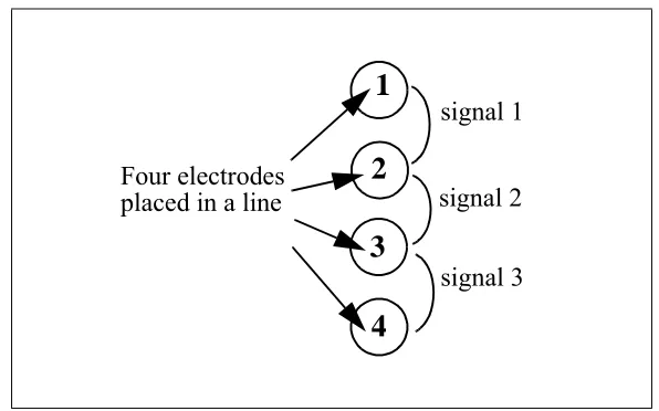
A method for identification of sEMG signal characteristics associated with elec-trodes located over an innervation site was described by Queisser, Blüthner, Bräuer & Seidel (1994). Queisser et al. (1994) investigated the location of electrode pair placement with respect to innervation zones of four muscles, including the semispinalis capitis and splenius capitis. To collect sEMG data in adjacent muscle areas, an electrode was placed on either side of the original electrode pair, such that there were four electrodes in a straight line. The criteria used for determining that the primary electrode pair was over an innervation zone were based on visual inspection of the three sEMG signals obtained from the four electrodes. Signals 1 and 3 were from the outer pairs of electrodes while signal 2 was from the original, central electrode pair (Figure 3).
Note how the electrode pair in the drawing on the left is located over the innervation site and will be collecting electrical activity from action potentials (represented by the arrows) moving in opposite directions. The drawing on right depicts an electrode pair location in which the action potential will move under the pick-up area of the first electrode in the pair and then the second.
When the signals were visually inspected from a two second test contraction at a 30% maximum voluntary contraction, a pronounced reduction of the amplitude from sig-nal 2 and a phase shift of nearly 180° in signals 1 and 3 were taken as indications of loca-tion over an innervaloca-tion site. These authors noted that the original electrode pair was located over the innervation zone for 3 of the 12 subjects for the semispinalis capitis and 5 of the 12 for the splenius capitis. They commented on the lack of data published about innervation zones in the neck musculature and the potential for changes from skin move-ment and muscle tension offsetting each other. Skin movemove-ment is discussed later in this section.
The second factor affecting the myoelectric signal collected by surface electrodes is crosstalk. Crosstalk is a term used to refer to the sEMG signal that contains activity from nearby muscles, as well as the one of interest. Data containing crosstalk requires careful interpretation; results from such signals can be misleading. At best EMG data con-taining crosstalk artifact reflects activity from a group of muscles working synergistically and at worst contains both agonist and antagonist activity. Accuracy of electrode place-ment and interelectrode distance are the two primary eleplace-ments that will influence the amount of crosstalk that will be collected in an sEMG signal. Accuracy of the electrode location refers to the degree of certainty regarding electrodes actually being placed over the muscle of interest. An accurate electrode location is critical when working with small
Figure 3: Four electrodes used to create three sEMG signals
signal 1
signal 2
signal 3 Four electrodes
placed in a line
1
2
3
niques are necessary for maintaining accuracy levels between experimental sessions as well as for performing validation studies. The specific location of electrode pairs has been documented to varying degrees of precision in the literature. Good documentation efforts provide the distance from a bony landmark to one or both electrodes and the interelectrode distance. The most precise descriptions also include the distance from the midline of the body.
sEMG was much larger than the intramuscular EMG area. Since the subject’s head was stationary with weights hung from different angles off the heads, the difference in muscle activation cannot be attributed to movement of the skin relative to the muscle. In the case of the splenius, it would seem that crosstalk could explain the increased activation area.
It has been suggested that surface electrodes should not be used when studying the splenius capitis (Mayoux-Benhamou, Revel & Vallee, 1995). Using computerized tomog-raphy (CT) scans to identify sampling locations in order to compare surface and intramus-cular EMG recordings from the splenius capitis, Mayoux-Benhamou et al. (1995) state that surface electrodes for the splenius collect crosstalk from the sternocleidomastiod. Although using CT scans to locate the electrodes over the splenius is an accurate approach, two aspects of the study’s methods weaken the argument for eliminating the use of sEMG when studying the splenius. First, the findings are based on the data from only one subject. Second, the location of the surface electrodes for the splenius were close to the sternocleidomastoid. Since the location chosen in the study was not identified as the only appropriate location, other positions along the splenius capitis may also be “open”, as seen using CT scans, but further from the sternocleidomastoid. It should also be noted that two of the five exertions in which there was evidence of crosstalk for the single sub-ject were not typical of working postures. Those exertions include flexing and extending the neck when the head was extended past a neutral position. Mayoux-Benhamou et al. (1995) also note that “the discrepancies were slight during slow movements” and in iso-metric testing the crosstalk from the sternocleidomastoid only “became obvious when this muscle was highly recruited.” There are few working postures in which the sternocleido-mastoid is highly recruited; therefore, this crosstalk issue may be more relevant to maxi-mum exertions for normalization purposes.
distances were documented. In order to compare these studies’ methods with others’, researchers need to document which edges on the electrode are being referenced (i.e., superior edges, inferior edges, edges closest together, or outside edges) and size of the electrode so that the center-to-center interelectrode distance can be calculated. Even though the interelectrode descriptions are important, 65% and 60% of the experiments reviewed for this research which collected neck extensor and splenius activity data, respectively, failed to document the interelectrode distance. Where documentation is pro-vided, differences in choices for placement and interelectrode distances were seen between studies, but the reasons for those choices are rarely provided. Also rarely addressed are issues pertaining to signal reliability. Only one study mentioned testing for innervation zones or ‘dead spots’ (Queisser et al., 1994).
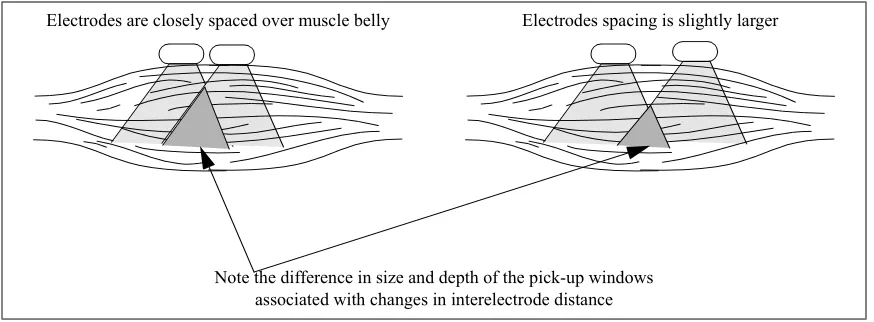
The third factor affecting the quality or accuracy of the signal being collected is the movement of the electrodes away from the muscle of interest. This movement of the electrodes impacts the ability to collect sEMG from the same muscle location throughout the experiment. In an investigation on arm position and trapezius activity, Mathiassen and Winkel (1990) emphasize that the changes in arm position inevitably change the sample of motor units recorded by surface electrodes, due to sliding of the skin over the muscle sur-face and changes in the shape of the underlying muscle belly. Although Mathiassen and Winkel (1990) referred to issues associated with arm position, these issues can be trans-lated to corresponding problems associated with sEMG data collection and changes in head posture including tilt, translation, and rotation.
Electrodes are closely spaced over muscle belly Electrodes spacing is slightly larger
Note the difference in size and depth of the pick-up windows associated with changes in interelectrode distance
2.3.1.1 Electrode location in sEMG studies of the primary neck extensors
Both muscle terminology and electrode location vary among sEMG studies of the posterior neck musculature. This inconsistency can make both the study interpretation and comparisons with other studies difficult. Twenty-three studies have reported collect-ing data from this region uscollect-ing surface electrodes (Table 3). Thirteen of the twenty-three studies investigated the external influences on the subjects; ten investigated theoretical issues. The studies which addressed the external environment on muscle activity included the impact of ergonomic aids (Schüldt, Ekholm, Harms-Ringdahl, Németh & Arborelius, 1987b), keyboard styles (Fernstrom, Ericson & Malker, 1994), interaction of cycle time and dynamic movements (Sundelin & Hagberg, 1992), break type (Sundelin & Hagberg, 1989), and workstation arrangement (Bauer & Wittig, 1998; Hamilton, 1996; Kumar & Scaife, 1979; Lannersten & Harms-Ringdahl, 1990; Saito, Miyao, Kondo, Sakakibara & Toyoshima, 1997; Sommerich, Joines & Psihogios, 2001; Turville, Psihogios, Ulmer & Mirka, 1998; Villanueva, Jonai & Saito, 1998; Villanueva, Jonai, Sotoyama, Hisanaga, Takeuchi & Saito, 1997). The theoretical studies included those measuring muscular response to loading of the head in any single direction (Keshner et al., 1989), investigating which test contractions maximally activated neck and shoulder muscles (Schüldt & Harms-Ringdahl, 1988a), investigating the EMG-torque relationship of neck muscles and force and cervical spine position (Queisser et al., 1994), the influence of neck posture on muscle activity (Harms-Ringdahl, Ekholm, Schüldt, Nemeth & Arborelius, 1986; Schüldt & Harms-Ringdahl, 1988b; Schüldt & Harms-Ringdahl, 1988c), the influence of whole trunk posture on neck muscle activity (Schüldt, Ekholm, Harms-Ringdahl, Arborelius & Németh, 1987a; Schüldt, Ekholm, Harms-Ringdahl, Nemeth & Arborelius, 1986), fatigu-ability of cervical paraspinal muscles (Gogia & Sabbahi, 1990), and the influence of arm posture on neck muscle activity (Mathiassen and Winkel, 1990).
muscles sampled (including CES) as “over the belly of the muscle or where it was most superficial.”
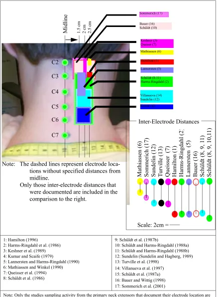
The other studies documented five distinct electrode locations (Figure 5): at the C1/C2 level (Keshner et al., 1989; Mathiassen and Winkel, 1990; Queisser et al., 1994), at the C2 level (Hamilton, 1996), at the C2-C3 level (Bauer and Wittig, ; Lannersten and Ringdahl, 1990; Schüldt et al., 1986; Schüldt et al., 1987b; Schüldt and Harms-Ringdahl, 1988a; Sommerich et al., 2001, Schüldt, 1988b; Sundelin and Hagberg, 1989; Villanueva et al., 1997), between C2 and C6 (Harms-Ringdahl et al., 1986; Schüldt et al., 1987a; Schüldt et al., 1987b), and at the C5/C6 level (Kumar and Scaife, 1979).
The studies examining the primary neck extensors use five names to refer to mus-cles sampled in primarily the same location: one refers to cervical paraspinal musmus-cles, four refer to neck extensors, two refer to semispinalis capitis, three refer to the trapezius pars descendens in the cervical region, and thirteen refer to cervical erector spinae. Figure 5 depicts the studies grouped by the label used to identify the muscle of interest and the doc-umented location of the electrodes. There is little difference in the coverage when com-paring the lower two blocks in Figure 6: trapezius pars descendens and cervical erector spinae. Although the area is more limited in the upper left block for the “neck extensors” that area is inconsistent between the studies and still covers areas sampled for the trape-zius. Although the two studies reporting sEMG data from the semispinalis are consistent and restricted, suggesting good precision, it is evident that seven or more studies may have sampled the same location.
1: Hamilton (1996)
2: Harms-Ringdahl et al. (1986) 3: Keshner et al. (1989) 4: Kumar and Scaife (1979)
5: Lannersten and Harms-Ringdahl (1990) 6: Mathiassen and Winkel (1990) 7: Queisser et al. (1994) 8: Schüldt et al. (1986)
9: Schüldt et al. (1987b)
10: Schüldt and Harms-Ringdahl (1988a) 11: Schüldt and Harms-Ringdahl (1988b) 12: Sundelin (Sundelin and Hagberg, 1989) 13: Turville et al. (1998)
14: Villanueva et al. (1997) 15: Schüldt et al. (1987a) 16: Bauer and Wittig (1998) 17: Sommerich et al. (2001)
Note: Only the studies sampling activity from the primary neck extensors that document their electrode location are included in this figure.
Figure 5: Location of Superior Electrode for Primary Neck Extensors
C2 C3 C4 C5 C6 C7 Keshner (3) Queiser (7) Mathiassen (6) Hamilton (1) Lannersten (5) Schüldt (8-11) Harms-Ringdahl (2) Villanueva (14) Sundelin (12) Kumar (4) Mathiassen ( 6) Som m er ich (17)
Sundelin (12) Tur
vill
e (
13)
Que
isse
r (7)
Ha m ilton (1) Ha rms-Ri ngda hl ( 2) La
nnersten (5)
Baue
r (16)
Schüldt (8, 9, 1
1)
Schüldt (8, 9, 10,1
1)
Note: The dashed lines represent electrode loca-tions without specified distances from midline.
Only those inter-electrode distances that were documented are included in the comparison to the right.
Scale: 2cm =
Mid
line
1.
5 cm
2 cm 2.5 cm
Bauer (16) Schüldt (10)
Inter-Electrode Distances
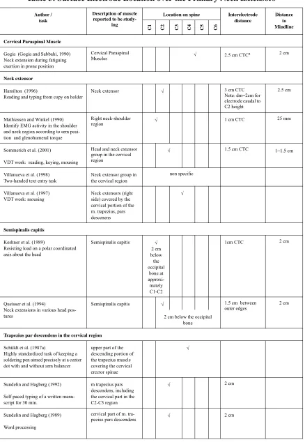
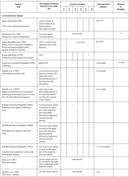
Table 3: Surface Electrode Location over the Primary Neck Extensors
Author / task
Description of muscle reported to be
study-ing
Location on spine Interelectrode distance
Distance to Mindline
C1 C2 C3 C4 C5 C6
Cervical Paraspinal Muscle
Gogia (Gogia and Sabbahi, 1990) Neck extension during fatiguing exertion in prone position
Cervical Paraspinal
Muscles √ 2.5 cm CTCa 2 cm
Neck extensor
Hamilton (1996)
Reading and typing from copy on holder
Neck extensor √ 3 cm CTC
Note: dm=2cm for electrode caudal to C2 height
2.5 cm
Mathiassen and Winkel (1990) Identify EMG activity in the shoulder and neck region according to arm posi-tion and glenohumeral torque
Right neck-shoulder
region √ 1 cm CTC 25 mm
Sommerich et al. (2001)
VDT work: reading, keying, mousing
Head and neck extensor group in the cervical region
√ 1.5 cm CTC 1−1.5 cm
Villanueva et al. (1998) Two-handed text entry task
Neck extensor group in the cervical region
non specific
Villanueva et al. (1997) VDT work: mousing
Neck extensors (right side) covered by the cervical portion of the m. trapezius, pars descenens
√
Semispinalis capitis
Keshner et al. (1989)
Resisting load on a polar coordinated axis about the head
Semispinalis capitis √
2 cm below the occipital bone at approxi-mately C1-C2
1cm CTC 2 cm
Queisser et al. (1994)
Neck extensions in various head pos-tures
Semispinalis capitis √ 1.5 cm between
outer edges 2 cm
2 cm below the occipital bone
Trapezius par descendens in the cervical region
Schüldt et al. (1987a)
Highly standardized task of keeping a soldering pen aimed precisely at a center dot with and without arm balancer
upper part of the descending portion of the trapezius muscle covering the cervical erector spinae
√
Sundelin and Hagberg (1992) Self paced typing of a written manu-script for 30 min.
m trapezius pars descendens, including the cervical part in the C2-C3 region
√ 2 cm
Sundelin and Hagberg (1989) Word processing
cervical part of m.
Cervical Erector Spinae
Bauer and Wittig (1998) VDT work: reading and mousing
where cervical m. erector spinae & m. trapezius pars descended overloap
√ 3cm CTC
Fernstrom et al. (1994) Typing on a variety of keyboards
M erector spinae cervicalis sinister
non specific √
Harms-Ringdahl et al. (1986) Sitting with cervical spine straight or flexed and thoraco-lumbar spine upright, inclined or reclined
CES covered by the uppermost part of the trapezius
√ (C2-C6) 3cm CTC
Kumar and Scaife (1979)
Detailed assembly using a microscope
CES √
Lannersten and Harms-Ringdahl (1990) Cashiering
Right CES √ 3 cm caudal 1.5-2cm
Schüldt et al. (1986) Seated light assembly work
over the cervical erector spinae covered by the upper part of the descending portion of the trapezius, pars descendens
√ 3 cm caudal
Schüldt et al. (1987b)
Highly standardized task of keeping a soldering pen aimed precisely at a center dot with and without arm balancer
upper part of the descending portion of the trapezius muscle covering the cervical erector spinae
√ 3 cm caudal
Schüldt and Harms-Ringdahl (1988c) Extending neck against a stationary sling
Cervical erector spinae covered by the upper part of the trapezius pars descendens referring to deep neck muscles plus the splenius
√
Schüldt and Harms-Ringdahl (1988b) Extending neck against a stationary sling
Cervical erector spinae covered by the upper part of the trapezius pars descendens referring to deep neck muscles plus the splenius
√
Schüldt and Harms-Ringdahl (1988a) Isometric test contractions of the neck and shoulder muscles
Cervical erector spinae covered by the upper part of the trapezius pars descendens
√ 3- 3.5 cm caudal
Saito et al. (1997) Word processing
erector spinae cervica-lis covered by the right side of upper part of the trapezius muscle
non specific
Turville et al. (1998) VDT work: reading
cervical erector spinae non specific 2 cm
a. CTC: center to center
Table 3: Surface Electrode Location over the Primary Neck Extensors
Author / task
Description of muscle reported to be
study-ing
Location on spine Interelectrode distance
Distance to Mindline
“Neck Extensors” Semispinalis capitis
Trapezius pars descendens “in the cervical region”
Cervical Erector Spinae
1: Hamilton (1996)
2: Harms-Ringdahl et al. (1986) 3: Keshner et al. (1989) 4: Kumar and Scaife (1979)
5: Lannersten and Harms-Ringdahl (1990)
6: Mathiassen and Winkel (1990) referred to “neck” pair of the Upper Trapezius 7: Queisser et al. (1994)
8: Schüldt et al. (1986)
9: Schüldt (1987b)
10: Schüldt and Harms-Ringdahl (1987b) 11: Schüldt and Harms-Ringdahl (1988b) 12: Sundelin and Hagberg (1989) 13: Turville et al. (1998) 14: Villanueva et al. (1997) 15: Schüldt et al. (1987a) 16: Bauer and Wittig (1998) 17: Sommerich et al. (2001)
Figure 6: Location of Superior Electrode for Neck Extensors Muscles C2 C3 C4 C5 C6 C7 Mathiassen (6) Hamilton (1) Villanueva (14) Sommerich (17) C2 C3 C4 C5 C6 C7 Keshner (3) Queiser (7) C2 C3 C4 C5 C6 C7 Schüldt(15) Sundelin (12) C2 C3 C4 C5 C6 C7 Lannersten (5)
Schüldt (8, 9, 11) Kumar (4) Bauer (16) Schüldt (10)
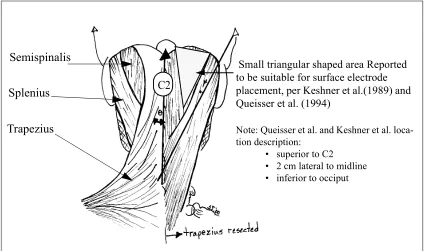
Identifying the proximity of the electrode pair to the midline is an important factor for the semispinalis for two reasons. First, the dimension aids in electrode location repeat-ability. Second, but of equal importance, the distance from the midline is also an impor-tant factor in reducing crosstalk from the splenius, as illustrated in Figure 7. Keshner et al. (1989) commented on the sparse number of fibers in the trapezius above C4, suggesting that crosstalk from the trapezius would not be a problem if the electrode pair was located superior to C4. However, the recommendation is further restricted by the proximity of the splenius (Keshner et al., 1989; Queisser et al., 1994). Hence, the description of an area (Keshner et al., 1989; Queisser et al., 1994) reported to be suitable for electrode location to minimize crosstalk is small. From Figure 7 it is evident that precise location of the electrode is necessary which includes the documentation of the distance from the midline.
Figure 7: Small Area Reported to be Suitable for Electrode Placement for Semispinalis Capitis
Small triangular shaped area Reported to be suitable for surface electrode placement, per Keshner et al.(1989) and Queisser et al. (1994)
Note: Queisser et al. and Keshner et al. loca-tion descriploca-tion:
• superior to C2 • 2 cm lateral to midline • inferior to occiput
Semispinalis
Splenius
Trapezius
2.3.1.2 Electrode location in EMG studies of splenius capitis
The previous section focused on the primary neck extensors; however, the splenius is both a neck extensor and rotator. The splenius originates from the spinous processes of C5-C7 and inserts on the occipital bone and the mastoid process. The splenius is superfi-cial to the primary neck extensors near the spinal column. In order to minimize crosstalk in the splenius signal, the splenius electrode location must be lateral to the primary neck extensors. Only nine studies (two applied, and seven lab-based) were found containing data collected from the splenius muscle using surface electrodes (Table 4). Two studies of the external environment examined problems in dentistry (Finsen, 1999; Finsen et al., 1997). The theoretical studies included investigations into pain and muscle activity (Bansevicius, Westgaard & Jensen, 1997), the effects of optical correction on sEMG activity (Lie & Watten, 1987), muscle coactivity during single direction head loading (Keshner et al., 1989), sEMG torque relationships of neck extensors in various postures of the cervical spine (Queisser et al., 1994), and the methods for maximal muscle activation (Schüldt and Harms-Ringdahl, 1988a; Schüldt and Harms-Ringdahl, 1988b; Schüldt and Harms-Ringdahl, 1988c).
No specific electrode locations were provided in two studies. Lie and Watten (1987) described the electrode pair location as recording activity “from the muscle in the upper neck” including the splenius. In the other studies three distinct electrode locations were documented at the C2 level (Bansevicius et al., 1997), at the C2-C3 level (Finsen, 1999; Finsen et al., 1997; Schüldt and Ringdahl, 1988a; Schüldt and Harms-Ringdahl, 1988b; Schüldt and Harms-Harms-Ringdahl, 1988c), and at the C4 level (Keshner et al., 1989; Queisser et al., 1994). Where electrode descriptions were provided, three dif-ferent interelectrode distances were reported. Figure 8 identifies the range over which the splenius has been documented to have been sampled. Given the parameters documented in two of these studies (Keshner et al., 1989; Queisser et al., 1994), for subjects with small neck length and circumference as shown in Figure 8, the electrode pair would not be posi-tioned over the splenius.
The second electrode was located 20 mm directly below the first. Schüldt and Harms-Ringdahl (1988a, 1988c) defined the splenius electrode location at the C2 (C3) level between the uppermost parts of the trapezius and sternocleidomastoid. Keshner et al. (1989) located the splenius electrode pair “by measuring 6 cm rostral to the bony promi-nence at C7 (approximately the C4 level), 6-8 cm lateral, and palpating for the muscle belly.” The muscle was palpated between the sternocleidomastoid and the trapezius while resisting “head extension and lateral rotation in the same direction as the muscle.” Figure 8 and Table 4 clearly indicate that the studies are inconsistent in at least their docu-mentation and probably their position along the length of the muscle and the depth of the muscle being sampled. Again, a correct and well documented method would provide a common ground for locating electrodes over the splenius.
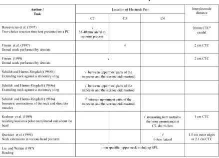
Table 4: Electrode Location over the Splenius
Author / Task
Location of Electrode Pair Interelectrode distance
C2 C3 C4
Bansevicius et al. (1997)
Two-choice reaction time test presented on a PC 35-40 mm lateral to √ spinous process
20mm CTCa
caudal
a. CTC: center to center Finsen et al. (1997)
Dental work performed by dentists √
2 cm CTC
Finsen (1999)
Dental work performed by dentists √
2 cm CTC
Schüldt and Harms-Ringdahl (1988b)
Extending neck against a stationary sling trapezius and the sternocleidomastoid√ between uppermost parts of the Schüldt and Harms-Ringdahl (1988c)
Extending neck against a stationary sling trapezius and the sternocleidomastoid√ between uppermost parts of the Schüldt and Harms-Ringdahl (1988a)
Isometric contractions of the neck and shoulder muscles
√ between uppermost parts of the trapezius and the sternocleidomastoid
Keshner et al. (1989)
resisting load on a polar coordinated axis about the head
√ measuring 6cm rostral to the bony prominance at
C7, dm=6-8cm
1 cm CTC
Queisser et al. (1994)
Neck extensions in various head postures 6-8cm lateral√
1.5 cm outer edges or 2.1 cm CTC Lie and Watten (1987)
Reading
1: Bansevicius et al. (1997) 2: Bauer and Wittig (1998)
3: Finsen (1999); Finsen et al. (1997) 4: Keshner et al. (1989)
5: Queisser et al. (1994)
6: Schüldt et al. (1988a, 1988b, 1988c)
*Note: this location is so far from the mid-line that it would wrap around the side of the subject’s neck which would not be clearly visible in this figure.
Figure 8: Location of Superior Electrode in Pair for Splenius
C2 C3 C4 C5 C6 C7
Bansevicius (1)
Finsen (3) Bauer (2) Schüldt (6)
Keshner (4)* Queisser (5)*
Keshner
(4)
Fi
nsen (3)
B
ans
evic
ius (
1)
Queisser (
5)
B
auer (
2)
Note: Dashed lines represent locations without speci-fied distances from midline
Inter-electrode distances not depicted were not
specified Scale: 2cm =
Mi
dline
2 cm 3 cm 4 cm 6 cm
6 cm
2.3.1.3 Repeatability
The previous sections addressed the need for a consistent methodology for elec-trode location in terms of accurate placement over the muscle of interest and the quality of the signal (i.e., not locating over an innervation site, minimizing crosstalk, etc.). This sec-tion will demonstrate the need for considering repeatability of electrode placement when collecting sEMG in the cervical spine. Repeatability needs to be addressed in terms of the experimenter’s ability to locate the electrode on the same site during different data collec-tion sessions (inter-session repeatability) and the ability of different experimenters to place electrodes in a consistent manner (inter-experimenter repeatability). Inter-session repeatability is extremely important for maintaining consistent conditions for experiments that must be run over several days. Inter-experimenter repeatability is important for experiments run by several experimenters, or when trying to replicate previous research. Documentation of the repeatability associated with electrode placement is limited. In a study of reproducibility of test contractions, Veiersted (1991) evaluated the reproduc-ibility of EMG measurements and electrode placement. Electrodes were positioned over the trapezius halfway between two bony landmarks and repositioned using a tracing paper map of the subject. Bony landmarks and skin characteristics such as birthmarks were marked on the paper map. Electrodes were repositioned to within +/- 2.5 mm longitudi-nally and +/- 1.5 mm transversely from their original location with a probability of 90%. These locations were verified by using transparent plaster corresponding to three fixed points on the skin. One aspect of this study included moving the electrode location longi-tudinally and transversely over the trapezius muscle in 3 mm increments. The results of the electrode position test (Veiersted, 1991) confirmed the findings of Vigreux (1979) that EMG amplitude can be greatly influenced by small changes in electrode position.
(see Figure 9) improved after removing the data collected by the electrodes at the level of C1/C2 and 25 mm from the spinal column from the analysis. The unilateral variable (see Figure 9) reached the statistical significance level of p<0.01 after removing the data col-lected by the electrodes at the level of C1/C2. Thus the variation associated with the data from the electrode pair located at the level of C1/C2 was high. This variation may have been a function of inconsistent electrode placement in the cervical region, changes in the portion of the muscle being sampled, or changes in antagonistic muscle activity levels.
A method for describing and/or marking the electrode location would improve repeatability, removing some of the subjective interpretation currently necessary when placing electrodes.
2.3.2 Normalization
LeVeau (1992) defined the goal of collecting sEMG data to be the comparison of muscle activity, individuals, and tasks — not the quantification of the myoelectric signal. The quantification and processing of the sEMG provides an indirect measure of the mus-cle contraction force necessary to perform an activity of interest. Unprocessed sEMG data (in microvolts) are influenced by electrode location, changes in tissue properties, and tem-perature (LeVeau, 1992). Since the relationship between muscle contraction force and EMG activity are influenced by these other factors, a standard reference value (referred to as normalizing data) is useful in comparing results. Several different methods of normal-ization appear in the literature:
• isometric maximal voluntary contractions (% of MVC); • submaximal isometric contractions (% of subMAX);
Rbil=EMGB ---EMG R
Runi=EMG---L EMGR
EMGB: EMG activity collected while torque was generated with both arms
EMGR: EMG activity collected while torque was generated with the right arm
EMGL: EMG activity collected while torque was generated with the left arm
• reference task (% of RT);
• reference contractions (% of RC); • isotonic contractions (% of IC); and
• normalization of task in terms of time (% of ITT).
For all the papers relating to sEMG activity in the neck, no studies used the isotonic con-tractions or normalization of task in terms of time. One study normalized the data to a ref-erence task of “sitting relaxed” which could be viewed as a submaximal isometric contraction; however, no level of exertion was defined (Wells, Norman, Shannon, Cole, Woo & Bao, 1998b). One study utilized reference contractions to normalize their data (Mathiassen and Winkel, 1990), while another used levels obtained during maximum exertions (Turville et al., 1998).
In several studies, data were normalized by selecting a maximum value for each muscle (MVC) from a series of pre and post experiment test contractions (Harms-Ringdahl & Ekholm, 1986; Lannersten and Harms-(Harms-Ringdahl, 1990). The maximum value for each muscle was used as a reference value. The level of muscle activity during the experiment was divided by the reference level for the specific muscle. The result is expressed as a percentage of the time-averaged myoelectric potential (%TAMP) during the reference contraction or normalized sEMG level (Ekholm, Arborelius, Fahlacrantz, Larsson & Mattsson, 1979; Jonai, Villanueva, Sotoyama, Hisanaga & Saito, 1997; Lan-nersten and Harms-Ringdahl, 1990; Villanueva et al., 1998; Villanueva et al., 1997). The methods of normalization used for the primary neck extensors and splenius are summa-rized in Table 5 and Table 6, respectively.
2.3.2.1 Posture during exertions for normalization purposes
The head and neck posture maintained during test contractions is of concern for several reasons. In other parts of the body the posture during maximal exertions has been
Table 5: Methods of Normalization Used with Primary Neck Extensors
Studies Normalization Methods
Not Normalized %MVC %sub MAX %RT
Hamilton (1996), Harms-Ringdahl et al. (1986)a, Keshner et al.
(1989)b, Kumar and Scaife (1979), Lannersten and Harms-Ringdahl
(1990),c Schüldt et al. (1986, 1987b), Schüldt and Harms-Ringdahl
(1988c)d, Schüldt and Harms-Ringdahl (1988a), Sommerich et al.
(2001), Villanueva et al. (1997, 1998), and Wells et al. (1998)
X
Turville et al. (1998),e X X
Queisser et al (1994) and Sundelin and Hagberg (1989) X X
Wells et al. (1998) X X (50%)
Mathiassen and Winkel (1990)f X (60%)
Saito et al. (1997) reports all results in non normalized, arbitrary
units which may have corresponded to micro-Volts
?
a. Harms-Ringdahl et al. (1986) described the posture in which maximal exertions (against manual resistance) were per-formed as a neutral posture
b. Keshner et al. (1989) also collected all data with the head and neck in a neutral posture.
c. Lannersten and Harms-Ringdahl (1990) referred to Schüldt and Harms-Ringdahl (Schüldt and Harms-Ringdahl 1988) when documenting their data collection
d. Schüldt and Harms-Ringdahl (1988c) referred to the positioning during maxes as an upright posture. e. Turville et al. (1998) referred to Schüldt (1988) when documenting their data collection
f. Mathiassen and Winkel (1990) reported data as a percentage of a submaximal exertion (60%MVC)
Table 6: Methods of Normalization Used with Splenius
Studies Not Normalized %MVC %sub MAX
Bansevicius et al. (1997) and Queisser et al. (1994)a X X
Finsen et al. (1997); Finsen (1999), (Schüldt and
Harms-Ringdahl 1988a, 1988c), and Keshner et al. (1989)b
X
Hautekiet et al. (1997)c X (resting)
a. Queisser et al. (1994) collected MVCs in a prone position which was the posture of interest or working posture. b. all maxes were collected in the upright posture
data to a maximal exertion performed in a posture different from the posture in which data were sampled, questions arise as to the capability of the person to exert a similar maxi-mum in the sampled posture. Therefore, normalization effectively scales the data to a value that is potentially different than the individual’s capability in that posture. Also, the portion of the muscle being sampled may have changed owing to the skin (and electrode) movement over the muscle, causing the sEMG signal to be normalized by data from a dif-ferent portion of the muscle.
The postures typically assumed during maximum exertions of neck muscles are based on a study by Schüldt and Harms-Ringdahl (1988b). For several muscles, Schüldt and Harms-Ringdahl (1988b) sought to identify the postures where consistent maximal exertions could be performed. MVC exertions were collected with the trunk stabilized in a sitting position while the subject performed various isometric exertions against resistance. Based on their results, Schüldt and Harms-Ringdahl (1988b) recommended a reduced set of test contractions designed to maximally activate the muscles of interest, making studies more manageable and reducing the effect of fatigue. However, the recommendations did not take into account the issues surrounding normalization of sEMG data in the posture in which the data for the task of interest were collected.
Schüldt and Harms-Ringdahl (1988b) found that three test contractions consis-tently provided consistent maximum activation for the right splenius, levator scapulae, sternocleidomastoid, and erector spinae. The three contractions are lateral flexion of head and cervical spine to the right with resistance against the temple, pushing head and upper cervical spine forward with resistance against the forehead, and extension of cervical spine with resistance at occiput and the chest stabilized. Schüldt and Harms-Ringdahl (1988b) found that the position of the lower-cervical-upper-thoracic spine impacted the level of activation during isometric neck extensions. Specifically, peak activity levels in the cervical erector spinae were obtained in a slightly flexed position. However, the upper-cervical spine position did not influence the level of activation during isometric maximum neck extensions. Several other test contractions were studied by Schüldt but were shown to be not as effective (Schüldt and Harms-Ringdahl, 1988b).