ORIGINAL ARTICLE Iran J Allergy Asthma Immunol December 2019; 18(6):649-654.
A Randomized Control Trial Study to Determine the Effect of Melatonin on
Serum Levels of IL-1β and TNF-α in Patients with Multiple Sclerosis
Masoomeh Yosefifard1, Gholamhassan Vaezi2, Ali Akbar Malekirad3,4, Fardin Faraji5, and Vida Hojati1
1
Department of Biology, Damghan Branch, Islamic Azad University, Damghan, Iran
2
Department of Biology, Science and Research Branch, Islamic Azad University, Tehran, Iran
3
Department of Biology, Payame Noor University, Tehran, Iran
4
Toxicology and Diseases Group, Pharmaceutical Science Research Center, Tehran University of Medical Sciences, Tehran, Iran
5
Department of Neurology, School of Medicine, Arak University of Medical Sciences, Arak, Iran
Received: 8 April 2019; Received in revised form: 15 May 2019 ; Accepted: 20 May 2019
ABSTRACT
Multiple sclerosis (MS) is the most common neurological disease that happens at a young
age. MS is an inflammatory disease; associated with the demyelination of the central nervous
system. Therefore, some inflammatory factors are effective in the mechanism and
progression of the disease. Melatonin, as a multi-effect substance including
anti-inflammatory effects, can reduce symptoms of MS in patients with a change in their
inflammatory factors level.
In this study, 50 MS patients who were referred to the MS Society of Markazi Province
were randomly selected. All patients were treated with routine MS treatment (interferon) and
were divided into control (25 placebo recipients) and treatment (25 recipients of 3 mg
melatonin per day for 24 weeks) groups. Anthropometric data of patients including height,
weight, and age were determined. Blood samples were collected after fasting in order to
determine serum levels of interleukin 1 beta (IL-1β) and tumor necrosis factor-alpha
(TNF-α). Then, samples were immediately centrifuged for serum separation and sera were
transferred to a freezer at -80°C and serum levels of these factors were determined; using
ELISA kit.
The results of this study showed that there was no significant difference between the
control and treatment groups in terms of serum levels of TNF-α. However, the level of
IL-1β was significantly reduced in the treatment group compared to the control group,
indicating that melatonin decreases this inflammatory substance.
Our findings suggest a valuable strategy in the treatment of patients who suffer from MS.
Keywords
: Interleukin 1 beta; Multiple sclerosis; Melatonin; Tumor necrosis
factor-alpha
Corresponding Authors: Gholamhassan Vaezi, PhD;
Department of Biology, Science and Research Branch, Islamic Azad University, P.O.Box 14515/775, Tehran, Iran. E-mail: [email protected]
Ali Akbar Malekirad, PhD;
INTRODUCTION
Multiple sclerosis (MS ( is one of the most common neurological diseases that can happen at a young age, commonly seen in people aged 15-50 years old. Although reported in some children, the incidence is not common in this age group. The highest known incidence of MS (250 cases per 100,000 people) is in northern Europe; while its incidence in the northern regions of the United States and Canada is relatively high. In contrast, its incidence in Japan (6 cases per 100,000 people), other parts of Asia, tropical Africa, and the Middle East is low. The possible justification of the latitude effect on the incidence of MS can be exposed to sunlight and its protective effects. Some infectious agents also contribute to the onset of MS. Human herpesvirus 6 (HHV6) and Chlamydia pneumonia can cause the onset of MS in some people. In addition, genetics is also a major contributor to MS. White people are inherently more susceptible to MS than Africans or Asians, even when residing in the same environment. The genetic locus of major histocompatibility complex (MHS) on chromosome 6 is a good candidate for determining the susceptibility to MS.1,2 MS is an inflammatory disease associated with the demyelination process in the central nervous system. Therefore, some inflammatory factors are effective in the mechanism and progression of the disease. One of the most important inflammatory factors in this field is tumor necrosis factor-alpha (TNF-α) which plays an important role in the damage to oligodendrocytes and myelin as well as the progression of the disease.3,4 TNF-α is known as a pro-inflammatory factor that interferes with the immune response.5,6 Responses of this factor in signaling pathways are involved in myelination, apoptosis, and inflammation7,8 Pathogenic functions of this factor include triggering inflammation, stimulation of vascular endothelium, immune system proliferation, and tissue damage.9,10 Interleukin-1 (IL-1) is one of the major pro-inflammatory cytokines and is involved in the treatment of many inflammatory diseases. According to studies in animals model with MS, the role of this factor has been clarified.11,12 IL-1 is expressed in peripheral tissue monocytes and macrophages and is also observed in most of the brain cells including neurons, astrocytes, oligodendrocytes, and endothelial cells. In addition, active microglia and invasive macrophages may be an important source of
IL-1; especially after damage to the brain or failure of the blood-brain barrier.13 The expression of interleukin-1 beta (IL-interleukin-1β) in the brain is low; while it increases significantly after the damage. Controversial reports indicate that this factor is expressed in the brain cortex, hippocampus, and basal.14
Melatonin or 5-methoxy-n-acetyl-tryptamine is a hormone secreted from the pineal gland and is synthesized from the neurotransmitter serotonin. Melatonin receptors in the brain can be seen in the prefrontal cortex, the cerebellum, the hippocampus, the basal ganglia, substantia nigra, nucleus accumbens, the retina, and also in different parts of the hypothalamus. In addition, these receptors are visible in peripheral tissues such as the digestive tract, adipose tissue, pancreas, ovaries, skin, lungs, heart, and lymphocytes.15,16 This hormone can be effective in regulating cytokines such as TNF-α,
interleukin-8
(IL-8),
interleukin 6
(IL-6), and other inflammatoryparameters.17,18 The performance of the matrix metalloproteinases which is involved in the damage to the brain and spinal cord as the secondary pathogenesis by the destruction of the blood-brain barrier and the development of inflammation is also regulated by melatonin. It reduces the activity of these enzymes and subsequently suppresses the production of certain inflammatory factors such as TNF-α.19,20 Researches published in recent years has suggested melatonin as a therapeutic supplement in many neurodegenerative diseases.17 In patients with MS, melatonin levels are reduced due to the deposition of calcium in the pineal gland and hypothalamic dysfunction.21,22 The melatonin plays a moderating role in the mechanisms of signal transfer by changing the ion flow and various ion channels. On the other hand, melatonin plays a protective role in the maturation and treatment of damaged white matter (myelinated pathways) in the rat model. Melatonin reduces the microglial activity and the oligodendroglia maturation, thereby it helps myelination and treatment of the axon.23,24 In this study, the serum levels of IL-1β and TNF-α were investigated in patients with MS who were treated with melatonin and placebo.
MATERIALS AND METHODS
approved at the ethics committee of Arak Medical University (IR.ARAKMU.REC.1394.9). The target population of this study was 50 patients with MS who were referred to the Arak MS society and Vali-Asr Hospital in Arak from April 2015 to August 2017. The sample size is calculated by the Cochran formula. Patients were selected based on inclusion criteria (patients with relapsing-remitting MS based on McDonald 2 criteria, individuals aged 18-50 years, those with an Expanded Disability Status Scale (EDSS) between 0-5.5, completion of the testimonial, and willingness and ability to cooperate in the study). Exclusion criteria included being affected with autoimmune disease and the use of non-interferon medications, vitamin B12, and alcohol. Consent letters have been received from all of the patients. All patients were treated with routine MS treatment (interferon) and were randomly divided into control (25 placebo recipients) and treatment (25 recipients of 3 mg melatonin per day for 24 weeks) groups. The blood samples of both groups were collected by a nurse in Venoject tubes in sterile conditions. Then, samples were immediately centrifuged for serum separation and sera were transferred to a freezer at -80°C. Samples were transferred to the Immunology Laboratory of Tabriz Medical University in frozen conditions and were defrosted for further evaluations at room temperature. Then, serum levels of IL-1β (E0143Hu) and TNF-α (E0082Hu) were determined; using Enzyme-linked immunosorbent (ELISA) tests. All kits were produced by Bioassay Technology Laboratory (china) and samples were evaluated according to the methodology of each kit. Finally, samples were
measured by the ELISA reader device at a wavelength of 450 nm (optical density).
Anthropometric data of patients including height, weight, and age were determined. The independent sample t-test was applied to compare these data. The normality of data distribution was evaluated by Kolmogorov-Smirnov and Man-Whitney non-parametric tests; using SPSS version 21.
RESULTS
The demographic data of the patients participating in each group were compared. Age, gender, weight, and height were matched together and there was no significant difference between them (Table 1).
The Results of the IL-1β Analysis
The serum level of IL-1β is expected to increase in patients with MS. In this study, we measured the serum levels of IL-1β in patients who were treated with melatonin as well as those receiving the placebo. The results indicated that the IL-1β level decreased significantly after 24 weeks in patients receiving melatonin (p<0.039) (Figure1).
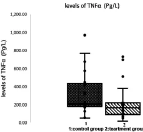
The Results of TNF-α Analysis
The serum level of TNF-α is expected to increase in patients with MS. In this study, we measured the serum levels of TNF-α in patients with MS that were treated with melatonin, as well as those receiving the placebo. As shown in Figure 2, there was no significant difference between the mean of TNF-α in the control and treatment groups (p<0.08).
Table1.Demographic information
Groups Demographic Factors
Control (n=25)
Case
(n=25) Probability Value (p- Value)
Age(Years)(Means±SE) 37.96±6.88 37.76±6.45 0.91
Male (n=5) Female (n=45)
2
23
3
22
0.64
Weight 73.63±10.71 74.33±9.20 0.80
Figure 1. Comparing the mean of Interleukin-1 beta (IL1β) in the control and treatment groups. There is a significant difference between the mean of IL-1β in the control and treatment groups
Figure 2. The mean of tumor necrosis factor-alpha (TNF-α) was compared in two groups. There is no significant difference between the mean of TNF-α in the control and treatment groups.
DISCUSSION
The results of this study indicated that there is a significant difference in the serum levels of IL-1β as an inflammatory factor between the control and treatment groups. In the patients receiving melatonin, melatonin led to a significant decrease in the level of IL-1β. Therefore, it can be argued that the reduced level of this factor is caused by receiving melatonin. Mason et al (2001) and Chih and et al (2017) showed that IL-1β levels increased significantly in patients
during brain damage.25,26 Liu et al (2017) reported that IL-1β increased in infectious diseases and injuries to the neural tissues.27 Liu et al found that the administration of exogenous melatonin reduced the inflammation of adipose tissue along with inhibiting the expression of the factor associated with inflammation of NLRP3 (which is also an apoptotic-based protein), and subsequently inhibiting the production of IL-1β.
treatment groups in the levels of the TNF-α. It is shown that melatonin has no effect on the expression of TNF-α. Comparing these results with the results of Arnasson et al, Sharif et al., (1991), and Kerstin et al (2018), it was found that TNF-α produced by dendritic cells and macrophages, had a significant increase in patients compared to healthy subjects. This increase is associated with the progression of the patient's disability.28,29 Farhadi et al (2014) and Tan et al (2018) emphasized the role of the pineal gland in responding to the inflammatory factors in patients. They reported that the level of melatonin was associated with the serum level of TNF-α in patients with MS and in the control group. The results of this study showed that the levels of melatonin in patients with MS are low and the levels of cytokines such as TNF-α are high.22,30 In this study, we tried to investigate the relationship between inflammatory factors and melatonin in patients with MS. Studies have shown that this hormone could be used to regulate the production of cytokines such as TNF-α and interleukins. The results of the present study also showed that the levels of IL-1β were significantly reduced in patients receiving melatonin. Therefore, melatonin could be used as an auxiliary, safe, and affordable drug with minimal side effects and a low toxicity risk which could reduce the symptoms of the disease. Several studies have also shown that melatonin has anti-inflammatory properties and leads to the elimination of fatigue states in MS patients.31 Jamshidifard et al (2013) and Wurtman (2017) showed that melatonin is effective in treating the visual impairment in MS patients.32,33 Other functions of this hormone include the regulation of the function of the metalloproteinase matrix which is involved in the damage to the brain and spinal cord in the secondary pathogenesis by damaging the blood-brain barrier and developing inflammation, as well as reducing the activity of the metalloproteinase matrix and subsequently suppressing the production of some inflammatory agents such as TNF-α.19
The level of melatonin is decreased in MS patients compared to normal people. This hormone plays a protective role in MS and several biological processes such as immune responses, and the circadian clock. This hormone has anti-inflammatory, immunomodulatory, and antioxidative effects and acts as an antioxidant in reducing the oxidative-nitrosative stress and inflammation and could reduce the inflammatory factors in MS patients. So, the protective
effects of these hormones could play an important role in MS pathogenesis. In this study, melatonin caused a reduction in the serum levels of IL-1β, therefore it might be a valuable therapeutic strategy in managing MS.
ACKNOWLEDGEMENTS
The authors would like to thank Arak’s Multiple sclerosis Society and Vali-Asr hospital located in Arak.
REFERENCES
1. Stein EM, Gennuso KP, Ugboaja DC, Remington PL. The epidemic of despair among White Americans: trends in the leading causes of premature death, 1999–2015. Am J public health 2017; 107(10):1541-7.
2. Oksenberg JR, Barcellos LF. The complex genetic aetiology of multiple sclerosis. Journal of Neurovirology 2000; 6(2):10.
3. Ontaneda DH, Cohen MJ. A Multiple sclerosis: new insights in pathogenesis and novel therapeutics. Annu Rev Med 2012; 63:389-404.
4. Mahad DH, Trapp BD, Lassmann H. Pathological mechanisms in progressive multiple sclerosis. Lancet Neurol 2015; 14(2):183-93.
5. Pröbstel AK, Sanderson NSR, Derfuss T. B cells and autoantibodies in multiple sclerosis. Int J Mol Sci 2015; 16(7):16576-92.
6. Martins GR,Gelaleti GB,Moschetta
MG,Maschio-Signorini LB,Zuccari DA. Proinflammatory and anti-inflammatory cytokines mediated by NF-κB factor as prognostic markers in mammary tumors. Mediators Inflamm J 2016; 2016:9512743.
7. Caminero A, Comabella M, Montalban X. Tumor
necrosis factor alpha (TNF-α), anti-TNF-α and
demyelination revisited: an ongoing story. J
Neuroimmunol 2011; 234(1-2):1-6.
8. Martin JP, Janette B, Etheresia p. The inflammatory effects of TNF-α and complement component 3 on coagulation. Sci Rep 2018; 8(1):1812.
9. Lim S, Cris SC. TNF-α: A paradigm of paradox and complexity in multiple sclerosis and its animal models. OpenAutoimmunJ 2010; 2(1):160-70.
10. Akash MSH, Rehman K, Liaqat A. Tumor Necrosis Factor-Alpha: Role in Development of Insulin Resistance and Pathogenesis of Type 2 Diabetes Mellitus. J Cell Biochem 2018; 119(1):105-10.
Gambotto A. immunity mediated by the cytokine IL-1 homologue 4 (IL-1H4/IL-1F7) induces IL-12-dependent adaptive and profound antitumor immunity. J Immunol 2003; 170(1):107-13.
12. Dinarello CA. Overview of the IL-1 family in innate inflammation and acquired immunity. Immunol Rev2018; 281(1):8-27.
13. Paré A,Mailhot B, Lévesque SA, Lacroix S. Involvement of the IL-1 system in experimental autoimmune encephalomyelitis and multiple sclerosis: Breaking the vicious cycle between IL-1β and GM-CSF. Brain Behav Immun2017; 62:1-8.
14. Mendiola AS, Cardona AE. The IL-1β phenomena in neuroinflammatory diseases. J Neural Transm (Vienna) 2018; 125(5):781-95.
15. Hardeland R, Cardinali DP, Srinivasan V,Spence
DW,Brown GM,Pandi-Perumal SR. Melatonin—a
pleiotropic, orchestrating regulator molecule. Prog Neurobio 2011; 93(3):350-84.
16. Liu J, Clough SJ, Hutchinson AJ, Adamah-Biassi EB,Popovska-Gorevski M,Dubocovich ML. MT1 and MT2 melatonin receptors: a therapeutic perspective. Annu Rev Pharmacol Toxicol 2016; 56:361-83.
17. Russel JR, Lucien C M, Dun XT. free radical
mechanisms and melatonin protection. Curr
Neuropharmacol 2010; 8(3):194–210.
18. Sun H and et al. Melatonin Treatment Improves Insulin Resistance and Pigmentation in Obese Patients with Acanthosis Nigricans. J Endocrinol 2018; 2018:46-7. 19. Esposito E, Cuzzocrea S. Antiinflammatory activity of
melatonin in central nervous system. Curr
Neuropharmacol 2010; 8(3):228–42.
20. Nabavi SM and et al. Anti-inflammatory effects of Melatonin: A mechanistic review. Crit Rev Food Sci Nutr 2018; 14:1-13.
21. Akpinar Z, Tokgöz S, Gökbel H, Okudan N, Uğuz F,Yilmaz G. The association of nocturnal serum melatonin levels with major depression in patients with
acute multiple sclerosis. Psychiatry Res 2008;
30:161(2):253-7.
22. Tan DX, Xu B, Zhou X, Reiter RJ. Pineal calcification,
melatonin production, aging, associated health
consequences and rejuvenation of the pineal gland. Molecules 2018; 23(2):301.
23. Sugden D, Davidson K, Hough KA, Teh MT. Melatonin,
melatonin receptors and melanophores: a moving story. Pigment Cell Res 2004; 17(5):454-60.
24. Wen J and et al. Efficacy of N-acetylserotonin and melatonin in the EAE model of multiple sclerosis. J Neuroimmune Pharmacol2016; 11(4):763-73.
25. Mason JL, Suzuki K, Chaplin DD, Matsushima GK.
Interleukin-1β promotes repair of the CNS. J
Neurosci 2001; 21(18):7046-52.
26. Chih ChL, Brain TE. New Insights into the Role of IL-1β in Experimental Autoimmune Encephalomyelitis and Multiple Sclerosis. J Immunol 2017; 198(12):4553-60. 27. Liu Zh and et al. Melatonin alleviates inflammasome
induced pyroptosis through inhibiting NF‐ κB/GSDMD
signal in mice adipose tissue. J Pineal Res2017; 63(1):12414.
28. Sharief MK, Hentges R. Association between tumor necrosis factor-α and disease progression in patients with multiple sclerosis. N Engl J Med1991; 325(7):467-72. 29. Göbel K, Ruck T, Meuth SG. Cytokine signaling in
multiple sclerosis: Lost in translation. Mult Scler 2018; 24(4):432-39.
30. Farhadi N,Oryan S, Nabiuni M. Serum levels of melatonin and cytokines in multiple sclerosis. Biomed J 2014; 37(2):90-2.
31. Alimoradian A, Shamsi M, Faraji F, Ahmadi M, Sayyed Sayyedi E, Evaluation of the Serum Melatonin Levels in the Treatment of Patients with Multiple Sclerosis. J Evaluation 2018; 21(131):55-64.
32. Jamshidi fard A, RH Faraji F, Ahmadi M, Sayeddi SE. Effects of melatonin on visual functioning of patients with multiple sclerosis.J Arak Uni Med Sci 2018; 21(2):55-64.