Tissue Engineering for Immature Teeth with Apical Periodontitis
Nozomu Yamauchi
A thesis submitted to the faculty of the University of North Carolina at Chapel Hill in partial fulfillment of the requirements for the degree of
Master of Science in the School of Dentistry (Endodontics).
Chapel Hill 2010
Approved by
Fabricio Teixeira, D.D.S., M.S., Ph.D. Eric M. Rivera, D.D.S., M.S.
Dedication
To my parents, to whom I dedicate my thesis.
You have guided me throughout my life and professional career in a positive direction. Your continual love and support for your children has taught me
the most important lessons in life. I am the luckiest son in the world.
To the rest of my family: Naoshi, Debbie, Alex, Sasha, Blake, Casey, Megu and Ben. Thanks for all the love and support. I am the luckiest brother and uncle in the world. Family is
everything.
To my grandparents: Thank you for being such wonderful influences in my life. To Grandma, who is a role model to me and inspires me to be the best endodontist I can be.
To my wife, Erica: You are my best friend and soul mate.
Your endless love and support inspires me every day. There are no words to describe how blessed I am to have you in my life. I am the luckiest husband in the world.
Abstract
Nozomu Yamauchi: Tissue Engineering for Immature Teeth with Apical Periodontitis (Under the direction of Dr. Fabricio Teixeira, Dr. Eric Rivera, and Dr. Mitsuo Yamauchi)
The purpose of this study was to explore novel strategies to engineer a vital support
structure within a root canal space by induced blood clot, exposure of dentin matrix and a
cross-linked collagen scaffold. Radiographically, there was significant difference in periradicular
healing and root wall thickening for scaffold groups. Two mineralized tissues were formed. A
dentin associated mineralized tissue (DAMT) and bony islands (BI). Histomorphometric analysis
showed more mineralized tissue formation in the scaffold groups. Exposure of the dentin matrix
appeared to increase the adherence of DAMT to dentin. Histochemical and
immunohistochemical characterization show DAMT is distinct from dentin or bone. Although
DAMT resembled cementum, it showed a more immature and woven collagen matrix. BI
resembled a bone matrix. The use of cross-linked collagen scaffold and exposure of dentin
matrix combined with blood clot may provide an efficient approach for the treatment of
Table of Contents
Part
1. Background and Introduction………....………...…...….1
1.1 Traditional Treatment of Immature Teeth With Non-Vital Pulps and Apical Periodontitis, and Its Problems……….…….1
1.2 Revascularizaton………...………...…..2
1.3 New Approach To Treatment of Immature Teeth With Non-Vital Pulps and Apical Periodontitis….………...………...…4
Disinfection….………..………...…4
Induced Bleeding/Blood Clot….………...…………..………...…5
1.4 Regeneration/Tissue Engineering……….……..………..……...…6
Stem Cells……..………..……...……….……6
Dentin Matrix as Source of Growth/Signaling Factors, Etc.…………....…….……7
Scaffold/Cross-Linked Collagen Scaffold.…………..……...……….8
1.5 Histological Characterization………..…………...………8
1.6 References………...………..………...11
2. Efficient Tissue Engineering Strategies For Immature Teeth With Apical Periodontitis………...…....………..………...…..…...15
! 2.1 Abstract………...……...…………..………….……….………...15
2.2 Introduction………...………..……...…...………...16
2.3 Materials and Methods………..………...17
Tooth Treatments………...………..……...………18
Tissue Preparation and Histological Evaluation………….………..20
Summary of Treatment and Control Groups………...21
2.4 Results…...………..……..………...21
Radiographic Analyses………...…..……..………...21
Histological Analyses………..……..………....…21
Mineralized Tissue Formation On Inner Dentin Walls and Mineralized Tissue Islands In Lumen………..……..…...…….……...22
Apical Development………..……..………22
! Effect of Demineralization and Exposure of Dentin Matrix………23
2.5 Discussion………..………..………23
Improvement of Root Thickening and Lengthening: Use of A Collagen Scaffold……….……...24
Effects of Demineralization and Exposure of Dentin Matrix………...………26
Characteristics of the Mineralized Tissue Formed………….………...………27
Further Characterization of the Newly Formed Tissues………….………...30
2.6 Conclusions………...…..………....…..………....30
2.7 List of Figures………..……….……….………..31
2.8 References……...………..………...42
3. Histochemical and Immunohistochemical Characterization of Mineralized Tissues After Tissue Engineering Treatments On Immature Teeth With Apical Periodontitis……..………45
3.1 Abstract………..…………...……….………...……….……...45
3.2 Introduction……....…………..…………...………...……….…..…….……...46
Histological Processing…...….……..……….….………….…...….…...48
3.4 Results…...………...…………..……….………..………....…49
H and E: General Observations of Dentin Associated Mineralized Tissue and Bony Islands………….………...….…..49
! Picrosirius Red Staining………….…...………..49
Immunohistochemistry………..50
3.5 Discussion………...………..……….……...…...………...51
3.6 Conclusions………...………..……….…….……...……..…54
3.7 List of Figures………...………..……….…………...…….55
3.8 References………...………..……….………63
PART ONE
Background and Introduction
!1.1.Traditional Treatment of Immature Teeth With Non-Vital Pulps and Apical
Periodontitis, and Its Problems
! Pulp necrosis of an immature tooth poses potential complications and challenges for treatment. Use of a standard cleaning and shaping protocol with endodontic files is not advised
due to the thin dentinal walls. Instrumentation of already thin walls would likely weaken the
tooth. Therefore, traditionally, only light filing, or no filing has been recommended. More
emphasis has been placed on irrigants and medicaments to disinfect the root canal space.
The obturation aspect of the treatment can be difficult to obtain a predictable apical seal
due to the openness of the apex. The open apex provides no barrier to contain the root canal
filling, thus having direct contact with the periodontal tissues. Treatment techniques for either
stimulating formation of a hard tissue barrier (i.e., long-term calcium hydroxide apexification) or
using materials to create an artificial barrier (i.e., MTA apexification) have shown success (1-4).
Long-term calcium hydroxide treatment may take up to 18months to form a hard tissue barrier,
requires long-term patient compliance, and may potentially weaken the dentin structure (5). The
use of MTA to create an artificial barrier has advantages of being biocompatible with
periodontal tissues, creating an apical seal, and being able to provide treatment in one or two
appointments. However, the MTA can be difficult to control and place and like the long-term
calcium hydroxide treatment, does not address the fact that the dentinal walls remain thin and
immature. There is no prospective long-term outcome study, which compares long-term calcium
In the treatment of an immature tooth, the major clinical problem is that the remaining
thin dentinal walls leaves the tooth weak and with an increased risk of subsequent fracture. It has
been shown that approximately 30% of immature teeth can fracture during or after root canal
treatment (2). Root reinforcement techniques have been introduced for the treatment of
immature teeth. These techniques have been shown to internally strengthen the teeth; however,
these pose problems for retreatment (6).
1.2. Revascularization
Trauma studies have shown that revascularization of a necrotic pulp is possible. This was
shown in immature teeth with open apices, which were avulsed and replanted. Ohman et al.
showed that replantation of immature human premolars results in the in-growth of tissue (7).
Skoglund et al. using microangiography showed that pulp revascularization occurred in dog teeth
which were replanted and autotransplanted (8). When an avulsed immature tooth is replanted,
new tissue has the capability of growing into the space and partially filling the space with
mineralized tissue. As long as the pulp is not infected, it may act as a scaffold into which the new
tissue can grow and eventually fill the space.
Hitchcock et al. (1985) showed that un-infected teeth in monkeys subjected to vital
apicoectomies were revascularized (9). Initially, the remaining pulp within the canal underwent
necrosis, but remained aseptic and with time, vital tissue replaced the pulp tissue. Most likely the
remaining pulp tissue acted as a scaffold for the new tissues to migrate and replace. They
observed that the source of the new tissue was likely periodontal ligament, which migrated
across the cut dentin surface and along the internal dentin wall. The new tissue contained
cemetoblasts, cellular cementum, fibroblasts, and dense collagen bundles. In some cases, there
transection or apicoectomy effectively created a sufficient space so that there can be cell
migration and subsequent tissue in-growth.
Laureys et al. investigated the in-growth and replacement of tissue between mature
apicoectomized teeth versus immature teeth (10). In this study, dog teeth were either
apicoectomized and autotransplanted, or autotransplanted while the teeth still had open apices
(i.e., immature). They concluded that there was no difference in the in-growth of new pulpal
tissue between mature apicoectomized teeth and immature teeth.
Kling et al. found 18% revascularization in 154 immature human avulsed teeth that were
replanted post trauma. They found that in teeth with apical foramen less than 1mm
mesio-distally there was no revascularization (11). Similarly, Cvek and others (1990) reported on a study
of experimental avulsions of monkey teeth. They showed that teeth with mature apices had no
revascularization occur (12).
Davis et al. showed that tissue in-growth into an empty canal space is possible if the apex
diameter was opened to a size #80 (13). In this study, teeth were filled several millimeters short
of the apex and viable tissue devoid of inflammation grew into the remaining canal space. The
authors concluded that adequate diameter and access of the foramen was more important than
formation of a blood clot within the canal.
Yanpiset and Trope showed there was an increase in revascularization rates when topical
doxycycline was used to decrease the concentration of bacteria in and around the pulp (14).
Ritter et al. demonstrated that when topical minocycline was placed on avulsed teeth, there was a
significant increase in revascularization rates (15).
These studies were based more on trauma-induced cases and not on immature teeth,
indicate that in these cases, revascularization and regeneration of vital tissues in and around the
roots do occur if the space within the apical third of the canal is available.
1.3. New approach to treatment of immature teeth with apical periodontitis
More recently, revascularization, or regenerative endodontic procedures in the treatment
of immature teeth with necrotic pulps and apical periodontitis has been reported (16-19). The
term revascularization, revitalization and regeneration have been poorly defined terms which
have been used interchangeably. Studies and!cases such as these have been recently considered
or defined to be regenerative endodontics (20).
The basis for regenerative endodontic procedures is the utilization of tissue engineering
therapies with the goal to replace, repair, maintain, and/or enhance tissue function (20).
Although there are many different definitions of regenerative medicine, in practice the term has
come to represent applications that repair or replace structural and functional tissues (21).
It appears that based on the cases that have shown promising outcomes, there are
several approaches to treatment that yield similar positive results clinically. The frequent
pretreatment diagnosis of the teeth is necrotic pulps and apical periodontitis with immature root
formation. There is a key disinfection process that must take place (i.e., with calcium hydroxide
or triple antibiotic paste). Many cases induce bleeding into the root canal space to form a blood
clot. Then a coronal tight seal is placed to ensure that bacteria do not re-enter the root canal.
The following discusses some of the key components that have been attributed in aiding
successful regenerative outcomes.
Disinfection
A key step in the process of regenerative treatment is proper disinfection of the root
canal system. Studies have been published demonstrating that adequate disinfection of the root
have shown poor success rates of revascularization if the teeth were necrotic with apical
periodontitis (24, 25). These studies were likely not able to predictably disinfect the root canal
system due to the unavailability of antibiotic paste/other techniques, thus leading to failure of
revascularization/regeneration. The use of a triple antibiotic paste of ciprofloxacin,
metronidazole, and minocycline has shown to effectively kill common endodontic pathogens in
vitro (26, 27) and in vivo (28) and aids in revascularization of avulsed and necrotic teeth (14-16).
By application of a treatment protocol using this triple antibiotic paste in vivo, the
necrotic infected immature teeth can be rendered bacteria free, thus creating an environment
conducive to revascularization. Using calcium hydroxide instead of triple antibiotic paste in
order to get revascularization to occur has been shown (29). Although both calcium hydroxide
and triple antibiotic paste can help promote further development of the pulp-dentin complex,
the teeth treated with triple antibiotic paste may produce greater root wall thickness (30). It
appears that provided that there is proper disinfection and no permanent damage to the
periradicular tissues, this creates an environment where regenerative type treatments can be
successfully performed.
Induced Bleeding/Blood Clot
Use of a blood clot appears to aid in revascularization (16). Nygaard-Ostby and Hjortdal
performed a study (24) in which bleeding was induced in the canal to create a blood clot. Files
were passed through the apex of experimental canals to induce bleeding, while controls were
maintained blood-free. Some of the teeth showed complete repair with fill of vital tissue. The
authors concluded the blood clot to be beneficial in aiding in-growth of new tissue. Horsted and
Nygaard Ostby (31) also conducted a human study in which bleeding was induced into unfilled
portions of a root canal. There was new connective tissue present in the unfilled canal space
Myers and Fountain (25) used whole blood and/or blood substitutes (i.e., Gelfoam with
blood) in infected, open apex teeth to try and aid dental pulp regeneration. Results showed that
regeneration or in-growth of connective tissue into the root canals was not enhanced by the
blood and/or blood substitutes. However, in the cases with healing present, these teeth showed
signs of continued root development with attempts at apical closure or apical bridging. The
reason for the high failure rate may be associated with persistent infection due to inadequate
disinfection.
More recently, Thibodeau et al. showed that a blood clot used in immature teeth with
apical periodontitis increased the success rates of revascularization (16). The study, using a dog
model, showed that after disinfection with triple antibiotic paste and induction of a blood clot,
revascularization with subsequent growth and development of the root canal system was
possible. They also tested if the addition of a collagen solution to the blood clot group aided in
revascularization. However, the results showed no statistical difference.
The blood clot may serve as a scaffold for the periapical cells including mesenchymal
stem cells to migrate into the root canal and eventually induce new tissue formation in the space.
It also contains many growth and differentiation factors, which are critical for wound healing
(32, 33) and tooth development.
1.4. Regeneration/Tissue Engineering
Regenerative/tissue engineering treatments in medicine have shown promising results
(34, 35). Biological basis for these treatments involve three main components of stem cells,
addition of growth factors/molecules, and a scaffold.
Stem Cells
Postnatal stem cells have been sourced from umbilical cord blood, umbilical cord, bone
teeth (37). By inducing bleeding from the periapical tissues, this is likely to contain a source of
mesenchymal stem cells.
Dentin Matrix As Source of Growth/Signaling Factors
There are two major limitations from the Thibodeau study which may have resulted in
rather modest effect on the new tissue formation: one, the lack of additional and specific
growth/differentiation factors and two, the lack of stable scaffold. The current study is designed
to address these limitations. At present, it is not possible to identify the proper concentrations of
various factors required for pulp revascularization and the formation of dentin-pulp complex
upon the induction of a blood clot. Thus in this study, we proposed to partially expose the
dentin matrix prior to induction of a blood clot by treatment with 17% EDTA. EDTA has been
shown to remove smear layer to expose the dentin matrix (38, 39).
Dentin matrix is composed of collagen (primarily type I) representing around 90% of the
matrix and non collagenous molecules which make up the remaining 10% (40). Type I collagen
serves as a three-dimensional (3D) template for mineralization but also is the major reservoir for
growth factors and signal molecules that are critical for dentinogenesis. Those factors can be
stored in the collagen matrix directly and/or via the collagen-binding small leucine-rich
proteoglycans that directly bind growth factors such as TGF-!. In addition, there are some
dentin-specific signaling molecules such as phosphophoryn (a cleavage product of dentin
sialophosphoprotein, DSPP) and DMP1 (41, 42) in dentin matrix. Thus, the exposure of dentin
matrix would make those critical factors/molecules available for cells to differentiate to
effectively induce dentin formation. As described above, since the optimal conditions for various
factors (types, concentrations, combination, etc.) and their effective delivery are not known at
Scaffold/Cross-linked Collagen Scaffold
Scaffolds need to be biodegradable, porous to allow for cell migration and proliferation,
and must keep its integrity for a sufficient period of time for the newly formed tissue to replace
it. In replantation studies described previously, it appears that the pulp tissue may have acted as a
scaffold for the in growing tissues to proliferate coronally and eventually replace. The induction
of bleeding and generation of a blood clot may also serve a similar purpose. Regarding the
scaffold, Thidodeau et al. (2007) used a collagen solution gel (16). While the use of collagen was
a laudable idea to further support cell migration and proliferation in the root canal, a collagen
solution may not have been an adequate matrix since it can quickly undergo resorption.
Cross-linked collagen scaffolds have been used in 3-D cell culture and have shown to
stay intact and stable longer than its non cross-linked counterpart. Nevins et al. (1998) did a
study in which cross-linked collagen gel was utilized within the root canal space (43). There was
no quantitative analysis to show any differences in the linked collagen gel or the non
cross-linked collagen gel. Also, this study used the collagen on vital pulp tissue, where our study used
the scaffold on necrotic, immature teeth.
1.5 Histological Characterization
Studies and clinical case reports of regenerative endodontic procedures have shown
promising results, but there is still a lack of biologic evidence to support an efficient method in
the treatment of immature teeth with apical periodontitis. In order to obtain insights into the
biological basis for the efficient tissue regeneration, the nature of the newly formed tissues
should be characterized at molecular, biochemical, and histological levels by using a proper
animal model.
Most recently, using the hematoxylin and eosin (H&E) stained sections from a previous
identified two types of mineralized tissues formed within the canal space after revascularization
treatment, which included induction of a blood clot after disinfection. A cementum-like tissue
was formed on the inner dentin wall, which they referred to as intracanal cementum (IC) and
bone-like tissue was found in the lumen space termed intracanal bone-like tissue (IB) (44).
Although this study gives an insight into what these tissues could be, further characterization
such as quantitative assessment of the mineralized tissue formation, the nature of mineralized
matrix (i.e. collagen matrix, some of the tissue-specific marker proteins), gene expression
pattern, etc., are warranted.
Bone and teeth contain collagen, the predominant matrix component, as well as several
non-collagenous proteins. The SIBLING family (Small Integrn-Binding Ligand N-linked
Glycoproteins) is one of the well-characterized group of noncollagenous proteins in mineralized
tissues and it includes dentin sialophosphoprotein (DSPP), osteopontin (OPN), and bone
sialoprotein (BSP).
Although they have been recently found in other non-mineralized tissues such as kidney
and salivary glands (45, 46), these SIBLINGs are mostly found in bones and teeth and have been
shown to be associated with the mineralization process. DSPP is currently one of the main
markers used to differentiate dentin matrix from other mineralized tissues including bone matrix
(47, 48). Although osteoblasts have been shown to express DSPP, the level of expression is 400
times lower (49). It has been demonstrated that DSPP is critical for dentin mineralization and
DSPP gene mutation leads to dentin defects (50). BSP and OPN are also members of SIBLING
family relatively abundant in bone and cementum matrix (51, 52).
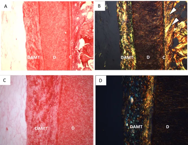
In this study, in order to characterize these newly formed mineralized tissues, we
relative abundance of three non-collagenous matrix proteins, i.e. dentin sialophosphoprotein
(DSPP), bone sialoprotein (BSP), and osteopontin (OPN) using immunohistochemstry.
In Part 2 of this study, the purpose was to examine the use of the tooth’s natural dentin
matrix combined with induced bleeding and a stable, cross-linked collagen scaffold in the
treatment of immature teeth with necrotic pulps and apical periodontitis. The null hypothesis
was that after disinfection of an infected immature dog root canal system, the exposure of the
dentin matrix and a cross-linked collagen scaffold will not improve deposition/thickening of the
root walls and in-growth of vital tissue into the lumen space. In Part 3 of this study, further
characterization of the newly formed mineralized tissues was studied using histochemical and
PART ONE 1.6 References
1. Cvek M. Prognosis of luxated non-vital maxillary incisors treated with calcium hydroxide and filled with gutta percha. A retrospective clinical study. Endod Dent Traumatol 1992: 8: 45-55 2. Kerekes K, Heide S, Jacobsen I. Follow-up examination of endodontic treatment in
traumatized juvenile incisors. J Endod 1980;6(9):744-748.
3. Giuliani V, Baccetti T, Pace R, Pagavino G. The use of MTA in teeth with necrotic pulps and open apices. Dent Traumatol 2002;18(4):217-221.
4. Witherspoon DE, Small JC, Regan JD, Nunn M. Retrospective analysis of open apex teeth obturated with mineral trioxide aggregate. J Endod 2008;34(10):1171-1176.
5. Andreasen JO, Farik B, Munksgaard EC. Long-term calcium hydroxide as a root canal dressing may increase risk of root fracture. Dent Traumatol 2002: 18: 134-137.
6. Katebzadeh N, Dalton BC, Trope M. Strengthening immature teeth during and after apexification. J Endod 1998;24(4):256-259.
7. Ohman A. Healing and sensitivity to pain in young replanted human teeth. An experimental, clinical, and histological study. Odontol Tidskr. 1965 Apr 15;73:166-227.
8. Skoglund A, Tronstad L, Wallenius K. A microangiographic study of vascular changes in replanted and autotransplanted teeth of young dogs. Oral Surg Oral Med Oral Pathol 1978;45(1):17-28.
9. Hitchcock R, Ellis E, 3rd, Cox CF. Intentional vital root transection: a 52-week
histopathologic study in Macaca mulatta. Oral Surg Oral Med Oral Pathol 1985;60(1):2-14. 10. Laureys W, Beele H, Cornelissen R, Dermaut L. Revascularization after cryopreservation and
autotransplantation of immature and mature apicoectomized teeth. Am J Orthod Dentofacial Orthop 2001;119(4):346-352.
11. Kling M, Cvek M, Mejare I. Rate and predictability of pulp revascularization in
therapeutically reimplanted permanent incisors. Endod Dent Traumatol 1986;2(3):83-89.
12. Cvek M, Cleaton-Jones P, Austin J, Lownie J, Kling M, Fatti P. Effect of topical application of doxycycline on pulp revascularization and periodontal healing in reimplanted monkey incisors. Endod Dent Traumatol 1990;6(4):170-176.
13. Davis MS, Joseph SW, Bucher JF. Periapical and intracranial healing following incomplete root canal fillings in dogs. Oral Surg Oral Med Oral Pathol 1971;31(5):662-675.
15. Ritter AL, Ritter AV, Murrah V, Sigurdsson A, Trope M. Pulp revascularization of replanted immature dog teeth after treatment with minocycline and doxycycline assessed by laser Doppler flowmetry, radiography, and histology. Dent Traumatol 2004; 20:75-84 16. Thibodeau B, Teixeira F, Yamauchi M, Caplan DJ, Trope M. Pulp revascularization of
immature dog teeth with apical periodontitis. J Endod 2007;33(6):680-689.
17. Ding RY, Cheung GS, Chen J, Yin XZ, Wang QQ, Zhang CF. Pulp revascularization of immature teeth with apical periodontitis: a clinical study. J Endod 2009;35(5):745-749.
18. Jung IY, Lee SJ, Hargreaves KM. Biologically based treatment of immature permanent teeth with pulpal necrosis: a case series. J Endod 2008;34(7):876-887.
19. Chueh LH, Ho YC, Kuo TC, Lai WH, Chen YH, Chiang CP. Regenerative endodontic treatment for necrotic immature permanent teeth. J Endod 2009;35(2):160-164.
20. Murray PE, Garcia-Godoy F, Hargreaves KM. Regenerative endodontics: a review of current status and a call for action. J Endod 2007;33(4):377-390.
21. Rahaman MN, Mao JJ. Stem cell-based composite tissue constructs for regenerative medicine. Biotechnol Bioeng 2005;91(3):261-284.
22. Kakehashi S, Stanley HR, Fitzgerald RJ. The Effects of Surgical Exposures of Dental Pulps in Germ-Free and Conventional Laboratory Rats. Oral Surg Oral Med Oral Pathol
1965;20:340-349.
23. Moller AJ, Fabricius L, Dahlen G, Ohman AE, Heyden G. Influence on periapical tissues of indigenous oral bacteria and necrotic pulp tissue in monkeys. Scand J Dent Res
1981;89(6):475-484.
24. Nygaard-Ostby B, Hjortdal O. Tissue formation in the root canal following pulp removal. Scand J Dent Res. 1971; 79(5): 333-49
25. Myers WC, Fountain SB. Dental pulp regeneration aided by blood and blood substitutes after experimentally induced periapical infection. Oral Surg Oral Med Oral Pathol 1974;37(3):441-450.
26.Hoshino E, Kurihara-Ando N, Sato I, Uematsu H, Sato M, Kota K, Iwaku M. In-vitro antibacterial susceptibility of bacteria taken from infected root dentine to a mixture of ciprofloxacin, metronidazole and minocycline. Int Endod J. 1996 Mar; 29(2):125-30.
27. Sato I, Ando-Kurihara N, Kota K, Iwaku M, Hoshino E. Sterilization of infected root-canal dentine by topical application of a mixture of ciprofloxacin, metronidazole and minocycline in situ. Int Endod J 1996;29(2):118-124.
29. Cotti E, Mereu M, Lusso D. Regenerative treatment of an immature, traumatized tooth with apical periodontitis: report of a case. J Endod 2008;34(5):611-616.
30. Bose R, Nummikoski P, Hargreaves K. A retrospective evaluation of radiographic outcomes in immature teeth with necrotic root canal systems treated with regenerative endodontic procedures. J Endod 2009;35(10):1343-1349.
31. Horsted P, Nygaard-Ostby B. Tissue formation in the root canal after total pulpectomy and partial root filling. Oral Surg Oral Med Oral Pathol. 1978 Aug; 46(2):275-82
32. Maeda H, Wada N, Nakamuta H, Akamine A. Human periapical granulation tissue contains osteogenic cells. Cell Tissue Res 2004;315(2):203-208
33. Marx RE. Platelet-Rich Plasma: Evidence to support its use. J Oral Maxillofac Surg. 2004; 62(4):489-96.
34. Atala A, Bauer SB, Soker S, Yoo JJ, Retik AB. Tissue-engineered autologous bladders for patients needing cystoplasty. Lancet 2006;367(9518):1241-1246.
35. Pham C, Greenwood J, Cleland H, Woodruff P, Maddern G. Bioengineered skin substitutes for the management of burns: a systematic review. Burns 2007;33(8):946-957.
36. Gimble J, Guilak F. Adipose-derived adult stem cells: isolation, characterization, and differentiation potential. Cytotherapy 2003;5(5):362-369.
37. Tsukamoto Y, Fukutani S, Shin-Ike T, Kubota T, Sato S, Suzuki Y, Mori M. Mineralized nodule formation by cultures of human dental pulp-derived fibroblasts. Arch Oral Biol 1992;37(12):1045-1055.
38. Smith AJ, Leaver AG. Non-collagenous components of the organic matrix of rabbit incisor dentine. Arch Oral Biol 1979;24(6):449-454.
39. Meryon SD, Tobias RS, Jakeman KJ. Smear removal agents: a quantitative study in vivo and in vitro. J Prosthet Dent 1987;57(2):174-179.
40. Linde A, Goldberg M. Dentinogenesis. Crit Rev Oral Biol Med. 1993; 4(5):679-728 41. Almushayt A, Narayanan K, Zaki AE, George A. Dentin matrix protein 1 induces
cytodifferentiation of dental pulp stem cells into odontoblasts. Gene Ther. 2006; 13(7):611-20.
42. Narayanan K, Srinivas R, Ramachandran A, Hao J, Quinn B, George A. Differentiation of embryonic stem cells to odontoblast-like cells by overexpression of dentin matrix protein 1. Proc Natl Acad Sci U S A. 2001; 98(8):4516-21.
44. Wang X, Thibodeau B, Trope M, Lin LM, Huang GT. Histologic characterization of regenerated tissues in canal space after the revitalization/revascularization procedure of immature dog teeth with apical periodontitis. J Endod 2010;36(1):56-63.
45. Shiraga H, Min W, VanDusen WJ, Clayman MD, Miner D, Terrell CH, et al. Inhibition of calcium oxalate crystal growth in vitro by uropontin: another member of the aspartic acid-rich protein superfamily. Proc Natl Acad Sci U S A 1992;89(1):426-430.
46. Ogbureke KU, Fisher LW. Expression of SIBLINGs and their partner MMPs in salivary glands. J Dent Res 2004;83(9):664-670.
47. Ritchie HH, Shigeyama Y, Somerman MJ, Butler WT. Partial cDNA sequencing of mouse dentine sialoprotein and detection of its specific expression by odontoblasts. Arch Oral Biol 1996;41(6):571-575.
48. D'Souza RN, Bachman T, Baumgardner KR, Butler WT, Litz M. Characterization of cellular responses involved in reparative dentinogenesis in rat molars. J Dent Res 1995;74(2):702-709.
49. Qin C, Brunn JC, Cadena E, Ridall A, Tsujigiwa H, Nagatsuka H, Nagai N, Butler WT. The expression of dentin sialophosphoprotein gene in bone. J Dent Res 2002;81(6):392-394.
50. Yamakoshi Y. Dentinogenesis and Dentin Sialophosphoprotein (DSPP). J Oral Biosci 2009;51(3):134.
51. Ganss B, Kim RH, Sodek J. Bone sialoprotein. Crit Rev Oral Biol Med 1999;10(1):79-98.
PART TWO
Tissue Engineering Strategies for Immature Teeth
With Apical Periodontitis
Nozomu Yamauchi, DDS,1 Shizuko Yamauchi, DDS, MS,1 Hideaki Nagaoka, DDS, PhD,2 Derek Duggan, DDS, MS,1 Sheng Zhong DDS1, Sun Min Lee, Fabricio B. Teixeira, DDS, MS, PhD,3 and Mitsuo Yamauchi, DDS, PhD2
1Department of Endodontics, University of North Carolina School of Dentistry. 2NC Oral Health Institute, University of North Carolina School of Dentistry.
3Department of Endodontics, University of Texas Health Science Center at San Antonio. !
2.1 Abstract
Introduction. Regenerative endodontic treatment on immature teeth with apical
periodontitis is promising but still not well established. The purpose of this study was to explore novel strategies to efficiently engineer a vital support structure within a root canal space by a combination of induced blood clot, exposure of dentin matrix and a cross-linked collagen scaffold.
Methods. Apical periodontitis was induced in six dogs with immature teeth (N=64).
After disinfection with triple antibiotic paste, the following groups were randomly assigned: blood clot (BC) alone, BC with a cross-linked collagen scaffold (CCS), BC with exposure of dentin matrix by EDTA treatment, and BC combined with CCS and EDTA. Positive (infected only) and negative controls (untreated) were included. The dogs were followed up for 3.5 months and sacrificed. With observers blinded, periradicular healing, and root wall thickening were radiographically analyzed using Friedman’s Chi-Square test. The dogs’ jaws were then fixed, demineralized, and stained with H&E for histological analyses. Histomorphometric analysis was performed to quantify the newly formed mineralized tissues in the canal space and evaluated using Kruskal-Wallis test and pair-wise comparison.
Results. Radiographically there was significant difference in periradicular healing and
root wall thickening (p<.05). Histomorphometric analysis showed significantly more mineralized tissue formation in the groups containing the scaffold (p<.05). Exposure of the dentin matrix by EDTA appeared to increase the adherence of the newly formed mineralized tissue to the root walls.
Conclusions. The use of cross-linked collagen scaffold and exposure of dentin matrix
combined with blood clot may provide an efficient approach to generate a vital support structure for the treatment of immature teeth with apical periodontitis.
2.2 Introduction
Recently, regenerative endodontic procedures for the treatment of immature teeth with
necrotic pulps and apical periodontitis have gained much attention due to some encouraging
results in basic as well as clinical studies (1-4). The advantage of regenerative endodontic
procedures over apexification procedures is that it allows potential root thickening and
lengthening (i.e., maturation) to continue via the generated vital tissue. With continued research
and improvement in regenerative endodontic procedures, these may soon become the standard
of care over apexification-type procedures and the similar approach could be applied to the
treatment of mature infected teeth (5, 6).
Tissue engineering is an approach consisting of a biocompatible, stable scaffold,
appropriate growth factors, and stem cells. It is a rapidly growing field of research that has been
well implemented in medicine (7, 8). However, its application has been limited in endodontics.
There have been promising results in bioroot/dentin-pulpal complex engineering (9, 10) and
strategies have been discussed (11), but its application to necrotic root canal spaces is still
lacking. Several case reports and a recent study by Thibodeau et al. indicated that induction of
blood clots is useful to generate mineralized tissues in the canal space that was previously
necrotic (3).
In this study, an attempt was made to explore an efficient tissue engineering strategy for
the treatment of immature teeth with necrotic pulps and apical periodontits. For this purpose, a
dog model (3) was employed and the efficacy of a combination of a cross-linked collagen
scaffold, partial exposure of the dentin matrix, and induced bleeding to generate new
mineralized tissues in the canal space was examined by radiographic and histological analyses,
2.3 Material and Methods
The animal protocol described in this study was approved by the Institutional Animal Care and Use Committee of the University of North Carolina at Chapel Hill. Sixty
double-rooted premolar teeth in six purpose-bred mixed breed canine model dogs (Alder Ridge Farms
Inc.) aged approximately six months were randomly divided into four treatment and control
groups (n=12 teeth/group). The teeth were randomly divided into 48 experimental and 12
negative (no treatment was performed) control teeth. Four positive control teeth (teeth were
infected only) were also prepared. Under general anesthesia induced by Pentothal
[AbbottLaboratories, NorthChicago, IL] 13.5 mg/kg intravenously and intubation and
maintenance with isoflurane [Halocarbon Laboratories, RiverEdge, NJ]) supplemented with
local anesthesia (bupivacaine plain 0.5%, Abbott Laboratories), the pulps of all experimental
teeth were mechanically exposed with a #2 round carbide bur (Brassler USA, Savannah, GA) in
a high-speed hand piece (Midwest, Mondovi, WI) under nonaseptic conditions. A sterile #20
stainless steel endodontic hand file (Dentsply Maillefer, Johnson City, TN) was used to disrupt
the pulp tissue in the canal spaces, without removing it from the canals. Supragingival plaque
scaled from the dogs’ teeth was mixed with sterile saline (0.9% sodium chloride; Hospira Inc.,
Lake Forest, IL) and this plaque suspension was soaked with sterile sponges (Dentsply Maillefer,
Johnson City, TN) and sealed temporarily in the pulp chambers of the teeth with IRM
(Intermediate Restorative Material, Dentsply Caulk). The animals were given analgesics
(Torbugesic 0.2 mg/kg; Butorphanol Tartrate, Fort Dodge Animal Health, Fort Dodge, IA)
postoperatively following this and all operative procedures, and were monitored by staff of the
Tooth Treatments
Before any interventions, the involved teeth were radiographed with radiograph
paralleling devices (Dentsply Rinn, Elgin, IL). These radiographic aids were used for all
subsequent radiographs and extreme care was taken to reproduce the same angle and
magnification. The teeth were monitored radiographically every week until there was
radiographic evidence of apical periodontitis (approximately three weeks). All previously infected
teeth were re-entered under aseptic conditions of rubber dam isolation with retractors and
surface disinfection with 0.12% chlorhexidine (Alpharma USPD, Baltimore, MD) and tincture of
iodine (Humco, Texarcana, TX) with the animals under general and local anesthesia. After
removal of the IRM and sponge, the canals were irrigated with 10 mL of 2.5% NaOCl (sodium
hypochlorite; Clorox,Oakland, CA) per tooth. No mechanical instrumentation was performed in
the canals. Canals were dried with sterile paper points (Dentsply Maillefer, Tulsa, OK) and
disinfected with a mixture of equal parts of metronidazole, ciprofloxacin, and minocycline mixed
with Macrogol and Propylene glycol (Central Compounding Center, Durham, NC) as described
by Hoshino (12). The paste was applied to the canal spaces with a sterile lentulo spiral (Dentsply
Maillefer, Johnson City, TN) in a slow speed handpiece (Midwest). The experimental teeth were
closed temporarily with a sterile sponge (Dentsply Maillefer) and IRM (Dentsply Caulk) for 2
weeks to allow disinfection of the canal spaces.
After two weeks, under the same conditions of asepsis and general and local anesthesia,
the temporary restorations and sponges were removed under rubber dam isolation from the
experimental teeth. The antibiotic mixture was irrigated from the canals with 10 mL of 2.5%
NaOCl (Clorox) and 10 mL of sterile saline (0.9% sodium chloride, Hospira Inc.) per tooth.
Experimental treatment groups were randomly assigned as follows: In Group 1, a sterile #20
terminus into the periapical tissues to induce bleeding to fill the canal space. The bleeding was
allowed to settle for several minutes before a double seal of ProRoot MTA (Dentsply Tulsa
Dental, Johnson City, TN) and glass ionomer (Fuji IX GC) was applied. In Group 2, 17%
Ethylenediaminetetraacetic Acid (EDTA, Vista Dental Products) was applied for two minutes in
the canal space prior to inducing bleeding and sealing. In Group 3, a cross-linked type I collagen
scaffold sponge (Nitta Gelatin Inc., Japan) was cut into small pieces, washed with phosphate
buffer saline and placed incrementally inside the canal system after bleeding was induced. The
sponge was soaked with blood and then was sealed. In Group 4, EDTA, induced bleeding, and
addition of the cross-linked collagen scaffold were applied before sealing. The 12 teeth randomly
assigned to Group 5 were negative controls. These teeth were left untouched to develop
naturally for comparison with the experimental teeth. Four teeth were assigned to positive
controls in which the teeth were infected and sealed. After the treatments, all of the teeth were
monitored radiographically on a monthly basis for three and a half months before the animals
were sacrificed.
Radiographic Analysis
All radiographs were exposed using a paralleling technique to best reproduce the
placement of radiographic film and angulation of the X-ray beam. Each radiograph was
randomly evaluated by two blinded evaluators. The evaluators were masked to dog and tooth
type, time of radiograph, and treatment status. Initial measurements from radiographs made at
the time of disinfection were compared with measurements made on the follow-up radiographs
after the treatment procedures. Each individual root was taken as a unit of measurement. Each
examiner assessed for the following: 1) presence or absence of radiographic healing of the
periradicular lesion, and 2) presence or absence of radiographic thickening of the root walls
discuss and come to an agreement. The data were analyzed using Friedman’s Chi-Square Test,
with the level of significance set at p<0.05.
Tissue Preparation and Histological Evaluation
The animals were sacrificed under general anesthesia provided by Socumb
(pentobarbital; Butler Company, Columbus, OH) at 30 mg/kg intravenously. The carotid
arteries were exposed and canulated. The animals were euthanized with additional pentobarbital
(Socumb, Butler Company) at a dose of 90 mg/kg intravenously. The animals were perfused
with 10% buffered formalin (Fisher Scientific, Fair Lawn, NJ). The jaw blocks with the involved
teeth were dissected and fixed in formaldehyde (Fisher Scientific).
After removal of all soft tissue and excess hard tissue from the jaw blocks, the specimens
were decalcified with Immunocal (Decal Chemical Corporation, Tallman, NY) for two months
with four changes of the solution over that period of time. The specimens were then washed
with distilled water, immersed in 70% ethyl alcohol, dehydrated through ascending gradations of
ethanol, embedded in paraffin, and sectioned on a Leica Jung RM2045 microtome. Five mm
sections were longitudinally cut through the roots and stained with hematoxylin and eosin
(H&E) and evaluated under light microscope (Olympus BX40)for the presence or absence of
healthy, vital tissue, newly formed mineralized tissues, and the attachment pattern of mineralized
tissue to dentin wall. Each individual root was taken as a unit of measurement. Under 4x
magnification, the apical half (approximately 4mm) of the root was analyzed. The newly formed
mineralized tissue area (M), and total canal space area (T) were measured by Image J software as
previously reported (13). Image J is an image measurement software written by Wayne Rasband
and freely available from the National Institutes of Health at http://rsb.info.nih.gov/ij/. The
percentage of mineralized tissue formation was calculated as M/T X 100, and compared using
Summary of Treatment and Control Groups
The groups were as follows:
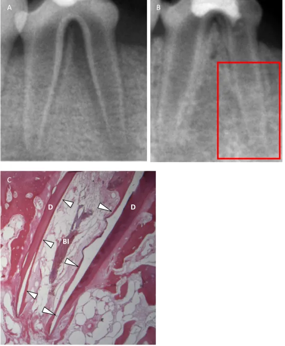
Group 1: 12 teeth: Infected! Disinfected! Induced Bleeding (Fig. 1) (B)
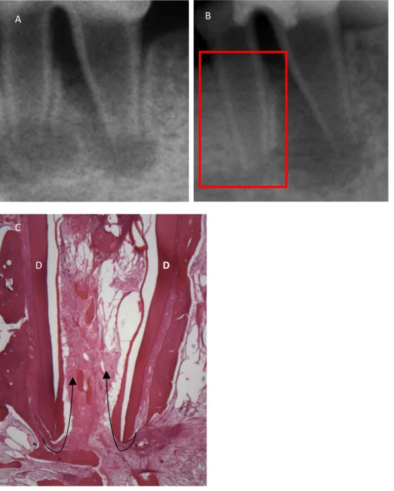
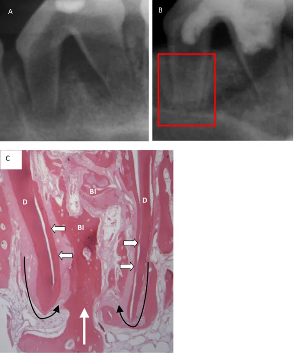
Group 2: 12 teeth: Infected! Disinfected! EDTA 17% ! Induced Bleeding (Fig. 2) (BE) Group 3: 12 teeth: Infected! Disinfected! Induced bleeding + Collagen Scaffold (Fig. 3) (BS) Group 4: 12 teeth: Infected! Disinfected! EDTA 17% + Induced Bleeding + Collagen
Scaffold (Fig. 4) (BES)
Group 5: 12 teeth – Negative control untouched teeth left to develop naturally for comparison Group 6: 4 teeth – Positive control infected with no treatment
2.4 Results
Three of the positive control teeth (infected and left with no treatments) had to be
extracted due to abscess formation and excessive mobility. The loss of these teeth did not cause
the animals any changes in behavior or eating habits. The extracted teeth were also fixed and
sectioned, and examined as described above.
Radiographic Analyses
Periradicular radiolucencies. Chi-square evaluation for the presence or
absence/improvement of periradicular radiolucencies showed a significant difference between
the groups (p=0.04) in which a scaffold was used (Fig. 5). After disinfection, BES had 87.5%
healing, BS had 79.2%, BE had 58.3%, and B had 56.52%.
Radiographic thickening of root canal walls.Chi-square evaluation between the groups
showed a statistically significant difference between the groups, which contained the scaffold
(p=0.03) (Fig. 6). After treatment, BES had 83.3% thickening, BS had 87.5%, BE had 54.2%,
and B had 65.2%.
Histological Analyses
The positive control teeth (infected only) showed no signs of newly formed tissues (i.e.,
no wall thickening or in growth of tissue into the lumen). The negative controls all showed
Mineralized Tissue Formation On Inner Dentin Walls and Mineralized Tissue Islands In Lumen
There were two types of newly formed mineralized tissues in the canal space: one
adhering to/detached from the inner dentin wall (dentin associated mineralized tissue) (DAMT)
(Fig. 7), another forming bony islands (BI) in the inner lumen independent of the dentin wall
(Fig. 7). DAMT appears as mineralized tissue with relatively uniform thickness devoid of
vasculature, and some portion anchored to the dentin wall while some detached from the wall.
There were relatively few cells embedded in the mineralized matrix. BI, on the other hand,
appeared to be distinct from DAMT. It appeared as mineralized matrix islands that contained
many embedded cells, blood vessels, and bone marrow-like tissues (Fig. 4D).
Kruskal-Wallis test revealed significant difference between the four experimental groups
in total mineralized tissue formation (p=.02) as well as in mineralized tissue wall formation on
the inner dentin walls (p=.04). Though the difference did not reach a significant level (p=0.056)
between the groups in bone-like mineralized tissue formation in the lumen, there was a clear
trend seen in which the groups with the scaffold had increased mineralized island tissue
formation. Figure 8 shows the mineralized tissue formation for each group with further
pair-wise comparison between each experimental group showing significantly more total mineralized
tissue formation in groups containing the scaffold (p<0.05).
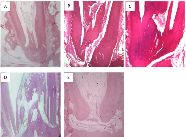
Apical Development
Several experimental groups showed continued apical growth and mineralized tissue
formation which closed the apex, while others remained open (Fig. 9). This was seen most often
in the groups containing the scaffold. The apical development appeared to be an
extension/growth of the cementum/DAMT structure and did not resemble dentin (i.e., devoid
of dentinal tubules). In other experimental groups there was no apical closure and only
(Fig. 9). One tooth that had the same treatment rendered could still show one root with apical
closure and the other with no closure (Fig. 3C; Fig. 3D).
Effect of Demineralization and Exposure of Dentin Matrix
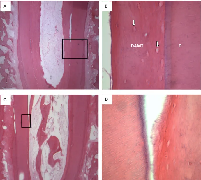
In several cases in which 17% EDTA was used to demineralize the dentin and partially
expose the dentin matrix, there were hair-like projections from the DAMT that appeared to be
mechanically embedding to the dentin wall (Fig. 10). This was not seen when the dentin was not
demineralized.
2.5 Discussion
Pulp necrosis of an immature tooth poses potential complications and challenges for
treatment. Use of standard cleaning and shaping protocol with endodontic files is not advised
due to the thin dentinal walls as instrumentation of already thin walls would further weaken the
tooth. The obturation may not obtain a predictable apical seal due to a large opening of the apex.
Though treatment techniques for either stimulating formation of a hard tissue barrier (i.e.,
long-term calcium hydroxide apexification treatment) or using materials to create an artificial barrier
(i.e., MTA apexification) have shown success (14-17), the long-term effects on the periodontal
tissues and tooth structure are still not well documented. Even if these treatments are
successfully performed, the remaining thin dentinal walls leaves the tooth weak and with an
increased risk of subsequent fracture. It has been shown that approximately 30% of immature
teeth can fracture during or after treatment (15). Root reinforcement techniques using composite
resin have been shown to internally strengthen the teeth, however, these pose problems for
retreatment (18). Synthetic materials that are used at present cannot replicate the durable and
dynamic collagen-based mineralized tissues such as dentin, bone, and cementum. Thus, an ideal
induce a mineralized structure within the canal space with the aid of surrounding tissues and
cells in order to strengthen these teeth.
It has been reported that in human patients, induction of bleeding (i.e., blood clots) in
the root canal space after proper disinfection facilitates root wall thickening and lengthening (2).
Based on radiographic evaluation, the authors speculated that the root and periapical structure
could have been further developed. A recent study by Thibodeau et al. demonstrated that, using
a dog model, blood clot induction indeed stimulated root thickening and in-growth of vital tissue
within the root canal space (3).
Although these results show promise for regenerative endodontic therapies, there are
still uncertainties and many questions that need to be addressed. For example:
1) How can the root thickening and possibly lengthening be improved and be a predictable treatment?
2) What are the characteristics of the mineralized tissues formed (e.g., quality of matrix)?
3) Can such teeth be mechanically comparable to naturally developed teeth?
Using a dog model, it was our attempt to address the first point in this report and the details of
the second in another publication. !
Improvement of Root Thickening and Lengthening: Use of A Collagen Scaffold
In the!current study, we demonstrated that the use of a cross-linked collagen scaffold
significantly increased formation of mineralized tissues. In the study by Thibodeau et al. (2007),
after disinfection, blood clots were induced with and without collagen solution (3). The authors
reported that while induction of blood clots helped mineralized tissue formation in the canal
space, an addition of collagen solution did not improve the tissue formation. This may be
because the “soluble” collagen was quickly resorbed, thus, not providing any effect. In our study,
irradiation and thermal dehydration. Therefore, it is less immunogenic and does not get resorbed
quickly allowing time for cells to migrate and proliferate. Nevins et al. (1998) did a study in
which cross-linked collagen gel was utilized within the root canal space (19). Although there was
no quantitative analysis to show any differences in the linked collagen gel or the non
cross-linked collagen gel, the study indicated that the cross-linking scaffold may improve treatment
outcomes. This study used the collagen on vital pulp tissue, while our study used the collagen
scaffold on necrotic, immature teeth. No other studies, to the best of our knowledge, have used
cross-linked collagen scaffolds in regenerative endodontic treatments in necrotic immature teeth.
The double-blinded radiographic examination indeed revealed the occurrence of
periapical bone healing and root thickening are both significantly greater in the groups with
collagen scaffold in comparison to those without it (Fig. 5). Improved healing of the apical
periodontitis in the collagen scaffold group was an interesting finding. It may have to do with
the osseoinductive properties of the scaffold. It is possible that some of the scaffold was pushed
out into the granulation tissue/PA tissues during treatment and may have affected healing
response, but this needs to be investigated further.
Histomorphometric analysis, the placement of the collagen scaffold significantly
increased the formation of new mineralized tissues in the canal space in comparison to the
groups without. This indicates that cross-linked collagen sponge, as compared to collagen
solution, is more suitable as a scaffold for the periapical mesenchymal or periodontal ligament
derived stem cells (PDLSC) to migrate and differentiate into mineralized tissue forming cells,
e.g., cementoblast/osteoblast-like cells. It is most likely that the porous, cross-linked
atelo-collagen sponge provided cells with a stable microenvironment allowing them to adhere and
In addition, from a clinical perspective, the use of the scaffold also allowed for easier and
more predictable outcome than MTA placement. When MTA is placed directly on the blood,
many times it can be pushed down the canal space and interfere with the generation of new
tissues.
Effects of Demineralization and Exposure of Dentin Matrix
Furthermore, in the current study, the effect of dentin matrix exposure with 17% EDTA
on the mineralized tissue formation was examined. EDTA has been shown to remove smear
layer to expose the dentin matrix (21, 22) and also appears not to damage stem cells (23).Dentin
matrix was exposed to allow the mesenchymal stem cells to interact with dentin matrix that may
help differentiate into odontoblast-like cells. As stated above, replantation/autotransplantation
studies show that the original pulp and odontoblastic layer rarely remain. Our results indicated
that such exposure did not facilitate to form odontoblast-like cells and thus dentin-like structure
(Fig. 10). There could be several reasons for this including:
1) The matrix component was not sufficiently exposed with the condition employed. In
this study, a mild demineralization condition, i.e. 17% EDTA for 2 minutes was chosen
considering the potential adverse effects on periapical cells. However, the treatment
conditions may not be sufficient to expose dentin matrix as the rate of demineralization
with 17% EDTA is relatively slow when compared to dilute acids like citric acid (24).
2) Possibly, factors important for odontoblast differentiation could be extracted with
EDTA. For instance, phosphoproteins/phophophoryns are mostly extracted with
EDTA (25, 26).
3) Mesenchymal stem cells residing in periodontal ligament and periapical area could be
more prone to differentiate into cementoblast/osteoblast-like cells when contact with
Though the apparent dentin-like structure was not observed, adherence of the
mineralized tissue to dentin wall seemed enhanced with the EDTA treatment. Several of the
histological samples showed a cleavage plane between the newly formed cementum-like tissue
and the dentin wall, which was likely due to processing. In the cases treated with EDTA, there
were several cases that showed projections/hair-like structures from the newly formed
mineralized tissue which appeared to adhere or interlock to the demineralized dentin wall.
Removal of smear layers may enhance attachment of cells to the wall surface (11). Exposing the
matrix did not show any negative effects in our study and may help adherence and strengthening
of the root walls. However, more studies need to be done to investigate this issue. One of the
main goals of regenerative endodontic procedures in immature teeth is to improve root wall
strengthening and length. It is not yet known whether these regenerative endodontic treatments
improve wall strength.
Characteristics of the Formed Mineralized Tissue
Our treatments produced!mainly two types of newly formed mineralized tissues in and
around the root canal structure. DAMT formed along the dentin wall are distinct from those
formed in the canal space (Fig. 4C, 4D). The former is a mineralized tissue that was devoid of
vasculature, contained embedded cells, and lacked apparent dentinal tubules-like structure (Fig.
7D). Thus, it resembles cementum. The mineralized tissue formed in the lumen, on the other
hand, resembles bone because of the presence of marrow and vasculature. Very recently, using
the histological specimens on a dog study previously done by Thibodeau et al. (3), Wang et al.
did further histological analysis (27). They also showed that in cases of thickened walls, there was
a cementum-like tissue formed along the inner dentin wall termed as intracanal cementum-like
tissue (IC) and the lumen was often filled with bone-like tissue that was described as intracanal
odontoblastic layer present. It is unknown whether some pulp tissue may have survived from the
initial stage. We did not observe any cases of a pulp-like tissue with an intact odontoblast-like
cell layer. In cases of autotransplanted and replanted teeth, it is rare to see the odontoblastic
layer remain (28). These cases have an intact, uninfected pulp, which is immediately
replanted/transplanted, and yet the odontoblastic layer usually will not survive.
From our study as well as others, induction of bleeding produces DAMT and BI. The
induced bleeding and subsequent formation of blood clot may serve not only as a scaffold, but
as a source of osteogenic cells and stem cells from the granulation tissue, apical papilla (SCAP),
PDL (PDLSC), and/or from the bone marrow and peripheral blood (29-32, 6).
Platelets make up a large part of a blood clot. They contain and secrete active growth
factors, and contain a number of serum proteins including fibrin, fibronectin, and vitronectin,
that can act as cell adhesion molecules for osteoconduction. Platelets also serve as a matrix for
bone, connective tissue, and epithelial cell migration in wound healing (33). The blood clot may
serve as a scaffold for the periapical cells including mesenchymal stem cells to migrate into the
root canal and eventually induce new tissue formation in the space (3).
The periodontal ligament contains stem cells (PDLSC) and the potential of PDLSC to
differentiate into specific cell lineages producing cementum/PDL-like structures has been
shown (34). In our study, some histological sections showed PDL migrating into the canal space
(Fig. 2C; Fig. 4C) as Wang et al. reported (28). Thus, the PDL cells may be repopulating the
inner dentin walls and inducing DAMT formation (28). Vojinovic showed that periodontal cells
migrate into the root canal space after pulpectomy in immature vital teeth, which may be a
similar mechanism as indicated in our study (35). Studies have shown that PDL cells can
repopulate damaged or exposed cementum/dentin surfaces, and form cementum-like tissues
cementum-like tissue being formed on the inner dentin walls. However, it is interesting to note
that in many cases, the cementum-like tissue being formed is much thicker than the natural
cementum layer. It is unknown if given time, this newly formed cementum-like tissue would
continue to be deposited until there is complete closure of the lumen space. Bone-like tissue
seemed to grow up into the lumen space and at times appeared to become continuous with the
cementum-like tissue. It appeared in many specimens that the bone-like islands were growing in
separately into the lumen space, possible from the bone marrow in the perirapical area. Both the
PDL cells migrating along the dentin walls and the cells from the bone marrow are vying or
competing for space inside the root canal lumen. Some cases showed the PDL-like tissue
between the DAMT and the BI as it would be naturally occurring on the outer root wall. It is
possible that the different types of cells may be directed to their preferred microenvironment to
form their respective tissues. The mechanisms by which those cells are directed and
differentiated into specific mineralized tissue-forming cells are warranted for further studies.
Apical development in the experimental groups was an interesting finding. In some
cases, there was continued cementum-like tissue deposition and closure of the apex, while in
other cases the apex remained open. Even in the same tooth which had very similar root
dimensions and the same treatment rendered, there were cases in which one root may show
apical closure and the other root it remains open (Fig. 3C, 3D; Fig. 9). It is not clear exactly why
this occurs. In clinical human studies on regenerative endodontic treatments, it appears that
there are only a certain percentage of cases that show increase in root length (4). It is possible
that some of the cases in which the root appears to gain length is due to cementum-like tissue
formation and not continued natural root development. It is unlikely that Hertwig’s epithelial
root sheath (HERS) remains intact and allow for natural continued root development in cases
studies show that the root can continue to develop if the HERS remains intact (37). In these
cases there is no infection and thus the HERS may remain intact.
Further Characterization of the Newly Formed Tissues
In order to characterize the mineralized tissues formed within the root canal space, we
are currently performing different staining methods and immunohistochemical studies using
dentin sialophosphoprotein (DSPP) and bone sialoprotein (BSP), that will be a subject of
separate publication. DSPP is much more prevalent in dentin, while BSP in more prevalent in
bone and cementum. Staining the newly formed tissues will help to further identify them.
There is great potential for tissue engineering strategies in treatment of diseased teeth
(10). Continued research investigating the use of stem cells, scaffolds, growth factors/signals,
gene therapy, etc., in root canal therapy will hopefully provide a way to predictably
regenerate/engineer a vital support structure within necrotic root canal spaces.
2.6 Conclusion
In conclusion, in the dog model with incomplete root development and periapical
periodontitis, the use of cross-linked collagen scaffold combined with bleeding induction
significantly enhanced the mineralized tissue formation in the canal space and partial
demineralization of the dentin wall could help adherence of mineralized tissue to the inner
dentin walls. This may provide an insight into the development of an efficient strategy to treat
PART TWO 2.7 List of Figures
Figures
Figure 1. Example of group with induced bleeding only (group B)………...…32 Figure 2. Example of group with 17% EDTA treatment and induced
bleeding (group BE)………..…..33 !
Figure 3. Example of group with induced bleeding and cross-linked
collagen scaffold (group B)...………...………..…………...34 !
Figure 4. Group with 17% EDTA, induced bleeding, and cross-linked
collagen scaffold (group BES)...35 !
Figure 5. Percentage of roots with improvement of periapical radiolucencies
assessed radiographically………...36 !
Figure 6. Percentage of roots with thickening of root walls assessed radiographically…..……...37 Figure 7. DAMT and BI within the canal space.………...38 Figure 8: Newly formed mineralized tissues assessed by histomorphometry...39 Figure 9. Differences in apical development...40 Figure 10. Thickening of the root wall with DAMT and the effects of partial
Figure 1. Example of group with induced bleeding only (Group B). (A) Pre and (B) post
radiographs showing healing of periradicular area and some increase in radiopacity around the root walls and in the lumen space. (C) H and E section of the boxed in root from (B). Thin layer of DAMT present indicated by white arrows and BI is present.
"!
#!
$!
!"
Figure 2. Example of group with 17% EDTA treatment and induced bleeding (Group BE). (A) Pre and (B) post operative radiographs. Root on left shows healing and some radiographic thickening which is more noticeable around the apical area. Root on the right shows no healing and no increased thickening. (C) H and E section of the boxed in root. Thin DAMT formation is present along the inner dentin wall (D) with migration of PDL along the lumen side of the DAMT indicated by black arrows. Small bony island are present in the lumen space.
"! $!
#!
Figure 3. Example of group with induced bleeding and cross-linked collagen scaffold (Group BS).
(A) Pre and (B) post operative radiographs. (C) H and E section of the red box in root in (B). (D) H and E section of the yellow box in (B). A thicker DAMT layer is present indicated by white triangles. Several areas of bony islands are present within the lumen space. The tooth received the same treatment but showed different apical development and lumen fill.
"! $!
#!
!" !"
Figure 4. Group with 17% EDTA, induced bleeding, and cross-linked collagen scaffold (Group BES). (A) Pre and (B) post operative radiographs. (C) H and E section of boxed in root from (B). Thin black arrows indicate migration of PDL into the lumen space along the inner dentin walls (D). DAMT (indicated by the white arrows) is formed along the inner dentin walls. Bony islands are formed within the lumen space, and shows as a direct extension from the PA bone as indicated by white arrow.
"!
#!
$!
!" #$"
#$"
Figure 7. DAMT and bony islands (BI) within the canal space. (A) BE group showing DAMT and
BI. White arrows indicate BI. White triangles indicate DAMT. (B) BES group showing DAMT and BI. White arrows indicate BI. White triangle indicates DAMT.
Figure 9. Differences in apical development. (A) BES group with apical closure. (B) BS group. (C)
BES group. (D) BES group showing no apical closure and in growth of bony islands. (E) BES group showing partial closure of the apical end.
"!
&! %!
Figure 10. Thickening of the root wall with DAMT and the effects of partial exposure of the dentin
matrix with 17% EDTA. (A) H and E section of BES group at the mid-root level. DAMT is thick and uniformly attached to the inner dentin wall. (B) High magnification of boxed area from (A). DAMT appears tightly attached to the dentin (D). White arrows indicate the cells embedded in the DAMT matrix. (C) H and E section of BE group and the mid-root level at an area where there was a cleavage plane. (D) High magnification of boxed in area from (C) showing DAMT with hair-like projections appearing to embed themselves into the exposed dentin matrix.
#!
!%&'" !" "
"! $!