FLAWED PHOSPHOLIPID FORMATION OR FAULTY FATTY ACID OXIDATION: DETERMINING THE CAUSE OF MITOCHONDRIAL DYSFUNCTION IN HEARTS LACKING
ACSL1
Trisha J. Grevengoed
A dissertation submitted to the faculty at the University of North Carolina at Chapel Hill in partial fulfillment of the requirements for the degree of Doctor of Philosophy in the Department of Nutrition
(Biochemistry) in the School of Public Health.
Chapel Hill 2015
iii
ABSTRACT
Trisha J. Grevengoed: Fatty acid activation in cardiac mitochondria: The role of ACSL1 in phospholipid formation and remodeling, substrate switching, and autophagic flux
(Under the direction of Rosalind A. Coleman)
Cardiovascular disease is the number one cause of death worldwide. In the heart, mitochondria provide up to 95% of energy, with most of this energy coming from metabolism of fatty acids (FA). FA must be converted to acyl-CoAs by acyl-CoA synthetases (ACS) before entry into pathways of β-oxidation or glycerolipid synthesis. ACSL1 contributes more than 90% of total cardiac ACSL activity, and mice with an inducible knockout of ACSL1 (Acsl1T-/-) have impaired cardiac FA oxidation. The effects of loss of ACSL1 on mitochondrial respiratory function, phospholipid formation, or autophagic flux have not yet been studied.
Acsl1T-/- hearts contained 3-fold more mitochondria with abnormal structure and displayed lower
respiratory function. Because ACSL1 exhibited a strong substrate preference for linoleate (18:2), we investigated the composition of mitochondrial phospholipids. Acsl1T-/- hearts contained 83% less tetralinoleoyl-cardiolipin (CL), the major form present in control hearts. Modulating ACSL1 expression in cell lines confirmed that ACSL1 is necessary for linoleate incorporation into CL. To determine whether increasing content of linoleate in CL would improve mitochondrial respiratory function, control
and Acsl1T-/- mice were fed a high linoleate diet, which normalized amount of tetralinoleoyl-CL, but did
not improve respiratory function.
The metabolic switch from FA use to high glucose use activates mechanistic target of rapamycin complex 1 (mTORC1), which initiates growth by increasing protein and RNA synthesis and FA
iv
consumption, which was likely caused by lower ATP synthase activity present in both vehicle- and rapamycin-treated Acsl1T-/- hearts. The autophagic rate was 88% lower in Acsl1T-/- hearts. mTORC1 inhibition increased autophagy to a rate that was 3.1-fold higher than in controls, allowing clearance of damaged mitochondria. ACSL1 deficiency in heart activated mTORC1, thereby inhibiting autophagy and increasing the number of damaged mitochondria with impaired respiratory capacity.
v
ACKNOWLEDGEMENTS
I would like to thank Dr. Rosalind Coleman for helping me develop the skills needed to become an independent researcher. She has instilled the value of critical thinking in every aspect of science. Her support and guidance has been invaluable to my development as a scientist.
Rosalind has also provided me with a laboratory of people that I can learn from: Jessica Ellis’s support through my first years of graduate school was vital to that time. Daniel Cooper has gone through much of the process with me and has been a useful critic of experiments as well a sympathetic ear when experiments inevitably did not work. Others in the lab, including David Paul, Angela Wendel, Matthew Keogh, and Florencia Pascual have provided guidance and support that I could not have done without.
vi
PREFACE
vii
TABLE OF CONTENTS
LIST OF TABLES ... ix
LIST OF FIGURES ... x
LIST OF ABBREVIATIONS ... xi
CHAPTER 1: BACKGROUND ... 1
Partitioning of fatty acids and the formation of fatty acyl-CoAs ... 1
Fatty acid use by acyl-CoA synthetases ... 2
Acyl-CoA binding protein and fatty acid binding proteins ... 3
Complex lipid synthesis and degradation ... 5
Acyl-CoA synthetases and fatty acid transport proteins ... 10
Regulation of long-chain ACS isoforms ... 12
Channeling. ... 14
Knockout and knockdown of ACSL1 ... 15
Overexpression of ACSL1 ... 17
Role of acyl-CoAs in disease ... 18
Background Figures ... 26
Specific Aims ... 34
CHAPTER 2: ACYL-COA SYNTHETASE 1 DEFICIENCY ALTERS CARDIOLIPIN SPECIES AND IMPAIRS MITOCHONDRIAL FUNCTION ... 36
Introduction ... 37
Methods ... 38
Results: ... 44
Discussion ... 51
Figures ... 54
CHAPTER 3: LOSS OF ACSL1 IMPAIRS CARDIAC AUTOPHAGY AND MITOCHONDRIAL STRUCTURE THROUGH MTORC1 ACTIVATION ... 67
Introduction ... 68
Materials and Methods ... 69
Results ... 72
Discussion ... 77
viii
CHAPTER 4: SYNTHESIS ... 89
Overview of findings ... 89
Public Health Significance ... 90
Future Directions ... 91
ix
LIST OF TABLES
x
LIST OF FIGURES
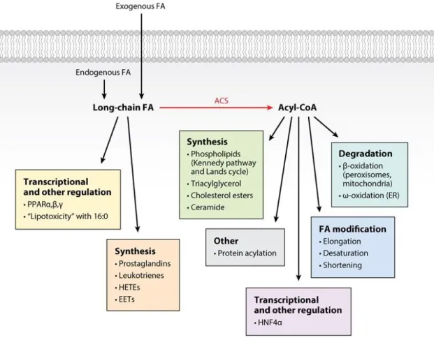
Figure 1.1 Metabolic fates of long-chain fatty acids. ... 26
Figure 1.2 Acyl-CoA metabolism. ... 27
Figure 1.3 Glycerolipid synthesis. ... 28
Figure 1.4. Cardiolipin remodeling in mammalian cells... 30
Figure 1.5 Cardiolipin in ETC function. ... 32
Figure 1.6. Mitochondrial quality control through fission and fusion, autophagy, or apoptosis. ... 33
Figure 2.1. ACSL1 is located on cardiac mitochondria and activates linoleate preferentially. ... 54
Figure 2.2. Loss of ACSL1 caused mitochondrial dysfunction. ... 55
Figure 2.3. Loss of ACSL1 alters acyl-chain composition of mitochondrial phospholipids. ... 58
Figure 2.4. Knockdown of ACSL1 impairs fatty acid oxidation and incorporation into lipids. ... 60
Figure 2.5. Overexpression of ACSL1 increases linoleate metabolism. ... 61
Figure 2.6. Pulse-chase and etomoxir treatment of H9c2 cells overexpressing ACSL1. ... 62
Figure 2.7. ACSL1 overexpression increased linoleate incorporation into CL in HEK-293 cells. ... 63
Figure 2.8. High linoleate diet partially normalized CL acyl-chain profile in Acsl1T-/- hearts but did not improve mitochondrial respiratory function. ... 64
Figure 2.9. Proposed pathway for how ACSL1 increases linoleate incorporation into cardiolipin (CL). .. 65
Figure 3.1. Loss of cardiac ACSL1 decreased fatty acid use and increased the use of glucose. ... 81
Figure 3.2. Two weeks of rapamycin treatment inhibited mTORC1 activation in Acsl1T-/- hearts. ... 82
Figure 3.3. mTORC1 inhibition improved mitochondrial structure in Acsl1T-/- hearts. ... 84
Figure 3.4. Rapamycin treatment normalized high mitochondrial number in Acsl1T-/-hearts. ... 85
Figure 3.5. Inhibition of mTORC1 activated autophagy in Acsl1T-/- hearts. ... 86
Figure 3.6. Rapamycin treatment partially normalized mitochondrial function in Acsl1T-/- hearts. ... 87
xi
LIST OF ABBREVIATIONS
ACBP Acyl-CoA binding protein ACC Acetyl-CoA carboxylase ACOT Acyl-CoA thioesterase ACS Acyl-CoA synthetase ACSBg ACS bubblegum ACSL Long-chain ACS ACSVL Very-long-chain ACS
AGPAT Acyl-CoA:1-acylglycerol-3-phosphate acyltransferase ANT Adenine nucleotide transferase
ASM Acid soluble metabolites Atg Autophagic protein
CDP-DAG Cytidine diphosphate-DAG
CEPT Diacylglycerol choline/ethanolamine phosphotransferase CMC Critical micellar concentration
CL Cardiolipin
CPT1 Carnitine palmitoyltransferase-1 DAG Diacylglycerol
DGAT Diacylglycerol acyltransferase ER Endoplasmic reticulum FA Fatty acid
FABP FA binding protein
FATP Fatty acid transport protein
xii HSL Hormone sensitive lipase
LC3b Microtubule associated protein light chain 3b mTOR Mechanistic target of rapamycin
mTORC1 Mechanistic target of rapamycin complex 1 p62/SQSTM1 Sequestome 1
OCR Oxygen consumption rate PA Phosphatidic acid
PC Phosphatidylcholine PE Phosphatidylethanolamine
PEMT Phosphatidylethanolamine N-methyltransferase PG Phosphatidylglycerol
PGPP Phosphatidylglycerophosphate phosphatase PGPS Phosphatidylglycerophosphate synthase PI Phosphatidylinositol
PIS Phosphatidylinositol synthase PS Phosphatidylserine
PSD Phosphatidylserine decarboxylase PSS Phosphatidylserine synthase SNP Single nucleotide polymorphism T3 Triiodothyronine
TAG Triacylglycerol
1
CHAPTER 1: BACKGROUND
Partitioning of fatty acids and the formation of fatty acyl-CoAs
Long-chain FAs derived from either de novo synthesis, dietary sources, or from the turnover of triacylglycerol, phospholipids, and cholesterol esters have multiple metabolic fates. These fates include the entry of FAs into pathways of degradation, incorporation or reincorporation into complex lipids, esterification to proteins, and the synthesis of eicosanoids. Long-chain FAs have additional roles in activating transcription factors, functioning as intracellular signals, and allosterically
modulating enzyme reactions (Fig. 1.1). Each of these outcomes except for some of those related to signaling and eicosanoid formation require the formation of a long-chain acyl-CoA by one of least 13 acyl-CoA synthetases (ACS) that use long-chain and very-long-chain FAs (ACSL, ACSVL, ACSBg). The 13 ACS isoforms are part of the 26-member ACS family, all of which contain related nucleotide (AMP/ATP) and FA binding motifs, and most of which include a transmembrane domain anchor at the N-terminus.
The long-chain ACS isoforms activate FA of 16-22 carbons (2,3) in an energetically costly two-step reaction that uses the equivalent of two high-energy bonds:
Fatty acid + ATP acyl-AMP + PPi Acyl-AMP + CoASH acyl-CoA + AMP
In addition to their uses in β-oxidation and glycerolipids synthesis (Fig. 1.2), acyl-CoAs are
critical signaling molecules as allosteric inhibitors of adenosine nucleotide translocase (ANT) (4,5), liver glucokinase (6), acetyl-CoA carboxylase (ACC) (7,8), HMG-CoA reductase (9),
2
micelles in aqueous solutions with the CoA groups exposed to the water phase (13). The measured critical micellar concentrations (CMC) for the most common long-chain acyl-CoAs, 18:1-CoA and 16:0-CoA are about 32 and 42 µM, respectively (14,15). However, within cells, acyl-CoAs are probably bound to proteins and membranes so that the concentration of acyl-CoAs would be too low to self-aggregate. Because of their amphipathic nature, acyl-CoAs can interfere with membrane integrity by acting as detergents; when high concentrations of acyl-CoAs are present, the permeability of membranes to small molecules like sucrose and citrate is altered (16). Myristoylation or
palmitoylation of proteins requires acyl-CoAs, but to our knowledge, no changes in protein acylation have been found in mice or cells with a deficiency of an ACS.
Because most mammalian cells contain several different long-chain ACS isoforms, it has been hypothesized that each isoform may partition or channel its long-chain FA substrates into specific downstream pathways. In addition, several of the isoforms have two different start sites, one of which lacks an N-terminal transmembrane anchor, or have alternative internal exons that result from differential splicing (17). Hypothesized differences in cell function include the use of the activated FA for pathways that synthesize glycerolipids and cholesterol esters, for pathways of FA elongation or desaturation, for degradative pathways in the mitochondria, ER, and peroxisomes, for protein acylation, and for transcriptional regulation.
Fatty acid use by acyl-CoA synthetases
FAs are carboxylic acids with long-chain hydrocarbon side groups. In animals, the
predominant long-chain FAs are those of 16 and 18 carbons with varying degrees of saturation. FAs of 20 carbons, like 20:4ω6 and 20:5ω3 form a small percent of the total FA content in animals, but
3
Alternatively, entry might be mediated by fatty acid binding protein (FABP) isoforms (18), or facilitated by the FA transport proteins (FATP) (19) that are themselves acyl-CoA synthetases.
Several groups, however, have shown that the rapidity of FA entry or “vectorial transport” is driven
by intracellular metabolism of the FA (18,20,21).
Understanding the process that channels FAs into specific metabolic pathways requires consideration of the physical chemistry of hydrophobic FAs which must move in an aqueous
environment. Further, in order to minimize futile cycles, synthetic and degradative pathways must be separated from one another both spatially and temporally. Cells overcome the problem of
hydrophobicity by converting the FA to an amphipathic molecule by the thioesterification of Coenzyme A to the carboxyl group. The ability of the cell to vectorially channel fatty acyl-CoAs towards or away from a metabolic pathway forms the basis of partitioning, and is likely to vary with cell type, intracellular location of carriers and enzymes, cellular energy status, and hormonal signals.
Acyl-CoA binding protein and fatty acid binding proteins
Selective partitioning of acyl-CoAs within cells requires methods of overcoming hydrophobicity, because the amphipathic fatty acyl-CoAs can move freely both in the aqueous cytosol and in
membrane monolayers. Two protein families, FABPs and acyl-CoA binding protein (ACBP), aid in FA and acyl-CoA movement within cells and are believed to protect cell membranes from the detergent effects of the acyl-CoAs. FABPs are isoforms of a 10 member intracellular lipid-binding protein family which reversibly binds hydrophobic ligands and, in theory, traffics them throughout the cytosol to various organelles (22). A recent comprehensive review of FABP isoforms identifies metabolic alterations in knockout models, but definitive functions have not been established (23). FABP isoforms are ubiquitously expressed, but differ in stoichiometry, affinity and specificity toward related ligands that include FAs, acyl-CoAs, eicosanoids, and peroxisome proliferator-activated receptor ligands. The amount of an FABP isoform in any tissue appears to reflect the tissue’s
4
specialize in lipid metabolism, FABPs make up 1–5% of all cytosolic proteins (24). Evidence for the importance of FABPs in lipid metabolism comes from loss-of-function studies in mice. FABP1, which is strongly expressed in liver and intestine, is the only isoform that binds both FA and fatty acyl-CoA; the other FABP isoforms bind only FA (24,25). Two independent Fabp1-/-mouse models have been generated but, despite the importance of lipid metabolism in liver and intestine, neither model has an overt phenotype (26,27). When mice are fed low fat chow, Fabp1-/-liver appears normal histologically, and serum TAG and total free FA levels are unchanged, although alterations in specific FAs are observed (28). In one of the Fabp1-/- models, the hepatic content of phospholipid, cholesterol, and cholesterol ester is greater than in the controls (28). Although the loss of Fabp1
reduces hepatic FA binding capacity, total liver lipid content, including TAG and free FA, is
unchanged. Only under extreme fasting conditions (48 hours) does the reduced FA binding capacity in the knockout mice cause a reduction in hepatic FA uptake, FA oxidation, and TAG levels (27). Although differences were observed in the effects of knockouts of the FABP1 and the intestinal FABP isoform, information related to acyl-CoAs was not provided (29). The adipose-type FABP4 (also known as aP2) is the major isoform in white and brown adipose tissue and macrophages (30,31). Because disruption of the Fabp4 gene in mice increases the cytosolic content of free FA, FABP4 is generally thought to facilitate FA transport between intracellular compartments for storage or export (32,33), however this model provides no evidence for mistargeted intracellular FA. The heart-type FABP3, which is most abundantly expressed in heart, skeletal muscle, and brain, is induced by acute cold exposure in rat brown adipose tissue (BAT) (34,35), by a 5-day cold exposure, or by a β3
-adrenergic receptor agonist in mouse subcutaneous white adipose as cells became “beiged” (36).
5
ACBPs bind medium- and long-chain acyl-CoAs with high affinity, but does not bind free FA, acyl-carnitine, or cholesterol (40). The affinity for acyl-CoAs is so much higher for ACBP than for liver-type FABP1 that it was suggested that ACBP is the major carrier of acyl-CoA in all cells including hepatocytes (41). ACBP expression and concentration are highest in liver, but ACBP is also present in high levels in the adrenal cortex, testis and epithelial cells. Because these tissues and cells specialize in secretion, they have high energy needs and may require ACBP to shuttle fatty acyl-CoAs towards energy producing oxidative pathways (42). Disruption of the ACBP homologue in yeast (ACB1), does not affect phospholipid synthesis or turnover, indicating that ACBP is not required for glycerolipid synthesis in yeast. However, yeast deficient in ACB1 have disordered plasma membrane structures as a result of aberrant and reduced sphingolipid synthesis (43). Highlighting the importance of ACBP in in vivo metabolism are studies from two separate Acbp
deficient mouse models. In the first model, the authors concluded that ACBP is an essential protein required for embryonic development because an implantation defect results in embryonic lethality (44). The second knockout was viable, but did not indicate a role for ACBP in trafficking acyl-CoAs, although liver acyl-CoA levels were ~40% lower than in controls; instead, the main effect of the knockout was an impaired skin barrier and the development of alopecia (45). In addition to their skin phenotype, Acbp-/- mice undergo a crisis around the weaning period, exhibiting weakness and poor weight gain (46). At this time point, SREBP maturation is impaired and SREBP target genes
involved in cholesterol biogenesis are not appropriately upregulated. It is unclear why two separately generated Acbp knockout models express these two disparate phenotypes, but these models indicate that ACBP and the acyl-CoAs they bind are essential for normal growth and development.
Complex lipid synthesis and degradation
6
ER ω-oxidation (48,49). Evolutionarily, the cell has developed organelles that perform each of these
processes, evidence of an additional level of fatty acyl-CoA partitioning. Further evidence for this type of partitioning lies in the organelle localization of synthetic and oxidative enzymes. However, although one generally thinks of each organelle as specializing in a single function, in fact, both glycerolipid synthesis and ω-oxidation occur in the ER, both alkyl lipid synthesis and β-oxidation take place in peroxisomes, and both FA synthesis and β-oxidation occur in mitochondria. It is not
known how FA and acyl-CoAs are independently directed into these separate pathways.
Glycerolipid Synthesis
The initial and committed step for the de novo synthesis of TAG and all glycerophospholipids is the acylation of sn-glycerol-3-phosphate with a fatty acyl-CoA to form 1-acyl-sn
-glycerol-3-phosphate (lysophosphatidic acid) catalyzed by glycerol-3--glycerol-3-phosphate acyltransferase (GPAT) (50). GPAT isoforms are present in the outer mitochondrial membrane (GPAT1 and -2) and in the endoplasmic reticulum (GPAT3 and -4) (51). Overexpression of GPAT1 in either isolated primary rat hepatocytes or in vivo in rats causes steatosis, confirming the important role of GPAT in initiating hepatic TAG synthesis (52,53). Mouse knockout models of the GPAT isoforms have provided clues as to the partitioning of acyl-CoAs towards synthetic or oxidative pathways. In studies comparing
Gpat1-/- and Gpat4-/- mice, for example, GPAT1, but not GPAT4, is required to incorporate de novo
synthesized FA into TAG and to divert FA away from oxidation (54). The ER GPATs are likely to channel exogenously derived acyl-CoAs towards TAG or phospholipid synthesis. It is possible that the location of GPAT1 at the outer mitochondrial membrane serves to divert de novo synthesized fatty acyl-CoAs away from carnitine palmitoyltransferase-1 (CPT1)-mediated entrance into the mitochondria where they would be oxidized. This hypothesis makes sense from a cellular
7
status. With low cellular energy, activated AMP-activated kinase inhibits GPAT1 and favors
mitochondrial β-oxidation, whereas dietary carbohydrate and insulin upregulate GPAT1 and promote
TAG synthesis (54,55).
After lysophosphatidic acid is synthesized, it is used in synthesis of TAG for storage or phospholipids for membrane synthesis (Fig. 1.3). In the next step, an acyl-CoA:1-acylglycerol-3-phosphate acyltransferase (AGPAT) will add a second acyl-CoA to form phosphatidic acid (PA). At this point, the PA can either be hydrolyzed by PA phosphatase (lipin) to form diacylglycerol (DAG) or be combined with cytidine triphosphate (CTP) to form cytidine diphosphate DAG (CDP-DAG). CDP-DAG is then combined with an inositol by phosphatidylinositol synthase (PIS) to form phosphatidylinositol (PI) at the ER. In the mitochondria, CDP-DAG is converted by
phosphatidylglycerophosphate synthase (PGPS) to phosphatidylglycerophosphate, which is then converted to phosphatidylglycerol (PG) by phosphatidylglycerophosphate phosphatase (PGPP). To form cardiolipin (CL), PG is combined with the phosphatidyl group of CDP-DAG by CL synthase in the mitochondrial matrix.
In the other branch of the synthesis pathway, DAG can be used by diacylglycerol acyltransferase (DGAT) to form TAG. DAG is also used to form phosphatidylserine (PS), phosphatidylcholine (PC), or phosphatidylethanolamine (PE). To make PC, the most abundant mammalian phospholipid, CDP-choline is combined with DAG by diacylglycerol
8
CL is highly remodeled after synthesis, typically to contain polyunsaturated fatty acids (Fig. 1.4). In heart, the predominant fatty acid is linoleate (18:2). Because CL synthase lacks a preference for phosphatidylglycerol or CDP-diacylglycerol species that contain linoleate (57,58), the acyl-chains of the nascent CL are more highly saturated than those of mature cardiac CL. CL is remodeled by successive removal of acyl-chains by a phospholipase, the identity of which is currently unknown, followed by replacement via transacylation from donor phospholipids, such as PC and PE, or by acyltransferase-mediated esterification of an acyl-CoA. Mutations in tafazzin cause Barth syndrome, an X-linked disorder characterized by skeletal muscle weakness and heart failure in childhood and low tetralinoleoyl-CL and high MLCL (59). In mammalian cells, two additional enzymes,
lysocardiolipin acyltransferase 1 (ALCAT1) and MLCL acyltransferase 1 (MLCL AT-1), can use acyl-CoAs to esterify MLCL (60). ALCAT1, however, is located on the ER, which would prevent its interaction with most CL (61), but MLCL AT-1 is found in mitochondria (62). Although
overexpressing MLCL AT-1 in tafazzin-deficient lymphoblasts increases both linoleate incorporation into CL and total CL content (62), the importance of MLCL AT-1 for normal CL remodeling in heart cells remains unclear.
Acyl-CoA degradation
The regulation of mitochondrial β-oxidation depends on cellular energy status. When ATP levels are low, acyl-CoAs are transported into the mitochondria by carnitine palmitoyltransferase-1 (CPT1). Mitochondrial β-oxidation of fatty acyl-CoAs is the major route of FA degradation, but
very-long-chain FAs and branched-chain FAs are poorly oxidized in mitochondria, and, instead, are degraded in peroxisomes. The β-oxidation capability of peroxisomes terminates at medium-chain
9
corresponding carnitine ester by one of two peroxisomal enzymes, carnitine acetyltransferase or carnitine octanoyltransferase (CRAT and CROT) (64).
Despite the high-energy cost of acyl-CoA synthesis, numerous acyl-CoA thioesterases (ACOT) reverse this reaction. Because several ACOTs are upregulated by PPARα under the same
conditions that promote acyl-CoA synthesis and oxidation, their physiological function remains unclear. The requirement for free CoASH within mitochondria is very high, reflecting the importance of CoASH in both the citric acid cycle and β-oxidation, so it is possible that ACOT operates to ensure
free CoASH sufficient to maintain optimal mitochondrial function.
Two distinct types of ACOT proteins (type I and II) have arisen by convergent evolution (65). Type I ACOTs (ACOTs 1–6) contain N-terminal β-sandwich and C-terminal α/β hydrolase domains. Type II ACOTs (ACOTs 7–13) use N-terminal hotdog-fold thioesterase domains (66). The organelle distribution is distinct for each type. Of the type I ACOTs, ACOT1 is located in the
cytosol, ACOT2 in mitochondria, and ACOT3-6 in peroxisomes. Of the type II ACOTs, ACOT8 is located in peroxisomes, ACOTs 7, -11, -12 and -13 are in the cytosol, and ACOT9, -10, and -13 are mitochondrial (66). Each of the ACOT isoforms has an acyl-chain length preference; recombinant ACOT3 prefers long-chain acyl-CoAs (12 - 18 carbons), whereas ACOT5 prefers medium-chain acyl-CoAs (C10-CoA) (67). ACOT8 uses acyl-CoA substrates ranging from 2 to 20 carbons, both saturated and unsaturated (63,68), and is strongly inhibited by CoASH (68). This broad substrate specificity and CoASH inhibition suggest that ACOT8 may sense CoASH content and regulate intra-peroxisomal acyl-CoA levels in order to ensure optimal flux through the cellular β-oxidation system. Because few knockout models have been reported, it is difficult to understand the specific roles of the ACOTs. However, ACOT13 (Them2) deficient mice fed a high fat diet are protected from weight gain, hepatic steatosis and glucose intolerance, implying that ACOT13 is important for hepatic
10
suggest that ACOTs modulate acyl-CoA flux through oxidative pathways. Thus, there may be a reciprocal relationship between the ACSLs and ACOTs to regulate the metabolic fates of acyl-CoAs via either mitochondrial or peroxisomal oxidation.
In hepatocytes, the ω-hydroxylation of medium and long-chain saturated FAs, mediated by the family of Cyp450 4A fatty acid omega hydroxylases, represents an important secondary pathway for FA metabolism in liver under conditions in which hepatocellular fatty acid flux rates exceed the capacities of the normally dominant esterification and mitochondrial β-oxidation pathways (49). This alternative pathway, which synthesizes dicarboxylic fatty acids, diminishes acyl-CoA flux through both the mitochondrial and peroxisomal β-oxidative pathways, perhaps preventing mitochondrial
dysfunction. The ω-oxidation of 20:4ω6 initiates the synthesis of the eicosanoid family of signaling molecules (70).
Acyl-CoA synthetases and fatty acid transport proteins
The 26 enzymes that comprise the ACS family have significant sequence homology with highly conserved domains that correspond to an ATP/AMP binding site and a FA binding site (2). Crystallization studies of bacterial and yeast acyl-CoA synthetases (71-73) suggest that the enzyme binds ATP, which induces a conformational change that opens a “gate” to the FA binding site (71). Once bound, the FA is converted to a FA-AMP intermediate. Coenzyme A (CoA) is then bound to the FA-AMP, and AMP is removed. Finally, the acyl-CoA and AMP are released, and the enzyme reverts to its original form.
11
the groups is common, and within each subfamily, individual isoforms have preferences for a specific chain length or saturation. The FA saturation and chain length preference of each ACS enzyme has been hypothesized to relate to the size and shape of the FA-binding site (71). Site-directed
mutagenesis of ACSL4 confirmed the FA-binding site and showed that specific amino acids in this site help to determine FA preference (75). In addition to FAs, certain ACSVL isoforms can use other molecules as substrates. ACSVL6 (FATP5) activates bile acids (76,77), and ACSVL1 (FATP2) activates 3α, 7α, 12α-trihydroxy-5β-cholestanoate (78).
Although it has been hypothesized that the subcellular location of each acyl-CoA synthetase determines acyl-CoA partitioning, several of the ACS isoforms have been found on multiple
membranes. For example, ACSL1 has been identified on the plasma membrane, ER, nucleus, mitochondria, peroxisomes, GLUT4 vesicles, and lipid droplets (79-84). Several explanations are possible for the abundance of putative subcellular locations. The location of ACSL1 may actually differ in different cell types, perhaps related to splice variants (17). Alternatively, the localization studies may not have examined purified organelles. With overexpression studies, the protein may have been mislocated. Finally, the ACS may move from one location to another under different physiological conditions. For example, FATP1 may translocate from ER to the plasma membrane after insulin stimulation (85).
Supporting the relationship between location and function, the endogenous ACSL1 in liver has been found on ER and mitochondria, corresponding to its effects on neutral lipid synthesis and FA oxidation (86), and cardiac ACSL1 has been identified on mitochondria, consistent with its large effect on FA oxidation (87). If one assumes that the identified location is accurate, one might suggest that ACSL3, which has been found on lipid droplets and ER, participates in FA uptake and
12
Because changing the expression level of intracellular ACSLs or FATPs alters cellular FA retention, FA uptake may be an exception to the idea that location dictates function (18,90,92). In 3T3-L1 cells, overexpression of FATP1 or FATP4 on the ER or ACSL1 on the mitochondria
increases FA uptake and retention by 40% (92). This result may be due to changing the concentration gradient as intracellular FAs are converted to acyl-CoAs or trapping of FAs in the cell with the addition of the CoA.
A mechanism by which substrates are sequentially channeled through a pathway is via multi-enzyme complexes (93). Thus, the location of ACSL1 may dictate where fatty acyl-CoAs are next directed by allowing the ACSL to interact with proteins involved in downstream processing of fatty acyl-CoAs. For example, ACSL1 co-immunoprecipitates with CPT1a and voltage-dependent anionic channel (VDAC) on the outer mitochondrial membrane (94). CPT1a catalyzes the conversion of acyl-CoA to an acyl-carnitine, which is required for transport into the mitochondrial matrix for oxidation (95). This complex of ACSL1, CPT1a, and VDAC could facilitate the transfer of the acyl-CoA product to VDAC and then to CPT1 which would convert it to an acyl-carnitine. Similar protein interactions could exist between ACSL1 or other ACS isoforms and acyltransferases on the ER. An alternative to a direct protein-to-protein transfer might be an ACS-mediated increase in the local concentration of its acyl-CoA product, thereby effecting a localized increase in the amount of substrate available for the downstream pathway.
Regulation of long-chain ACS isoforms
ACSL expression is highly regulated by both nutrient status of the cell and by the
13
different isoforms. A high fat diet increases the expression of liver Acsl1 (97,98). In liver, a 48-hour fast decreases the amount of ACSL1 on microsomes, whereas a fasting-refeeding regimen increases microsomal ACSL1 (82). A fasting–sucrose refeeding protocol increases Acsl5 mRNA in liver, but not in intestine (99). In hamster liver, Acsl3 expression decreases with high fructose feeding (100) and increases with high fat, high cholesterol diet (101).
ACSL activity in adipose is decreased by exercise and noradrenaline, stimuli which increase lipolysis (102,103). Fasting, a time of diminished TAG synthesis, decreases adipose ACSL activity 52% (97). PPARγ agonists increase Acsl1 expression in adipocytes (104). PPARγ is necessary for adipocyte differentiation, a time of high lipid accumulation, indicating that ACSL1 may play a role in early lipid accumulation in adipocytes. However, loss of ACSL1 does not prevent accumulation of TAG in adipocytes (105), indicating that the majority of TAG synthesis is not dependent on ACSL1.
Acsl1 gene transcription in adipocytes is increased by overeating, insulin, triiodothyronine (T3), and
PPARα and PPARγ agonists (102,104,106).
In the heart, peroxisome proliferator-activated receptor α (PPARα) increases the transcription
of Acsl1 (107). Incubation with either insulin or oleate also increases Acsl1 and Acsl3 expression in
14
decreases more than 2-fold between embryonic day 16 and post-natal day 7, indicating a potential importance in heart development (108).
Acsl3 mRNA is upregulated under disparate conditions, including induction by poliovirus
protein 2A infection of HeLa cells; the requirement of ACSL3 for viral proliferation appears to be related to the incorporation of activated FAs into phosphatidylcholine (109). ER stress via activated GSK-3b induces the expression of Acsl3 in the hepatocarcinoma cell line HuH-7 and in mouse liver, and knocking down Acsl3, but not Acsl1, with shRNA, blocks ER stress-related lipid accumulation (110).
Norepinephrine or glucagon treatment rapidly decreases ACSL activity in adipocytes, and insulin quenches the effect of norepinephrine on ACSL activity within minutes (103). This rapid change in ACSL activity suggests that post-translational modifications occur to modulate ACSL activity in response to nutritional status and other stimuli. Using mass spectrometry, 25
phosphorylation and 15 acetylation sites were identified on ACSL1 in liver and brown adipocytes. When seven of these sites were mutated to mimic phosphorylation or acetylation, the activity of ACSL1 decreased, confirming the importance of post-translational modifications in regulating ACSL1 activity (111). The phosphorylation of ACSL1 and ACSL4 is also altered by fasting and
ob/ob genotype in the liver (112), but how these changes in phosphorylation affect activity has not
been studied.
Channeling.
Evidence that acyl-CoAs are channeled or partitioned into different pathways was first obtained in Saccharomyces cerevisiae, which expresses three well-studied long-chain ACS isoforms (termed Faa1-3p). Analyses of null alleles showed that the ability to use exogenous FA required Faa1p, that Faa2p and Faa3p activate only endogenous FA, and that none of these Faa proteins channel FA towards β-oxidation (113). Replacing yeast Faa null mutants with rodent ACSL or FATP
15
in ACS-deficient Escherichia coli complementation studies showed that each of the 5 rat ACSL isoforms differs in its ability to channel FA into phospholipid synthesis and β-oxidation (116).
The differential effects of inhibiting FA incorporation into triacylglycerol and phospholipid in cultured rat hepatocytes and human fibroblasts also suggested the possibility of channeling in
mammalian cells. Thus, the FA acid analog triacsin C decreases [1-14C]oleic acid incorporationinto
TAG relative to phospholipid and oxidation products (117,118). Because triacsin C is a competitive inhibitor of ACSL1, ACSL3, and ACSL4 (119,120), inhibition studies could not identify specific roles for the individual ACSL isoforms.
Knockout and knockdown of ACSL1
One way to learn about function is to observe the effect on animal or cell physiology and biochemistry in the absence of a particular gene. Knockouts have been made for several of the genes that encode proteins able to activate long-chain fatty acids. Multiple caveats impede firm conclusions based on knockout models. Problems include the fact that many of the ACSL isoforms have splice variants or different start sites, that the expression or activity of other ACSL isoforms may increase to compensate for the absent enzyme, that the long-term absence of a particular enzyme may induce changes in the cell or animal that modify or distort the effect of the missing protein, and that an ACSL isoform may not only be located on several subcellular membranes, but its location and function may differ in different tissues. Thus, the interpretation of function derived from knockout animals remains tentative.
ACSL1, the most extensively studied isoform, is highly expressed in liver, heart, white and brown adipose, and skeletal muscle (107). Multi-tissue and tissue-specific knockouts indicate that ACSL1 has different functions in different tissues (Table 1.2). In liver, the knockout causes a 50% decrease in total ACSL activity and a 25-35% decrease in hepatic acyl-CoA content and a 20% decrease in the incorporation of [14C]oleate into TAG (86). Although incorporation of oleate into
16
contributes specifically to the incorporation of 18:0-CoA (86). Long-chain acyl-carnitines are 50% lower than controls, suggesting that trafficking of acyl-CoAs into both TAG and oxidation pathways is impaired by the knockout. These data could be interpreted as consistent with an enzyme that either does not target its acyl-CoA product into a specific pathway or that, because of its dual location on both the mitochondria and the ER, ACSL1 partitions its product into both synthetic and degradative pathways.
In contrast, tissue-specific knockouts of ACSL1 in highly oxidative tissues like heart (87) or white or brown adipose (105) strongly suggest that channeling towards β-oxidation is primary. In these tissues, the knockout causes an 80-90% decrease in total ACSL activity and profound decreases in the oxidation of long-chain fatty acids, without altering the incorporation of [14C]oleate into TAG
or phospholipid. In Acsl1-/- heart, the uptake of the FA analog Br-[14C]palmitate is lower than
controls, whereas uptake of 2-deoxy-[14C]glucose increases 8-fold. In ACSL1-deficient brown
adipose, the defect in FA oxidation impairs the ability of the mice to maintain a normal body temperature when placed at 4 ºC. In both heart and brown adipose, although Acsl3 mRNA is upregulated, this isoform is apparently ineffective in supplying acyl-CoA for oxidation and thermogenesis. Similarly, in white adipose, the loss of ACSL1 activity causes a 50% decrease in [14C]18:1 oxidation, but no alteration in FA incorporation into TAG or phospholipid; in fact,
compared to controls, white adipose depots are 40% larger (105). Interestingly, an shRNA-mediated knockdown of ACSL1 in 3T3-L1 adipocytes supported a role in FA re-esterification, suggesting that the function of ACSL1 in these cells may differ from that in mouse adipose tissue (121).
In macrophages from diabetic mice and humans, ACSL1 is upregulated; it increases the metabolism of 20:4ω6 and enhances inflammation and atherosclerosis (122). Unlike the deficiency in
liver, adipose, and heart, ACSL1 deficiency in macrophages did not impair either FA oxidation or the accumulation of neutral lipid (122). Surprisingly, the deficiency caused a reduction in the levels of 20:4ω6-CoA and prevented the increased production of PGE2 that is usually observed in mice with
17
activation of 20:4 and depletion of the membrane phospholipid pool available as a substrate for phospholipase A2 (123) or caused by lack of ACSL1-mediated activation of 18:2 as a substrate for the elongation and desaturation enzymes that convert 18:2-CoA to 20:4-CoA (124). In addition, when macrophages are activated by a variety of inflammatory signals, Acsl1 mRNA and protein increase markedly, and the absence of ACSL1 reduced lipopolysaccharide (LPS)-stimulated increase in 16:0-, 18:1-, and 20:4-CoA levels, diminished multiple acyl- and alkyl-PC species, and reduced the turnover of 20:4 in several phospholipids, but did not affect the LPS-stimulated increase in ceramide species (125). Similarly, the absence of ACSL1 in endothelial cells resulted in a >50% decrease in ACSL total activity, but no change in 16:0 oxidation (126). These data show clearly that the function of ACSL1 in macrophages differs fundamentally from its function in oxidative tissues and liver.
Overexpression of ACSL1
When a protein has been over-expressed, the interpretation of its function is problematic. The transfected protein may be located in a membrane or organelle with which it is not normally
18
accumulating acyl-CoAs to synthesize triacylglycerol and sequester the excess acyl-CoAs in cytoprotective lipid droplets (129).
In other studies in which ACSL and FATP/ACSVL isoforms are over-expressed in cultured cells, a common result has been to increase the incorporation of FA into glycerolipids. Thus, over-expression of FATP1 causes an increase in FA incorporation into TAG in in HEK293 cells (130) and skeletal muscle (131) and the overexpression of ACSL1 increases FA incorporation into TAG. These overexpression studies led to a radically different interpretation of function than subsequent studies, which showed that the absence of either FATP1 or ACSL1 impairs FA oxidation. For a
comprehensive review of ACSL over-expression studies, see (132).
Role of acyl-CoAs in disease
Cancer
A hallmark of tumorigenesis is the upregulation of genes that encode enzymes that synthesize FAs and complex lipids (133,134). Although lipids are required for enhanced membrane biosynthesis in rapidly proliferating cells, a role beyond that of simple cellular growth is suggested by the
upregulation of isoforms that are specific for lipids with specialized properties. For example, upregulated Acsl4 is particularly associated with hepatocellular carcinoma and aggressive cancers in breast, prostate, and colon (135-138). ACSL4 prefers to activate 20:4ω6 (139) and promotes tumor cell survival by two separate mechanisms. In colon cancer, ACSL4 overexpression may prevent apoptosis by depleting pro-apoptotic unesterified 20:4ω6 (140,141). In hepatocellular carcinoma, ACSL4 overexpression generates 20:4ω6-CoAs that might promote cell proliferation and growth by
19
proliferation, more studies will be needed to elucidate the mechanism by which 20:4ω6-CoA
enhances growth.
Obesity and type 2 diabetes mellitus
Obesity and type 2 diabetes mellitus are associated disorders that share the underlying features of insulin resistance and dyslipidemia. The dyslipidemia is characterized by hypertriglyceridemia, low HDL, and elevated free FA. Although the pathogenesis of the insulin resistance syndrome is
controversial, three factors are held in common: 1) hypersecretion of insulin by pancreatic β-cells; 2) increases in intra-abdominal adiposity, with high circulating levels of free FA; and 3) insulin
resistance in skeletal muscle. All three of these factors are associated with disordered FA metabolism and, secondarily, with disordered acyl-CoA metabolism.
In an attempt to mechanistically link the three commonly held factors, Prentki and Corkey hypothesized that elevated cytosolic long-chain acyl-CoAs cause the insulin resistance syndrome. This hypothesis is based on the work of McGarry and Foster who showed that malonyl-CoA, the “signal of plenty,” inhibits CPT1, thereby blocking acyl-CoA transport into the mitochondria for
β-oxidation (145,146). Prentki and Corkey hypothesize that with nutrient surfeit, glucose metabolism increases in pancreatic β-cells, liver, and muscle, causing cytosolic malonyl-CoA levels to rise, which then inhibits the mitochondrial β-oxidation of acyl-CoAs, allowing acyl-CoAs to accumulate (147).
The accumulation of cytosolic long-chain acyl-CoAs in β-cells can modify the acylation state of key regulatory proteins involved in the regulation of ion channels and exocytosis of insulin (148). Indeed, in both cultured β-cells and rodent pancreatic islets, adding exogenous FA and glucose
increases long-chain acyl-CoA content concomitantly with increased insulin secretion, basal
hyperinsulinemia, and reduced prandial insulin release (149-151). It is unclear whether the resultant hyperinsulinemia results from insulin resistance or is the driver of insulin resistance (152).
(IRS-20
1) and reduces the cells’ ability to respond to insulin (153). Despite an associative study linking
elevated hepatic long-chain-acyl-CoA content and plasma insulin levels (154), two knockout mouse models have not confirmed a direct link between elevated hepatic acyl-CoA content and hepatic insulin resistance. In mice with liver-specific deficiency of Acsl1, a 25-35% decrease in hepatic acyl-CoA content does not protect the mice from developing diet-induced insulin resistance (86), and in
Gpat1 deficient liver, which has a nearly 2-fold increase in acyl-CoA content, the mice are protected
from diet-induced insulin resistance (155). Thus, at least in liver, acyl-CoA accumulation does not necessarily result in insulin resistance.
Evidence for muscle acyl-CoA accumulation as a cause of insulin resistance is better supported by studies in both rats and humans, in which high fat feeding or direct lipid infusion increases intramuscular acyl-CoA levels and diminishes muscle uptake of glucose in response to insulin (154,156). Conversely, when morbidly obese subjects lose weight, insulin sensitivity improves together with a reduction in intramuscular acyl-CoA levels (157). This indirect evidence associates the accumulation of cytosolic long-chain acyl-CoAs in muscle with the development of insulin resistance.
These studies do not support a direct role for long-chain acyl-CoA accumulation in the development insulin resistance. While it is appealing to identify a single molecule as a unifying cause for the development of insulin resistance, it is more likely that long-chain acyl-CoAs are merely a marker for metabolically dysfunctional tissues.
Cardiovascular Disease
21
uptake is low (164,165). Diabetic cardiomyopathy is defined as contractile dysfunction that cannot be accounted for exclusively by arterial hypertension or coronary artery disease in individuals with diabetes (166). Diabetic cardiomyopathy is also associated with impaired insulin signaling and mitochondrial dysfunction (166). In heart, mitochondria produce up to 95% of the total ATP (167). In addition to energy production, mitochondria are a site of phospholipid synthesis (56), calcium uptake (168), and induction of apoptosis (168). Therefore, these organelles are critical to heart health.
Mitochondria are the site of both the TCA cycle and the electron transport chain. Whereas glycolysis can produce 2 net ATP from a glucose molecule, oxidative phosphorylation can produce up to 36 ATP molecules, showing the importance of mitochondria to energy production. The high efficiency of ATP production from glucose or fatty acids is especially important in the heart, which is constantly beating and thus in demand of continuous ATP. When one acetyl-CoA, the end product of both glycolysis (after conversion of pyruvate by pyruvate dehydrogenase (PDH)) and fatty acid breakdown, is used in the TCA cycle, 3 NADH and 1 FADH2 molecules are formed, which can be
used by the electron transport chain. The electron transport chain (ETC) consists of five complexes (I-V) on the inner mitochondrial membrane. These complexes form a proton gradient by transferring electrons to acceptors, such as oxygen, and coupling this gradient to ATP production in the complex V, which is also known as ATP synthase (Fig. 1.4).
Impairments to any part of the ETC can be catastrophic for energy production. Complex I receives 2 electrons from NADH, which are then transferred through iron-sulfur clusters to form ubiquinol and transports 2 hydrogen ions across the inner mitochondrial membrane to the
mitochondrial matrix. Deficiency of complex I is the most common childhood-onset mitochondrial disease and typically results in death in childhood (169). The disease can present with lactic acidosis, mitochondrial encephalomyopathy, hypertrophic cardiomyopathy, or an optic neuropathy (169). Complex II forms ubiquinol using energy from conversion of FADH2 to FAD+. Complex II
22
oxidizes ubiquinol to pump 4 hydrogen ions across the inner mitochondrial membrane. Deficiency of complex III can cause hearing loss, acidosis, liver disease, encephalopathy, and death (171). Complex IV transfers electrons to oxygen, which then combines with two hydrogen ions on the inside of the membrane to form water. Four hydrogen ions are pumped across the membrane, further contributing to the proton gradient. Complex IV deficiency is the second most common respiratory chain
deficiency and can present with severe myopathy, cardiomyopathy, liver failure, and encephalopathy (172). Complex V uses the proton gradient formed to produce ATP. Deficiency of complex V is found in about 1% of mitochondrial disorders, and this deficiency is associated with neuropathy, ataxia, and early death (173). Deficiency of any mitochondrial complex has the potential to decrease ATP synthesis and increase reactive oxygen species formation (174).
Cardiolipin in Mitochondrial Function
In the inner mitochondrial membrane, CL closely associates with the ETC complexes, and a small number of CL molecules are tightly bound to the complexes (175,176) (Fig. 1.4). Complexes III and IV are found in CL-rich portions of the inner mitochondrial membrane, and this high amount of CL is necessary for their normal function (175,177,178). Providing CL to the ischemic rat heart using liposomes can improve complex III activity (179). CL is necessary for cristae membrane curvature, dimerization of complex V, and normal ATP synthase activity (180,181). Mitochondrial complexes also aggregate in the membrane to form supercomplexes, and CL is necessary for their stability (182,183). In addition to ETC complexes, adenine nucleotide translocase (184), carnitine palmitoyltransferase-I (185), mitochondrial phosphate carrier (186), and carnitine acyltransferase (187) interact with CL and display increased activity when these enzymes are in CL-containing membranes compared to CL-deficient liposomes or membranes.
23
peroxides, or have oxygen added via action of lipoxygenases or peroxidases (189). Impaired ETC function, blocking electron flow, can increase formation of reactive oxygen species and oxidize CL (179). The peroxidation of CL may cause apoptosis (190) and excess formation of MLCL if not counteracted by remodeling (191). CL is especially prone to oxidative damage if linoleate acyl chains are replaced with arachidonate or DHA, such as occurs with heart failure and aging (189). Thus, the acyl chain composition may be important in CL’s function in mitochondria.
CL content and acyl chain composition are altered in several heart diseases. Total CL content decreases during regional ischemia (192). Linoleate content of CL is diminished with pressure overload in mice and in spontaneously hypertensive rats (193,194). Due to the high amount of linoleate in CL and its loss in disease states, it has been hypothesized that a specific acyl-chain composition is necessary for normal mitochondrial function. However, recent studies in
Saccharomyces cerevisiae in which CL is not remodeled nor converted to MLCL, contain acyl chains
with a mixture of lengths and degrees of unsaturation and retain normal mitochondrial function (195,196). These studies suggest that the respiratory defect in S. cerevisiae was caused by the accumulation of MLCL and/or the decrease in the mitochondrial content of CL, but not to changes in the saturation of the CL acyl chains.
Mitochondrial quality control
24
complex (mTORC1) binds unc-51-like protein (ULK1), preventing formation of the autophagosome (202) (Fig. 1.6). When mTORC1 is inactivated by AMPK or low nutrient status, ULK1 is
dephosphorylated and activated, allowing it to activate autophagy proteins (Atg) for autophagosome formation. LC3 is a protein involved in the formation of the autophagosome and in cargo recognition. LC3-I is inactive and must be cleaved and lipidated by the addition of a phosphatidylethanolamine (PE) to form LC3-II. The ratio of active to inactive LC3 is a commonly used measure of autophagic rate. Once the autophagosome is fully formed, the autophagosome fuses with lysosomes, forming the autophagolysosome, in which enzymatic degradation of proteins, nucleic acids, lipids, and
carbohydrates occurs (201).
When mitochondria are subjected to stress, CL translocates to the outer mitochondrial membrane and acts as a signal for mitophagy or fission (203,204). Mitochondrial fusion and fission allow the damaged portions to be combined and sequestered into one small mitochondria with a low membrane potential to be degraded by mitophagy. CL contributes to this process because it can strongly bind proteins necessary for these processes. For mitochondrial fission, CL on the outer mitochondrial membrane anchors dynamin-related protein 1 (DRP1), a fission protein normally found in the cytosol (205). Once on the outer mitochondrial membrane, DRP1 can be activated and initiate mitochondrial fission. CL also binds LC3, (203), and this CL-LC3 conjugation acts as a signal for degradation of the mitochondria (203). Under more extreme stress, oxidation of CL by Cytochrome c can induce the formation of a pore in the mitochondrial membranes to allow the release of
Cytochrome c and induces apoptosis (206).
25
and allows binding of the cytosolic autophagy adaptor p62 (sequestome 1, SQSTM1), which can then bind LC3 to recruit the autophagosome for degradation of the damaged mitochondria (207). Having two pathways for specifically targeting mitochondria for degradation allows the cell to clear out damaged mitochondria without removing healthy mitochondria, which could compromise the energy-producing capabilities of the cell.
In summary, acyl-CoA trafficking is necessary to maintaining cellular viability through providing substrate for both ATP production and glycerolipid synthesis. It has been hypothesized that each ACS isoform is able to target the acyl-CoA to a specific fate, potentially based on its subcellular location or fatty acid preference. By altering the amount of ACS isoforms in cells and tissues, we have begun to understand the function of these enzymes. The expression and activity of different ACS isoforms are altered in different disease states, prompting us to question the importance of ACS enzymes in the development of these diseases. This dissertation work focuses on how loss of ACSL1, the major ACSL isoform in the heart, impacts heart health with an emphasis on mitochondrial
26
Background Figures
27
Figure 1.2 Acyl-CoA metabolism. The major initial enzymatic steps in the metabolism of long-chain acyl-CoAs include 13 independent thioesterases to hydrolyze acyl-CoAs, fatty acyl-CoA reductase, which converts the acyl-CoA to a fatty alcohol that will be incorporated into ether lipids, and ATs, which incorporate fatty acids into complex lipids and acylated proteins. In the major degradative pathways, CPT1 converts acyl-CoAs to acylcarnitines that enter the mitochondria for β-oxidation, whereas very-long-chain acyl-CoAs begin to be oxidized in peroxisomes, releasing acetyl-CoAs, until they are chain-shortened to eight carbons, which complete their oxidation in the mitochondria.
28
Figure 1.3 Glycerolipid synthesis. Abbreviations: AGPAT - acyl-CoA:1-acylglycerol-3-phosphate acyltransferase; CDP-DAG - cytidine diphosphate-DAG; CEPT - diacylglycerol
choline/ethanolamine phosphotransferase; CL – cardiolipin; DAG – diacylglycerol; DGAT – diacylglycerol acyltransferase; MAM- mitochondria-associated membrane; PA – phosphatidic acid; PC- phosphatidylcholine; PE – phosphatidylethanolamine; PEMT - phosphatidylethanolamine N-methyltransferase; PG – phosphatidylglycerol; PGPP - phosphatidylglycerophosphate phosphatase; PGPS - phosphatidylglycerophosphate synthase; PI – phosphatidylinositol; PIS - phosphatidylinositol synthase; PS – phosphatidylserine; PSD - phosphatidylserine decarboxylase; PSS -
29 Table 1.1 Site of phospholipid synthesis.
Phospholipid Enzyme Precursor
Site of Synthesis
PC
CEPT CDP-choline + DAG ER
PEMT PE ER
(MAM)
PE
CEPT CDP-ethanolamine +
DAG
ER
PSD PS Mito
PS PSS PC + serine ER
PSS PE + serine ER
PG PGPP PGP Mito
CL CL synthase PG + CDP-DAG Mito
Tafazzin; MLCL AT1; ALCAT1 MLCL Mito
30
Figure 1.4. Cardiolipin remodeling in mammalian cells. After synthesis, CL contains shorter, more saturated fatty acids than are found in mature CL. To remodel CL, a lipase cleaves an acyl chain to form MLCL. Tafazzin can then transacylate CL using a different phospholipid as an acyl-chain donor. An acyl-CoA can be directly added to MLCL by MLCL AT-1 or ALCAT1. After several cycles, cardiac CL contains four linoleates (18:2). Abbreviations: CL- cardiolipin; MLCL- monolyso-cardiolipin; PC- phosphatidylcholine; PE- phosphatidylethanolamine; MLCL AT1- MLCL
31
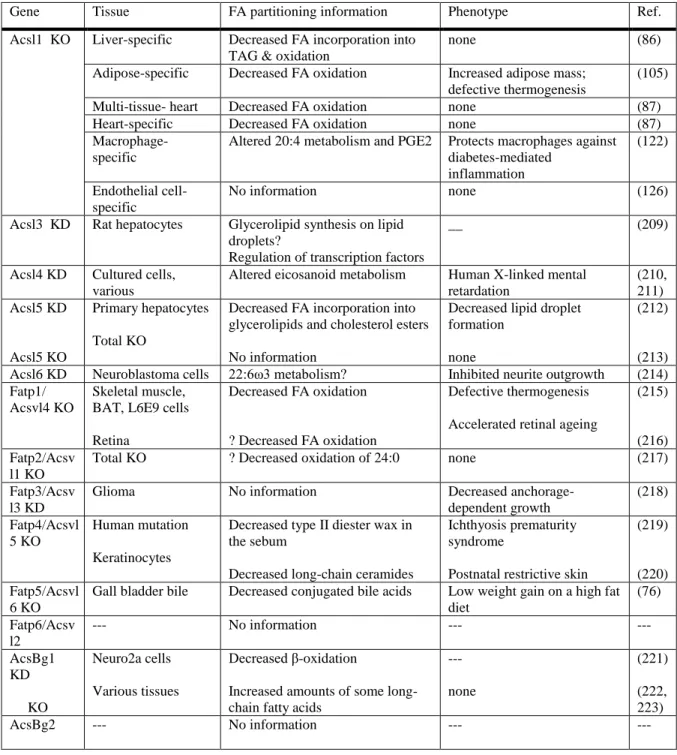
Table 1.2. Evidence for partitioning from loss of function studies Published in (1).
Gene Tissue FA partitioning information Phenotype Ref. Acsl1 KO Liver-specific Decreased FA incorporation into
TAG & oxidation
none (86)
Adipose-specific Decreased FA oxidation Increased adipose mass; defective thermogenesis
(105) Multi-tissue- heart Decreased FA oxidation none (87) Heart-specific Decreased FA oxidation none (87)
Macrophage-specific
Altered 20:4 metabolism and PGE2 Protects macrophages against diabetes-mediated
inflammation
(122) Endothelial cell-
specific
No information none (126)
Acsl3 KD Rat hepatocytes Glycerolipid synthesis on lipid droplets?
Regulation of transcription factors
__ (209)
Acsl4 KD Cultured cells, various
Altered eicosanoid metabolism Human X-linked mental retardation (210, 211) Acsl5 KD Acsl5 KO Primary hepatocytes Total KO
Decreased FA incorporation into glycerolipids and cholesterol esters No information
Decreased lipid droplet formation
none
(212) (213) Acsl6 KD Neuroblastoma cells 22:6ω3 metabolism? Inhibited neurite outgrowth (214) Fatp1/
Acsvl4 KO
Skeletal muscle, BAT, L6E9 cells Retina
Decreased FA oxidation ? Decreased FA oxidation
Defective thermogenesis Accelerated retinal ageing
(215) (216) Fatp2/Acsv
l1 KO
Total KO ? Decreased oxidation of 24:0 none (217) Fatp3/Acsv
l3 KD
Glioma No information Decreased anchorage-dependent growth (218) Fatp4/Acsvl 5 KO Human mutation Keratinocytes
Decreased type II diester wax in the sebum
Decreased long-chain ceramides
Ichthyosis prematurity syndrome
Postnatal restrictive skin
(219) (220) Fatp5/Acsvl
6 KO
Gall bladder bile Decreased conjugated bile acids Low weight gain on a high fat diet
(76) Fatp6/Acsv
l2
--- No information --- ---
AcsBg1 KD KO Neuro2a cells Various tissues Decreased β-oxidation
Increased amounts of some long-chain fatty acids
--- none
(221) (222, 223)
32
33
Figure 1.6. Mitochondrial quality control through fission and fusion, autophagy, or apoptosis. Damaged mitochondria are cleared by general autophagy, controlled by mTORC1 or by mitophagy controlled by the Pink/Parkin pathway or by CL externalization. Abbreviations: mTORC1-
34
Specific Aims
Mitochondria produce up to 95% of ATP made in cardiomyocytes, making these critical organelles to heart health. Loss of mitochondrial respiratory function is seen in many cardiac
diseases, such as heart failure, Barth syndrome, and aging (225,226), which could render these hearts less able to respond to stressors such as low energy availability or exercise. Understanding the causes and consequences of cardiac mitochondrial dysfunction can have a large impact on how heart disease is treated.
Long chain acyl-CoA synthetases (ACSLs) catalyze the addition of coenzyme A (CoA) to long chain fatty acids, the predominant dietary fatty acids, thereby activating and enabling them to enter into pathways of either oxidation or incorporation into complex lipids. ACSL1 is the major ACSL isoform in heart, contributing 90% of total ACSL activity. Loss of ACSL1 has profound effects on cardiac metabolism. In heart, loss of ACSL1 causes an 80-90% loss of fatty acid oxidation and an 8-fold increase in glucose use (105,227,228). With loss of 90% of ACSL activity, which is required for glycerolipids synthesis, hearts lacking ACSL1 may also have altered phospholipid and TAG synthesis. Alterations in saturation and chain length of fatty acids incorporated into membrane lipids can change membrane dynamics, movement of substrates and solutes across membranes, and lipid raft composition (229-231) . Loss of ACSL1 also causes activation of mTORC1, which can increase growth and inhibit autophagy (87). These changes to substrate use, membrane composition, cell growth, and autophagy can all affect mitochondrial function.
Preliminary experiments using transmission electron microscopy of hearts from multi-tissue Acsl1 knockouts (Acsl1T-/-) showed many swollen and vacuolated mitochondria with disrupted cristae. Compared to floxed littermate controls, the oxygen consumption response of Acsl1T-/- heart
35
susceptibility to apoptosis. Therefore, loss of ACSL1 causes mitochondrial dysfunction, which could predispose Acsl1T-/- hearts to failure if stressed.
Overall aim: Determine why loss of ACSL1 in heart causes mitochondrial dysfunction. Aim 1. Determine if loss of ACSL1 alters cardiac phospholipids.
1a. Determine whether ACSL1 determines acyl-chain composition of membrane phospholipids, specifically focusing on mitochondrial cardiolipin.
1b. Determine whether alterations to mitochondrial phospholipids alters mitochondrial function.
Aim 2. Determine if activation of mTOR in Acsl1T-/-hearts impairs mitochondrial function. 2a. Determine whether activation of mTORC1 in Acsl1T-/- hearts prevents clearance of damaged mitochondria through inhibition of autophagy.
36
CHAPTER 2: ACYL-COA SYNTHETASE 1 DEFICIENCY ALTERS CARDIOLIPIN
SPECIES AND IMPAIRS MITOCHONDRIAL FUNCTION
Trisha J. Grevengoed1, Sarah A. Martin2, Lalage Katunga3, Daniel E. Cooper1, Ethan J.
Anderson3, Robert C. Murphy2, Rosalind A. Coleman1
Summary
Long-chain acyl-CoA synthetase 1 (ACSL1) contributes more than 90% of total cardiac ACSL activity, but its role in phospholipid synthesis has not been determined. Mice with an inducible knockout of ACSL1 (Acsl1T-/-) have impaired cardiac fatty acid oxidation and rely on glucose for ATP production. In Acsl1T-/- mice, cardiac mitochondria were dysfunctional. Because ACSL1 exhibited a strong substrate preference for linoleate, we investigated the composition of heart phospholipids.
Acsl1T-/- hearts contained 83% less tetralinoleoyl-cardiolipin (CL), the major form present in control
hearts. A stable knockdown of ACSL1 in H9c2 rat cardiomyocytes resulted in low incorporation of linoleate into CL, as well as diminished incorporation of palmitate and oleate into other phospholipids. Overexpression of ACSL1 in both H9c2 and HEK-293 cells increased incorporation of linoleate into CL and other phospholipids. To determine whether increasing the content of linoleate in CL would improve mitochondrial respiratory function, control and Acsl1T-/- mice were fed a high linoleate diet; this normalized the amount of tetralinoleoyl-CL, but did not improve respiratory function. Thus, ACSL1 is required for the normal composition of several phospholipid species in heart. Although ACSL1 determines the acyl-chain composition of heart CL, a high tetralinoleoyl-CL content may not be required for normal function.
1 Department of Nutrition, University of North Carolina at Chapel Hill, NC 27599
37
Introduction
In order to metabolize long-chain fatty acids in pathways of β-oxidation or the synthesis of complex lipids, they must first be activated to acyl-CoAs by long-chain acyl-CoA synthetases (ACSL). Five mammalian ACSL isoforms have been identified, each with a specific substrate preference, subcellular location, and tissue distribution (232). In the heart, the ACSL1 isoform predominates, such that with deficiency, total ACSL specific activity and fatty acid oxidation decrease by more than 90% (87). Because ACSL activity is required for the incorporation of fatty acids into phospholipids, we asked whether the ACSL1 isoform is also required for the synthesis and remodeling of cardiac phospholipids, particularly cardiolipin (CL).
The mitochondrial phospholipid CL contributes to many aspects of mitochondrial function, including energy production through oxidative phosphorylation (233,234), mitochondrial fission and fusion (235,236), and cellular apoptosis (237). Tetralinoleoyl-CL is the predominant CL species in the mammalian heart (238), but the mechanism by which this species is formed is unclear. Because the enzymes of CL synthesis lack acyl-chain specificity, nascent CL contains a mixture of acyl chain lengths and degrees of unsaturation (58). To obtain mature CL, most remodeling occurs within the mitochondria by sequentially removing each acyl chain to form monolyso-CL (MLCL) and then replacing the missing fatty acid with linoleate (18:2), added by the transacylation from a donor phospholipid (239) or by direct incorporation of a linoleoyl-CoA (62,240).
38
Here we show that ACSL1, which has a distinct preference for linoleate, significantly contributes to CL remodeling. Because fatty acids must be converted to acyl-CoAs, both to be available for the initial steps in the synthesis of phospholipids, as well as to enter the mitochondrial matrix where CL remodeling occurs, we asked whether ACSL1 might be responsible for activating linoleate destined to be incorporated into CL. We found that hearts lacking ACSL1 are deficient in tetralinoleoyl-CL, but that normalizing the CL species cannot ameliorate the mitochondrial dysfunction in these hearts. These findings call into question the idea that the acyl-chains of CL are important for cardiac and mitochondrial respiratory function.
Methods
Animal care and diets: All protocols were approved by the Institutional Animal Care and Use Committee at University of North Carolina at Chapel Hill. Mice were housed under a 12 h light/dark cycle with free access to food and water. Unless otherwise specified, mice were fed a purified low-fat diet (Research Diets, DB12451B). A multi-tissue knockout of ACSL1 was achieved by mating mice with loxP sequences flanking exon one of the Acsl1 gene to animals expressing a tamoxifen-inducible Cre driven by a ubiquitous promoter enhancer (87). Between six and eight weeks of age, Acsl1T-/- and littermate control Acsl1flox/flox (control) mice were injected intraperitoneally on four consecutive days with 20 mg/mL (75 μg/g body weight) tamoxifen dissolved in corn oil. All studies were performed 20
39
fed a high-linoleate safflower oil diet (Research Diets, D02062104, 45% kcal fat (75% linoleate)) for 4 weeks.
ACSL activity assay: ACSL specific activity was measured in heart membranes and cell homogenates (87). Briefly, homogenized tissues were centrifuged at 100,000 x g for 1 h at 4°C to isolate total membrane fractions. Between 1 and 6 µg of protein was incubated with 50 µM [1-14C]fatty acid
(unless otherwise indicated), 10 mM ATP, 250 μM CoA, 5 mM dithiothreitol, and 8 mM MgCl2 in 175
mM Tris, pH 7.4 at RT° for 10 min. The enzyme reaction was stopped with 1 mL of Dole’s solution
(heptane:isopropanol:1M H2SO4; 80:20:1; v/v). Two mL of heptane and 0.5 ml of water were added
to separate phases. Radioactivity of the acyl-CoAs in the aqueous phase was measured using a liquid scintillation counter.
Mitochondrial function studies: Mitochondrial function was measured in permeabilized myofibers and in isolated mitochondria prepared from portions of the left ventricle and septum. After dissection, muscle samples were placed in ice-cold (4C) Buffer X containing (in mM): 7.23 K2EGTA,
2.77 CaK2EGTA, 20 imidazole, 20 taurine, 5.7 ATP, 14.3 phosphocreatine, 6.56 MgCl2-6H2O and 50
MES (pH 7.1, 295 mOsm). Fibers were delicately separated in ice-cold Buffer X using fine forceps under a dissecting scope. Cardiac fibers were then permeabilized in Buffer X with 50 µg/mL saponin for 30 min, then washed in ice-cold wash buffer Z (110 mM K-MES, 35 mM KCl, 1 mM EGTA, 5 mM K2HPO4, 3 mM MgCl2·6H2O, 5 mg/ml bovine serum albumin, pH 7.1, 295 mOsm) to remove
endogenous substrates. To prevent Ca2+ independent contraction of the permeabilized fibers, 20 M
blebbistatin was added to Buffer Z during wash and experiments. All mitochondrial O2 consumption
(JO2) measurements were performed at 30°C using the Oroboros O2K Oxygraph system (Oroboros
Instruments). The H2O2 and Ca2+ uptake measurements were performed in a spectrofluorometer