Address for correspondence Dr. Sabyasachi Banerjee Department of Dermatology, Malda Medical College, Darjeeling, West Bengal, India
Original Article
Current mycological profile of dermatophytosis
in a tertiary care set up in North Bengal
Sabyasachi Banerjee*, Kalyan Khan**, Palash Mandal**, Sanjay Kumar Mallick***
* Department of Dermatology, Malda Medical College, West Bengal ** Department of Pathology, North Bengal Medical College, West Bengal *** Department of Microbiology, North Bengal Medical College, West Bengal
Abstract Objective To determine the occurrence, distribution & mycological profile of dermatophytosis in North Bengal Medical College and Hospital, in Darjeeling, West Bengal, India.
Methods A total of 200 specimens were collected from clinically suspected dermatophytoses from February to April 2013. Samples of skin scrapings, hair shafts and nails were sent to laboratory from dermatology OPD for direct examination, fungal culture and identification.
Results Adult males outnumbered females in all cases of dermatophytosis except tinea corporis. Most cases showed high culture sensitivity except tinea unguium. Trichophyton rubrum was the most commonly isolated fungal organism.
Conclusion This study identifies the clinical distribution and predominant organisms causing dermatophytosis in North Bengal, which may be useful to ascertain the past and present trends in dermatophytosis and provide insight into future diagnosis and treatment.
Key words
Dermatophytosis, tinea, Trichophyton. Introduction
Dermatophytes are a group of keratinolytic ascomycetes which are similar by appearance, physiological properties, taxonomic position, antigenicity, growth requirements and nature of infectivity. They are aerobic fungi that produce proteases that digest keratin and allow colonization, invasion and infection of stratum
corneum of the skin, hair and nail.1
Dermatophytosis, also called ‘tinea’, refers to a superficial fungal infection of keratinized tissue caused by dermatophyte fungi. The infection can
produce dermal inflammatory response with intense itching.
The dematophyte species belong to three genera
of fungi: Trichophyton, Microsporum and
Epidermophyton. The wide variation in clinical presentation depends upon the strains and species of the fungus, site of body infected and immune status of the host. The clinical diagnosis of dermatophytosis is confirmed by direct
microscopy of KOH preparation and by culture.2
Methods
Consecutive patients including infants and children having clinical evidence of some type of dermatophytosis who attended outpatient department of Dermatology, North Bengal Medical College from February to April 2013 were taken up for the study. Detailed history was taken, general examination and morphology of lesions including site, number, distribution, border, surface, scaling were noted. Specimens of skin scrapings, hair and nail clipping were treated with 10% KOH solution over slide and examined under light microscope. Among them,
200 cases of microscopically confirmed
dermatophytosis were taken up for further analysis. Portion of each collected specimen was added in Sabouraud’s dextrose agar medium and incubated at 25°C and 37°C and were examined for evidence of growth up to three weeks. The culture isolates were further studied for colony morphology and microscopical examination of lactophenol blue mounts was done. Special tests like hair perforation test, urease production test and slide culture were carried out using standard
techniques3 wherever necessary for
identification of species.
Results
Table 1 shows age and sex wise distribution of
200 study cases. 61.5% of cases were male and 78% were adults. Sex and age-wise break up of clinical types of dermatophytosis follows in
Table 2.
Regarding sensitivity of cultural procedures in each individual type of dermatophytosis (compared to KOH examination), tinea barbae shows 100% sensitivity (all cases shows growth in culture), followed by tinea faciei 86%, tinea cruris 86%, tinea pedis 83% and tinea corporis 81%. Tinea manuum and tinea capitis showed sensitivity of 65% and 55%, respectively. In
case of tinea unguium, sensitivity of culture procedure was 32% only.
Table 3 shows the aetiological agents isolated by culture from different clinical types of tinea
infection. Overall, T. rubrum constituted
overwhelming majority (90%) of cultural
isolates. However, in tinea capitis, T. violaceum
was the commonest isolate (66.6%). Other
dermatophyte species isolated were T.
mentagrophyte (Figure 1 and 2), E. floccosum,
T. tonsurans (Figure 3) and T. terrestrae, in order of decreasing frequency. T. rubrum was isolated from a 55-year-old male, a patient of non-Hodgkin lymphoma who presented with disseminated tinea corporis.
Discussion
The present study conducted on dermatophytes revealed a number of interesting facts. In the present study the most susceptible age group was the adults, the incidence was 78% (Table
1). Maximum cases were in the 21-30 years
group. Similar results were obtained from the studies also.4
Present study showed a definite male
predominance over the females as evidenced by 127 male patients against 77 female patients. This fact was supported by other workers. This tilt towards males may be due to their more exposure to a favourable environment i.e. greater outdoor physical activity and increased sweating or the tendency of females to hide their diseases in covered parts of body (Table 2).
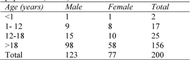
Table 1 Age- and sex-wise break-up of study population (n=200).
Age (years) Male Female Total
<1 1 1 2
1- 12 9 8 17
12-18 15 10 25
>18 98 58 156
Table 2 Clinical types of dermatophytosis and age and sex distribution (n=200).
Clinical diagnosis <1 year 1-12 12-18 years >18 years Total
M F M F M F M F
Tinea capitis 1 0 5 3 0 0 1 1 11
Tinea barbae 0 0 0 0 0 0 1 0 1
Tinea faciei 0 1 0 0 1 0 4 1 7
Tinea manuum 0 0 2 0 3 2 17 2 26
Tinea corporis 0 0 0 2 2 3 25 35 67
Tinea cruris 0 0 1 0 2 2 8 3 16
Tinea pedis 0 0 0 1 2 0 14 7 24
Tinea unguium 0 0 0 2 7 3 10 7 29
Combination lesions 0 0 0 0 2 4 8 4 18
Generalized 0 0 0 0 0 0 1 0 1
Total 1 1 8 8 19 14 80 60 200
Table 3 Cultural isolates from different clinical types of dermatophytosis (n=200). Individual
disease
Trichophyton rubrum
T. mentagr- ophytes
T. viola-ceum
T. tonsurans
T. terrestrae
Epidermophyton floccosum
Total
Tinea capitis 1 0 4 1 0 0 6
Tinea barbae 1 0 0 0 0 0 1
Tinea faciei 1 0 0 0 0 1 2
T. manuum 15 1 0 0 0 1 17
Tinea corporis 43 2 0 1 0 0 46
Tinea cruris 6 0 0 0 0 0 6
Tinea pedis 19 0 0 0 0 1 20
Tinea unguium 12 0 0 0 1 0 13
Combination lesions
18 0 0 0 0 0 18
Generalized 1 0 0 0 0 0 1
Total 117 3 4 2 1 3 130
On overall estimation, tinea corporis
outnumbered tinea unguium in the present study, followed by tinea manuum and tinea pedis
(Table 2). Tinea corporis came out as the single
largest clinical type in many other Indian studies.5
Tinea capitis was found to be the commonest
dermatophytosis among the children
(below 12 years.), occurrence being 50%. Looking the other way round, 9 out of 11 patients of tinea capitis were below 12 years. This prevalence of tinea capitis among children is also reported from India and abroad also.6
In the present study, tinea corporis was the only dermatophytosis which was distinctly commoner in females than in males (Table 2). This could be due to occlusive synthetic garments used by ladies in our country.
Tinea cruris, however, was much more prevalent
among the males than among the females (Table
2). Men, being more active than women and due
to peculiar male anatomy, groin remains warm, moist and subject to occlusion, resulting in male preponderance of tinea cruris.
There was a case of generalized dermatophytosis in the present study; a 55-year male patient
suffering from non-Hodgkin lymphoma and T.
rubrum was isolated from him. Similar cases
were reported by Kauffman.7
In the present study, only KOH proven cases
were selected. Among them, 65% cases (130 out of 200) showed growth on subsequent culture. Similar results were obtained by previous workers also.8 Regarding the success of culture
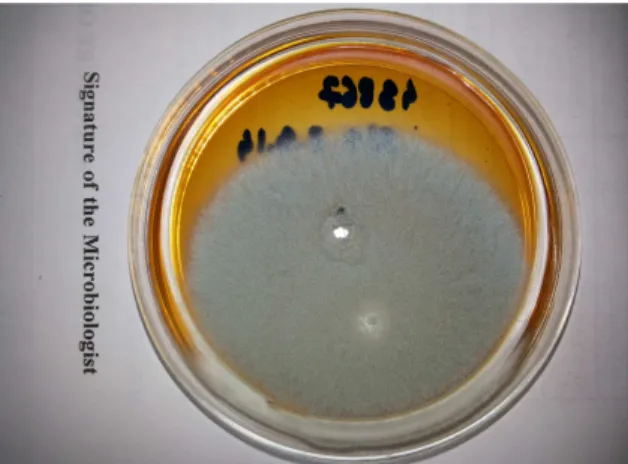
Figure 1 Reverse view of colony morphology of Trichophyton mentagrophyte 14 days after incubation in Saboraud’s dextrose agar.
Figure 2 Micromorphology of Trichophyton mentagrophyte on lactophenol cotton blue mount showing spiral hyphae, pyriform as well as spherical microconidia in clusters.
Figure 3 Macroscopic colony morphology of Trichophyton tonsurans, raised centre and radial grooves.
index of positivity was very high in cases of tinea barbae (100%), tinea faciei (86%), tinea cruris (86%), tinea pedis (83%) and tinea corporis (81%). On the other hand result of culture was positive in only 32% cases of tinea
unguium. A variability in culture isolation
ranging from 44.6% to 70.7% has been found in the Indian subcontinent.9
Trichophyton was the commonest genus
isolated, the other genus was Epidermophyton.
Overall, the Trichophyton genera dominated
with 90% of the isolates followed by
Epidermophyton (5%) and Microsporum (5%) in majority of Indian studies.10-12
The commonest species of the genus
Trichophyton obtained in the preset study was T. rubrum (90% of the culture positive cases),
followed by T. violaceum (3%), T.
mentagrophyte (2.3%) and T. tonsurans (1.5%). Single case of T. terrestrae was isolated from
one case of tinea unguium (Table 3). This
overwhelming majority of T. rubrum was
supported by study of Pandey and Pandey.8
In the present study, E. floccosum was reported
in 2.3% of the culture positive cases. Though its prevalence was reported to be quite high (up to
32.8%) in some studies, Ghannoum et al.13
reported lower figure (0.7%).
T. tonsurans was isolated from 1.5% of culture positive cases in the present study. Very close
figures came out from the study of Elewski.14
Any species of the genus Microsporum could
not be isolated from any case in the present study. Many authors found no cases of
Conclusion
The statistical data presented above lead to a
number of probable conclusions.
Dermatophytosis is mostly an adult disease with male preponderance. Tinea corporis is the most
frequent type. Trichophyton rubrum is the single
most common dermatophyte species isolated in the study. As always, mycological confirmation of tinea unguium through culture still possess a
challenge. The overall clinicomycological
pattern of dermatophytosis should be compared with past trends and scenario of other geographical areas to keep abreast with changing trend and regional variation of it and to improve diagnosis and treatment.
Acknowledgement
We sincerely acknowledge the contribution of Late Dr. Bidyut Deogharia while preparing the manuscript.
References
1. Gorbach SL, Barlett JL, Blacklow NR, eds. Infectious disease. 3rd ed. Philadelphia: Lippincort Williams and Wilkins; 2004. p.1162-80.
2. Kanwar AJ, Mamta, Chander J. Superficial fungal infections. In: Valia GR, ed. IADVL Textbook and Altas of Dermatology. 2nd ed. Mumbai: Bhalani Publishing House; 2001. p. 215-58.
3. Roberts GD. Laboratory methods in basic mycology. In: Ellen Baron JO, Peterson LR, Finegold SM, eds. Bailly and Scott’s Diagnostic Microbiology. 9th ed. St Louis: CV Mosby Company Ltd; 1990.p.715-24. 4. Peerapur BV, Inamdar AC, Pushpa PV,
Srikant B. Clinicomycological study of
dermatophytosis in Bijapur. Indian J Dermatol Venerol Leprol. 2004;22:273-4. 5. Patwardhan N, Dave R. Dermatomycosis in
and around Aurangabad. Indian J Pathol Microbiol. 1999;42:455-62.
6. Bennett M, Fleischer A, Loveless J. Oral griseofulvin remains the treatment of choice for tinea captitis in children. Pediatr Dermatol. 2000;17:304-9.
7. Kauffman CA, ed. Atlas of Fungal Infections, 2nd ed. Hong Kong, Springer Science and Business Media LLC; 2006. 8. Pandey A, Pandey M. Isolation and
characterization of dermatophytes with tinea infections at Gwalior (m.p.), India. Int J Pharm Sci Invent. 2013;2:5-8.
9. Bhagra S, Ganju SA, Kanga A et al. Mycological pattern of dermatophytosis ina and around Shimla hill. Indian J Dermatol. 2014;59:268-70.
10. Bindu V, Pavithran K. Clinicomycological study of dermatophytosis in Calicut. Indian J Dermatol Venerol Leprol. 2002;68:259-61. 11. Singh S, Beena PM. Profile of dermatophyte infection in Baroda. Indian J Dermatol Venrol Leprol. 2003;69:281-3.
12. Grover SC, Roy PC. Clinicomycological profile of superficial mycosis in a hospital in North East India. Med J Armed Forces India. 2002;59:114-6.
13. Ghannoum M, Hajjeh R, Scher R. A large-scale North American study of fungal isolates from nails: the frequency of onychomycosis, fungal distribution, and antifungal susceptibility patterns. J Am Acad Dermatol. 2000;43:641-8.
14. Elewski B. Prevalence of onychomycosis in patients attending a dermatology clinic in northeastern Ohio for other conditions. Arch Dermatol. 1999;133:1172-3.