636
THE EFFECT OF TiO
2NANOPARTICLES (DOPED OR NOT WITH Ag,
ENCAPSULATED OR NOT IN LYPOSOMES), ON THE SPLEEN
ULTRASTRUCTURE IN
Mus musculus
SPECIES,EXPOSED AT A STRESS
FACTOR
X-RAYS)
Mihaela CORNEANU1,Constantin CRĂCIUN2, Gabriel C. CORNEANU3,Ioan GROZESCU4,Daniel HĂDĂRUGĂ5,Carmen LAZĂU4,Septimiu TRIPON2
1
Banat’s University of Agricultural Sciences and Veterinary Medicine “King Michael the Ith”,
Timişoara, Romania; Calea Aradului 119, E-mail: [email protected]; 2
Babeş-Bolyai University, Electron Microscopy Centre, Cluj-Napoca, Clinicilor 5-7 Street, E-mail [email protected], Romania; [email protected];
0756-105521; 3University of Craiova, Agriculture Faculty, A.I. Cuza Street 13, Craiova-200585
3[email protected], Craiova, Romania;
4
I.N.C.-D.E.M.C., 300860-Timişoara, 1 Plautius Andronescu Street, E-mail:
[email protected] Romania; 5
Politehnica University, Industrial Chemistry Technology and Environment Engineering Dept., 6 Victoriei Square, 300006-Timişoara, E-mail: [email protected]; Romania.
Keywords.Spleen ultrastructure, TiO2 doped or not with Ag; lyposomes; X-rays sub-lethal dose
Abstract. The present experiment was performed on young females of Mus musculus, 22-24 g each, intraperitoneal injected with a suspension of titanium dioxide (five injections of 0.5 ml each, one at two days, with 0.01% TiO2 or TiO2-Ag suspension). The
TiO2 nanoparticles of anatase crystallization form, 10-20 nm size, wereconjugated or not
with 1% Ag, encapsulated or not in lyposome. A day after the third injection, half of theanimals received a sublethal dose of X-rays (2.58 Gy; the stress factor). A day after the last injection, the animals were sacrificed through the section of thecarotid artery. Ultrastructural investigations were performed at the spleen level. The analysis of the ultrastructural features from the spleen level enabledthe following observations: the single action of the TiO2 or TiO2-Ag nanoparticles induced an inflammatory process, but in the presence of X-rays, they manifested a slight protective effect; the TiO2-Ag nanoparticles, encapsulated in liposome, manifested a strong radioprotective effect, but the endocapsulation process is not optimal and has to be improved; the presence of a protective effect at the spleen level can suggest that the TiO2-Ag nanoparticles can be used to enhance the organism resistance in case of carcinogenic treatment (in animal or in homo).
INTRODUCTION
Initially, the TiO2 nanoparticles were considered an inert material. Subsequently, it was pointed that TiO2 nanoparticles can induce lesions inthe genetic matter (DNA and chromosomes), as well as at the ultrastructural level (Corneanu et al., 2012). The studies performed in different research centres established that they can migrate and accumulated in different organsandinduce oxidative stress and cell death. The biological investigations
pointed out different biological effects of the TiO2-Me nanoparticles, depending on the
637
it was analyzed the interaction ofTiO2 nanoparticles (single or doped with a metal), with the hepatic cell.
In the present paper, it was studied theeffect ofTiO2 nanoparticles, conjugated or not
with silver, encapsulated or not in liposomes, at the spleen level, toward a stress factor (a sublethal dose of X-rays). As analysiselement,were used the ultrastructural modifications at the spleenlevel. The obtained results underlinethat the TiO2-Ag nanoparticles,
encapsulated in liposomes enhanced the organism resistance to some stress factors and,
thus, can be used in the carcinogenic treatment.
MATERIAL AND METHOD
The experiment was performed on young Mus musculus females, 22-24 g each,
intraperitoneally injected (one at two days) with 0.5 ml from an aqueous solution of a 0.01% bioactive solution. The bioactive substance was a suspension of 0.01% TiO2 nanoparticles of 10-20 nm size, anatase form crystallization, doped (conjugated) or not with 0.01% Ag aqueous solution, encapsulatedor not in liposome. The animals from the Control variant did not receivthe injection or were injected with the same amount of distilled water.As stress factor, it was used a sublethal dose of x-rays (5.28 Gy). A day after the third injection, half ofthe animals fromtheexperimental variant were irradiated (the entire body), at a SEIFERT, ERESCO MF-1 installation (Germany), at the following parameters: 185 kV, 5 mA, 1 mm Al filter, dose debit 52.8 R/min, total doses being of 528 R (5.28 Gy). After the X-irradiation, there were administered (in the same conditions), two intraperitoneal injections. A day afterthe fifthinjection, all theanimals were sacrificed through the section of thejugular venous or carotid artery.
Thebiological matter, from different organswas sampled for the electron microscopy
analysis. In this experiment, the analyses were performed at spleen level. Small pieces of
about 1 mm3 of spleen (white and red pulp), from all theexperimental variants,
weresampledfor ultrastructural investigations. Theprocessingof the biological matter for the electron microscopy investigations was performedaccording tothe classical work protocol:prefixing in a 2.7% glutaraldehide solution (2 ½ h); postfixing in a 1% Millonig solution (1 ½ h); embedding and infiltration in Epon 812. The seriated sections of about 80-90 nm thickness were performed at ultratome, then contrasted with uranyl acetate and lead citrate. The examinationwas performed at an TEM JEM JEOL-1010 electron
microscope in the Electron Microscopy Centre, Babeş-Bolyai University (Cluj-Napoca).
Thechosen imageswere successively captured and preserved in the data basis,by meansof Megaview III Soft Image Analysissoftware. The obtained images were processed using the Wiever Imaging Analysis programme, being transformed in tiff images with a 16 pixels resolution.
RESULTS AND DISCUSSIONS
Spleen, electron microscopy analysis Control variant.
The white pulp presented a characteristic structure for normal mice. Itcontained lymphocytes of variable size, disposed around the periarterial lymphoid sheaths and splenic lymphaticfollicles. Many lymphocyteswerein mitotic division (Fig. 1), at this level being present proliferative processes. In the germinative centre of the lymphaticfollicle, there predominated big lymphocytes, with bulk nucleus and fine-granulated chromatin and evident nucleus. The cytoplasmwasrich in aggregate ribosomes, which weresimilar to
some polysomes. The mitochondria presented evidenced cristae and profiles of the
638
nucleoli and heterochromatin condensed in blocks, little cytoplasm but they wererich in cellular organelles that appeared in an incisure of the nucleus. The middle-sizedlymphocytes presented an intermediate structure between big and small lymphocytes. Between lymphocytes, there were stellated reticulate cells, with numerous prolongers. The marginal area of the pulpwas rich in lymphocytes and plasmocytes. The plasmocytes presenteda big nucleus, withperipheralheterochromatin blocks and awidelyrepresented endoplasmic reticulum. In the white pulp of the spleen, there were also presenteosinophils and basophils, as well as megakaryocytes, involved in the thrombocyte generation. In the marginal pulp area, there were present macrophages involved in the phagocytosis and removal of the antigenic rests. The red pulp from the young mice presented venous sinuses normally structured and interstitial spaces populated especially with haematies and, rarely, migrated macrophages, lymphocytes or plasmocytes.
Control, X-rays.
The mice irradiation with a unique sub-lethal dose of 5.28 Gy conducted to some deep adulteration at the spleen level. There took place an accentuated depletion, a rarefication of thelymphocytes and other cell components at the spleen level,whileother components presented an adulterated structure. The organism response was represented throughenhanced division rate of the cells. The adulteration and degeneration of the cellsfromthe white and red pulp was evident through the presence of the pyknotic nuclei, cytoplasm balonization and vacuolizationand the brokenplasmalemma lymphocyte, a/o (Fig. 2). Asthe lymphocyte population wasrarefied, the reticulate cells and their prolongation wereevident. The splenic macrophages were very solicited in the inclusion and digestion of the destroyed lymphocytes, to be destroyed also, their content being released in theextracell area. Some haematies from red pulp presented an adulterate structure and a diffuse shape. The megakaryocytes presented deformed nuclei and adulterated cytoplasm, the synthesis function and liberation of thrombocytes being adulterated.
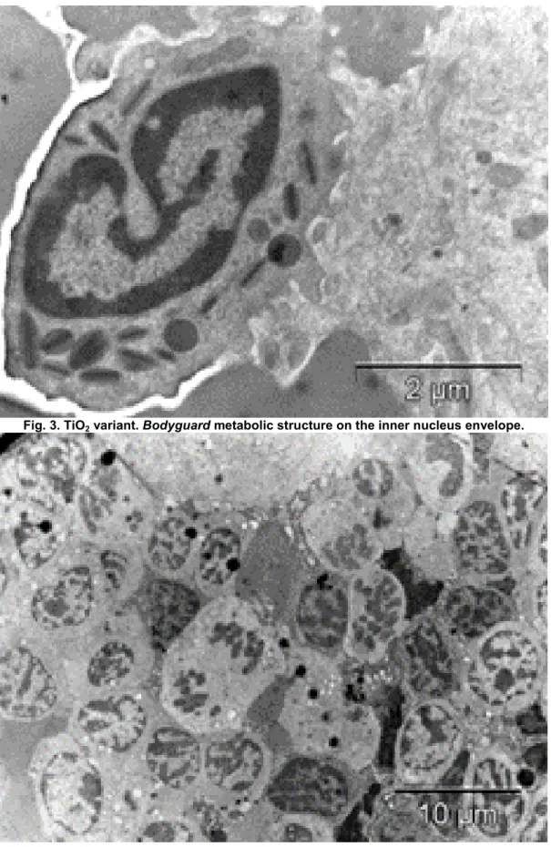
TiO2, undoped.
The undoped TiO2 administered at the animals from this experiment determined
theappearance of some metabolic reversible structures of the nucleus, as bodyguard(Fig.
3) at the spleen level. The bodyguard structurewas represented through constitutive heterochromatin, disposed on inner nuclear envelope, for theprotection of the gene involvedin cell survival. This structure was described by Hsu (1973), Corneanu etCrăciun
and al. (1999), a/o. The cell population, which supported a deletion, was characterized
through the reduction of the lymphocyte number. Moreover, the lymphocytes presented a polymorphism for their nuclei shape. The cells maintained their integrity, the destruction of the plasmalemma being observed especially at the red pulp level. At this level, the ageing cells aredestroyed, together with some haematies. The presence of theneutrophilsand eosinophils, at the red pulp level, indicateda reaction of theorganism similar with the presence of an inflammatory process, determined by the presence of an exogenous
substance (TiO2 nanoparticles). The presence ofTiO2 nanoparticles determined the
reduction of the mitotic rata of lymphocytes.
TiO2 undoped, irradiated.
The presence of TiO2 nanoparticlesat the time of mice irradiation,
639
The X-irradiation promoted the lymphocytes multiplication, numerous mitoses being present. In the red pulp, there were also present neutrophils and a part of the lymphocytes were balonized, especially at the cytoplasm level. Under X-irradiation influence, the red cells from the red pulp presented a diffuse structure. The protective effect of the TiO2 nanoparticles vs. -irradiationwas underlined also by the ultrastructural features of the megakaryocytes. Thus, in the variant irradiated in the absence of a radioprotective substance (TiO2 nanoparticles), the all megakaryocytes presented adulterate ultrastructure;when TiO2 nanoparticles were present during the X-irradiation time, in the spleen, there were present both megakaryocytes with adulterated ultrastructure and megakaryocytes with normal ultrastructure (Fig. 4).
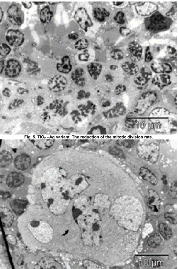
TiO2-Ag, unirradiated (TiO2 nanoparticles doped with silver).
The TiO2-Ag doped nanoparticles represented an exogenous substance, a stress factor and determined a reaction from the organism (Fig. 5). In the white pulp of the spleen, there were predominant the small lymphocytes, ageing, among which there were pyknotic lymphocytes, which can be destroyed. The lymphocytes of medium size and the big lymphocytes (the young lymphocytes), were in a small number, the small number of lymphocytes in the mitotic division being explicable. The majority of the lymphocytes presented an adulteration degree. In the red pulp, among the red cells, therewaspresent a small number of lymphocytes, neutrophils, as well as plasmocytes with dilated endoplasmic reticulum. The megakaryocytes presented a normal structure. TiO2-Ag induced a slowdown of the lymphocyte mitotic division. Thus, it took place the ageing of the existent cells, together with their structure adulteration. This processwas obvious at the nuclei level, which presented a tendency for picnotization.
TiO2-Ag, X-irradiation.
The concomitant action of the two exogenous factors (TiO2 nanoparticles and X-rays),led to the reduction of their negative action, recorded at their single action (Fig. 6). There occured the mitotic division of the young lymphocytes (of big size), which were more frequent, the lymphocytes of medium size having polymorphic nuclei and slightly adulterated ultrastructure. At the red pulp level, more heamaties were in different degradation stages and some plasmocytes and macrophages endured an adulteration process with vacuolization, having thus cells rarefaction. The normally structured megakaryocytes, delivered thrombocytes.
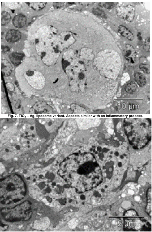
TiO2-Ag, encapsulated in liposome, without X-irradiation.
The encapsulation of the TiO2-Ag nanoparticles in the liposome presented effects of mosaic type: in some areas, it did not induce significant modifications, while in other areas, there were recorded some fragmentary effects. In the areas without significant modifications, the recorded aspects were similar with those recorded at theadministration of TiO2-Ag non-capsulated in the liposome (Fig. 7). In the white pulp, the small lymphocytes with polymorph nucleiwere predominant, as well as lymphocytes with pyknotic or degenerated nuclei. There were some areas the structure of which is normal and lymphocytes are in mitotic division.This process can occur due to a deficient encapsulation. The red pulp of the spleen presented a normal structure, between heamaties being present numerous small and medium lymphocytes, with normal structure. The presence of some polymorphonuclear leucocytes and eosinophils suggested the existence of aninflammatory process, determined (probably) by a deficient encapsulation of the TiO2-Ag nanoparticles. There were also present young megakaryocytes in development together with others in intense activity, hawing in the cytoplasm many mitochondria, endoplasmic reticulum, dense cytoplasmic matrix and vesicles with an electron-dense matter. These components will be in the thrombocytes structure.
TiO2-Ag, encapsulated in liposome, X-irradiation.
640
a zonal protective effect in the white pulp of the spleen (Fig. 8). In some areasof the white pulp, the small lymphocytes were predominant, accompanied by lymphocytes of medium size, usually with polymorph nuclei. Some lymphocytes presented a balonized cytoplasm and the others presented pyknotic nuclei, andin the reticulate cells, there were present vesiculations.In other areas, especially near the red pulp, lymphocytes of middle size were predominantwhile some big, normally structuredlymphocytes werein mitotic division. In the red pulp, between heamaties, therewasa small lymphocyte number, some in division. Others were in disintegration;the macrophages are very active, in cytoplasm being presents lysosomes and cell rests in the digestionphase, as well as polymorphonuclears in an intense activity.
CONCLUSIONS
*The animals from the Control variant presented normal features of the spleen.
*The singular action of a stress factor (a sublethal dose of X-rays) induced severe adulterations at the spleen level.
*The singular presence of the TiO2 doped or not with silver (TiO2-Ag or TiO2) determined a response from the spleen, similarly with the responseinduced by the presence of an inflammatory agent: neutrophils and eosinophils were present in the red pulp; cell population supported a depletion, the lymphocytes number being very small; inhibitory effect over the division of the lymphocytes, a/o.
*At the X-irradiation, TiO2 / TiO2-Ag attenuated the destructive effects of the X-rays, permitting a reduction of the lesions amount from the spleen.
*TiO2 – Ag nanoparticles encapsulated in liposomes presented strong radioprotective effect, but the capsulation processwas not optimal,requiring an amelioration.
*The actual method for the inclusion in liposomes of doped TiO2Ag (TiO2-Me) nanoparticleswas deficient, being affected areas especially from the white pulp. Probably, TiO2-Ag/liposomes presentedefficacy at the red pulp level and in the neighbouring areas from the white pulp.
*The irradiation of the micein the absence of protecting agent induced severe adulterations at the spleen level: rarefication of the cell organelles (especially in lymphocytes), enhancement of the mitotic division rata (a possibility explanation for their numeric reduction is related to destructions, adulterations and degeneration of some lymphocytes through cytoplasm balonization, vacuolization and rupture of plasmalemma, a/o). Moreover, the splenic macrophages can be also destroyed. Also, the megakaryocytes were not capable forthe deliverance of a normal amount of thrombocytes.
*The TiO2 undoped presence in the animal irradiation presented a protecting effect: the cell population was rarefied, but the induced changes were limited and reversible; irradiation induced the intensification of the mitotic activity at lymphocytesto compensate the destruction induced as a result of the X-rays action. The protected macrophages of TiO2 presence were in intense activity, removing the ageing and destroyed lymphocytes; partof the megakaryocytes presented a normal structure, as a result of the protection offered by TiO2 nanoparticles presence.
*TiO2-Ag induced a slowdown of the mitotic division in lymphocytes, with the consequence of ageing of the existent lymphocytes, concomitant with their structure adulteration (pyknosis tendency of their nuclei).
641
*The presence of TiO2-Agencapsulated in liposomes, in the white pulp, conducted to the protecting effect in some areas, while in other areas, the aspects are similar with the non-encapsulated TiO2 effect. This process is probably a result of deficient encapsulation of TiO2-Ag. The red pulp presented a normal structure, being present young megakaryocytes in growth, together with others in an intense activity, with many cell organelles in the cytoplasm, as well as vesicles with electron-dense content, components from the next thrombocytes. The presence of some polymorphonuclears leucocytes and eosinophils supports the existence of an inflammatory process, determined (probably) bya deficient encapsulation of the TiO2-Ag.
*In the white pulp, there were areas in which small lymphocytes were predominant together with average lymphocytes with reversible adulterations. In other areas, near the red pulp, the big and average lymphocytes with normal structure were predominant, some cells being in mitotic division. In the red pulp, among degraded haematies, there were lymphocytes with normal structure, in mitotic division, as well as somedegraded lymphocytes. The macrophages were in an intense phagocytosis activity, together with some polymorphonuclears.
Acknowledgements. These researches weresupportedby the IMUNONANOMAT Grant 70/2007, financed by the National Council for Programs Management, CNMP-Bucharest (Romania). The authors wish to thank Mrs. Cornelia Raţiu and Paula Sfârloagă (I.N.C.-D.E.M.C.-Timişoara) for their contribution to the preparation of TiO2 nanoparticles.
REFERENCES
1.Corneanu C.G., Crăciun C., Crăciun V., Corneanu M., 1998 – Nucleus ultrastructure in different genotypes of Lycopersicon esculentum Mill. and cell metabolic activity. Acta
Horti Bot. Buc., 27: 59-68.
2. Corneanu C.G., Crăciun C., Corneanu M., Lazău C., Grozescu I., Silosi I., Rogoz S., Prodan G. C., Barbu-Tudoran L., Mihali C., Ştefănescu I., Corneanu L.-M., 2007 – The
TiO2-Pt nanoparticles implication in the immune response and their interaction with the
animal cell. In: Progress in Nanoscience and Nanotechnologies, 14 (Eds.: M. Zaharescu,
L. Giurgiu, D. Dascălu), pp. 140-148. Edit. Academiei Române, Bucureşti.
3. Corneanu C.G., Crăciun C., Corneanu M., Lazău C., Grozescu I., Tripon S., 2012 –
The eukaryote cell interaction with doped TiO2 nanoparticles. In: Nanomaterials and
nanostructures for various applications (Eds. Gh. Brezeanu, H. Iovu, C. Cobianu, D. Dascălu). Edit. Academiei Române, Bucureşti, pp. 172-191.
4. Disdier C., 2016 – Evaluation of the e-ects of TiO2 nanoparticles exposure on the adult and vulnerable brains. Toxicology and food chain. Universite Paris-Saclay.
5. Doudi M., Setorki M., 2015 – Influence of titanium dioxide nanoparticles on oxidative
stress and pulmoinary disfunction. Zahedan J. Res. Med. 17 (9): e1062.
6. Horikoshi S., Serpone N., 2013 – Introduction to nanoparticles. In: Microwaves in nanoparticle synthesis - Fundamentals and Applications (S. Horikoshi, N. Serpone, Eds.), Wiley-VCH, Weinheim, Germany, 352 pp., Chapter 1, pp. 1-24.
7. Hsu T.C., 1973 – A possible function of constitutive heterochromatin. The bodyguard
hypothesis. Genetica 9: 139-142.
8. Iavicoli I., Leso V., Fontana L., Bergamaschi A., 2011 – Toxicological effects of titanium dioxide nanoparticles: a review of in vitro mammalian studies.
Eur. Rev. Med. Pharm. Sci., 15: 481-508.
9. Yonezawa T., Kawasaki H., Tarui A., Watanabe T., Arakawa R., Shimada T., Mafuné F., 2009 - Detailed investigations on the possibility of nanoparticles of various metal elements for surface-assisted laser desorption /ionization mass spectrometry. Analytical
642
Figures.
643
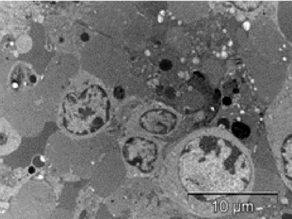
Fig. 2. Control, X-rays variant. Adulteration induced by X-rays in lymphocyte.
644
Fig. 4. TiO2 – X-rays variant. Protective effect on the lymphocyte division, induced by TiO2 presence
during the X-irradiation.
Fig. 5. TiO2 –Ag variant. The reduction of the mitotic division rate.
645
Fig. 7. TiO2 – Ag, liposome variant. Aspects similar with an inflammatory process.