Journal of Global Pharma Technology
Available Online at:
www.jgpt.co.in
RESEARCH ARTICLE
Anatomical and Ultra-sonographic Study of the Aortic Arch
Branches in the Native Goat (
Capra hircus)
Fatimah Swadi Zghair
1*,Rabab Abd Alameer Nasser
2,Hanaa Kareem Ali
Alshammary
2,Yassin Mahmood Rasheed
31. College of Veterinary Medicine, Al-Qadissiya University/Iraq.
2. Department of Anatomy and Histology, College of Vet. Medicine, Diyala, University/Iraq.
3. Department of Surgery and Obstetric, College of Vet. Medicine, Diyala, University/Iraq.
*Corresponding Author: Fatimah Swadi Zghair
Abstract
Five health adult goats of Iraqi local breed of both sexes, were collected from Diyala Governorate weighing range between (6-8kg±40.11), and their ages were between (1-3) months. Ultrasound Scanner technique was used to study the aortic arch branches; ultrasonic examinations were done with a real-time B-Mode scanner supplied with a 7.5 MHz-linear array rectal transducer. All these goats were euthanized. The aortic arch of the goats is extended cranially, dorsally, and turn caudally situated below the vertebra columns at the level of the third intercostal space, above the pulmonary trunk. The aortic arch gives two branches in which the short branch called left-subclavian artery and large brachiocephalic trunk. Moreover, the brachiocephalic trunk is a great vessel cranial branch that yields two large branches; right-subclavian artery and bicarotid trunk which divided into left-common carotid artery and right-common carotid artery.
Keywords: Anatomy, Aortic arch, Branches, Native goat, Ultrasonography.
Introduction
The goat subspecies; Capra aegagrus hircus,
is a domestic member of the Bovidae family that is sheep-like species of which both
belong to the sub-family of
goat-antelope, Caprinae. Interestingly, 300 of goat distinct breeds are present [1].Economically, goats are beneficial for the production of food meat and fiber-based materials plus help in
controlling vegetation restoration [2].
According to their adjustment to different climate conditions, goats are present global wide in continents such as Asia, Africa, Americas, and Europe [3].
Ultrasonography for organ inspection in male and female ruminants provides a detailed-real-time non-invasive exploration technique depending on type and the site of the probe plus the frequency of the operation [4, 7].The aims of the current study were to gain knowledge about abnormal branches rooted from the aortic arch, provide anatomical-based characteristics, determine the vascular architecture of the aortic arch in Iraqi goats.
The great arteries originated from the heart (left ventricles) include aorta that curved to right, forming an arch that its branches arise from aortic arch with different patterns according to the species of mammals [8].The superior mediastinum is its location. Usually, the arch is divided into three branches that include the brachiocephalic trunk, the left-common carotid artery, and left subclavian artery supplying head, neck, and upper limbs [9, 10].The aortic arch in most domestic animals and in rabbits joins the pulmonary trunk via ligamentum arteriosum, the remain of the fetal ductus arteriosus [9, 11].
aortic-arch cranial-branch is the brachiocephalic trunk that supplies blood to the hand limbs, head, neck, and ventral portion of chest.
The brachiocephalic trunk initiates the right subclavian artery and bicarotid trunk. In pig, cat, and dog, the left subclavian artery takes a separate, more distal origin from the arch of the aorta [9]. The bicarotid trunk in ruminants (cow, sheep, and goat) is a short common trunk, which originates from the common brachiocephalic trunk, extends cranially, and branches into the common carotid arteries, left and right [12]. The two branches of the brachiocephalic in canine supply caudal pole of thymus [13].
Some author mentioned the abnormal branches of the arch of the aorta and compared with the normal case in goat explained by other reporters in which the large branch originates from the arch of the aorta called brachiocephalic trunk. This trunk gives both left and right subclavian arteries. About 5 to 6 cm after the origin of these arteries, they feed the front half of the thoracic and the fore limbs [14].
Some researchers showed in human that the aortic arch provides two branches that arise from the surface of the upper convex that belongs to the arch; the brachiocephalic is the first branch, and the left subclavian artery is the second branch. Moreover, the root of the brachiocephlic trunk gives the left common carotid artery [8, 15].The anomalous in the distribution via the origins of the vessels belong to the large aortic arch in human may induce alterations in the cerebral hemo-dynamics generating disorders in brain [16].
Previously, studies showed different patterns of the aortic arch branching, classified of japans cadavers as three types; A, B, and C [15]. In human stated (91,4%) branches are directly originated from aortic usual three branches and variation were found about (9.6)% [16, 17] that is believed that a few anomalies of the aortic arch belongs to chromosome 22q11 deletion [18].
Information regarding arteries originated from aortic arch in domestic animals are limited; most studies conducted were on laboratory mammals as guinea pig [19], porcupines [20], rabbit [11, 21], mole-rat [22, 23], and red squirrels [24].
Materials and Methods
Five health adult goats of Iraqi local breed of both sexes, were collected from Diyala Governorate weighing range between (6-8kg±40.11), and their ages were between (1-3) months. Ultrasound Scanner technique was used to study the aortic arch branches; ultrasonic examinations were done with a real-time B-Mode scanner supplied with a 7.5MHz-linear array rectal transducer. All
these goats were euthanized.The types of the
scanner (Welld ultrasound, Shenzhen well. D. Medical Electronics).Ultrasound scanners were recorded using a Light wave record and play video, USB 2.0 TV BOX recorder for further analysis. The goats were scanned in lateral recumbency after shaving the site of examination (Fig. 2).
These goats were euthanized with intra muscular injection of xylazine (Rompum**) at 35m/kg B.W+(ketamine***) at 5mg/kg B.W. [24]. The opening of the chest was done by bone cutter and removing the ribs from second to six. After opening the thoracic cavity, cannula of plastic 0.5cm in diameter and 10-15 cm in length was inserted into the thoracic aorta, and colored latex was injected and left the carcasses for 24hrs. Later, they were kept in 10% formalin till the study of the patterns of arteries originated from aortic arch was initiated. A Vernier was used in the measurements of the arteries.
Results
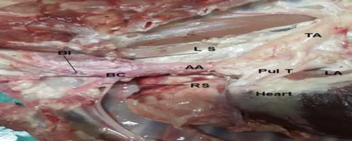
The aorta in the native goat was situated in thoracic cavity, the aorta up dive than curved which forms the aortic arch, and it was extended cranially, dorsally, and turn caudally situated below the vertebra columns at the level of the third intercostal space, above the pulmonary trunk and jointed with pulmonary by ligaments in which these results are similar to those in horse [9, 10] that exit from left ventricle. The course of the aortic arch ceaseless with ascending aorta, it usually gives two branches; small one called
left subclavian artery and large
brachiocephalic trunk (Fig. 1).
artery of the native goats supplied the head
and the structures of the neck.
The ultrasonography pictures taken to the large vessels of the aortic arch included the aortic arch that gives brachiocephlic trunk and subclavian artery. The brachiocephlic trunk was divided into two branches; left subclavian artery and common carotid artery
(Fig. 2) used ultra-sonographic (Fig. 3).The anatomical measurement of the diameter of the aortic arch, brachiocephalic and bicarotid arteries were 7.3mm, 5.8mm, and 4.9mm
respectively (Fig .1), while the
ultrasonagraphical measurements of the diameter of the aortic arch, brachiocephalic and bicarotid arteries were 5.32mm, 3.8mm, and 3.9mm respectively (Fig. 2) and (Table 1).
Table 1: Measurement of diameters of the arteries
Aortic arch branches (mm) Anatomical (mm) ultra-sonographic (mm)
Aortic arch 7.32±0.22 5±30.5
Brachiocephalic artery 5.8±0.3 3.8±0.37
Bicarotid artery 4.9 ±0.29 3.9±40
Discussion
The course of the aortic arch ceaseless with ascending aorta, it usually gives two branches small one called left subclavian artery and large brachiocephalic trunk (Fig .1). These results are similar to those mentioned by [5, 12], in most domestic animals such as in cattle [10] and in sheep and goat [12]; in guinea [19], and in rabbit [11], but the branches arise from aortic arch in native goat is not resemble the aortic arch branching pattern found in human that states the aortic arch yield into three branches innominate artery, left common carotid, and left subclavian artery [11, 16, 17].
Brachiocephalic trunk is a great vessel in which the cranial branch yields into two branches that include bicarotid trunk and right subclavian artery. The bicarotid trunk was divided into left common and right common arteries. These results are in agreement with that in dog [9] and cat [10]
but in disagreement with that in goat [14]. In porcupines, three arteries were observed that originated from aortic arch which include first brachiocephalic artery, second left carotid common artery, and third left subclavian artery, but the bicarotid trunk was absent [20]. The brachiocephalic yields into three branches in rabbit [21, 22], in Guinea pig [19], in red squirrels and mole rat [23, 24], and in rat [22].
The brachiocephalic artery of the native goat supplied the head and the structures of the neck, and this result was stated in canine [13] and the left subclavian artery supplied the hand limb. The ultrasonography pictures taken to the large vessels of the aortic arch included the aortic arch that gives brachiocephlic trunk and subclavian artery. The brachiocephlic trunk was divided into two branches; left subclavian artery and common carotid artery (Fig. 2) used ultra-sonographic (Fig. 3). The ultrasonography pictures of the vessels are very important during the surgery.
Fig. 2: Ultrasonographic pictures to aortic arch (AA) of the Iraqi goats and the main branches; brachiocephalic trunk (BC), Left subclavian artery (Rc) and right subclavian artery (Lc), left common carotid, Right common carotid (Rc)
Fig. 3: The ultrasonography pictures of the vessels
References
1. Nomura K, Yonezawa T, Mano S,
Kawakami S, Shedlock AM, Hasegawa M, et al (2013) Domestication process of the goat revealed by an analysis of the nearly complete mitochondrial protein-encoding genes. PLoS One [Internet]. [cited 2019 Mar 22];8(8):e67775. Available from: http://www.ncbi.nlm.nih.gov/pubmed/2393 6295
2. Shrestha JNB, Fahmy MH (2005)
Breeding goats for meat production: a review: 1. Genetic resources, management and breed evaluation. Small Rumin Res
[Internet]. 1 [cited 2019 Mar 22]:
58(2):93-106. Available from:
https://www.sciencedirect.com/science/artic le/pii/S0921448803001834
3. Solaiman SG (2010) Goat science and
production [Internet]. Wiley-Blackwell; [cited 2019 Mar 22]. 425 p. Available from:
https://www.wiley.com/en- us/Goat+Science+and+Production-p-9780813809366
4. Lazaridis LJ, Brozos CN, Kiossis EA
of the male-A review. J Hell Vet Med Soc [Internet]. 15 [cited 2019 22]: 63(3):217.
Available from:
https://ejournals.epublishing.ekt.gr/index.p hp/jhvms/article/view/15437
5. Danków JWŚM (2002) Ultrasound
measurements of goat’s mammary gland cisterns during lactation. (in Polish with
English summary). Med. Weter,
58(12):977-80.
6. Caja G, Such X, Ruberte J, Carretero A,
Navarro M (1999) The use of
ultrasonography in the study of mammary gland cistern during lactation in sheep. Wageningen Pers EAAP, 95: 91-3.
7. Ślosarz P, Wojtowski J, Gut A, Jelinska M
GM (2000) Preliminary results of application of ultrasound technique for estimation of milk yield in the sheep. Zesz Nauk Przegl. Hod., (63):113-8.
8. Layton KF, Kallmes DF, Cloft HJ, Lindell
EP, Cox VS (2006) Bovine aortic arch variant in humans: clarification of a
common misnomer. AJNR Am J
Neuroradiol [Internet]. 1 [cited 2019 Mar
22];27(7):1541-2. Available from:
http://www.ncbi.nlm.nih.gov/pubmed/1690 8576
9. Getty (1975) Sisson and Grossman’s the
Anatomy of the Domestic Animals. Philadelphia: Saunders; Equine (565-568), Ruminants (960-966), Pig (1306-1.
10.König HE, Liebich H-G, Bragulla H (2009)
Veterinary anatomy of domestic
mammals : textbook and colour atlas.
Schattauer, 453-457.
11.Dimitrov RK, Vladova D, Stamatova KD,
Kostov DB, Stefanov M (2012) Anatomical Computed Tomographic Study of The Heart and Some Mediastinal Vessels of
the Rabbit (OryctOlagus cuniculus)
[Internet]. [cited 2019 Mar 22]. Available from: https://www.semanticscholar.org/paper/An atomical-Computed-Tomographic-Study- of-The-Heart-Dimitrov-Vladova/7b22f3dad3870c5650eb237140b53 047e5e605dd
12.Rao NS, Kishore KS, Sujatha K, Rao
Himaja K (2016) Aortic Arch Arteries in
Man and Domestic Animals : A
Comparative Study [Internet]. [cited 2019
Mar 22]. Available from:
https://www.semanticscholar.org/paper/Ao
rtic-Arch-Arteries-in-Man-and-domestic-
Animals-%3A-Rao-Kishore/df456ca899fa0d6b1e137a4476ff65 8b9ed0ef65
13.Tipirdamaz Hy and S Materials and
methods. 1- Yalçin H Tipirdamaz S (2002) A macroanatomic Investig Arter Vessel canine thymus Rev Méd Vét, [Internet]. 2002 [cited 2019 Mar 22];153(3):173–175.
Available from:
https://pdfs.semanticscholar.org/aff4/9b6fb eb3671dede65f92c149ec23c362c8ee.pdf
14.L YB, A (2009) Abnormal subclavian
branching of the left aortic in a Native Goat. J. Anim. Vet. Adv., 8(3):554-6.
15.Jainapur S, ND an. M (2014) Left common
carotid(2) artery arising from
brachiocephalic trunk. Int. J. Ana. Res, 2(3):484-88 2321-4287.
16.WS AHA, R (2010) An anatomical study of
the aortic arch variations. JKAU: Med. Sci., 17(2):37-54.
17.Nayak SR, Pai MM, Prabhu L V, D’Costa
S, Shetty P (2006) Anatomical
organization of aortic arch variations in the India: embryological basis and review. J. Vasc. Bras. [Internet]. [cited 2019 Mar
22];5(2):95-100. Available from:
http://www.scielo.br/scielo.php?script=sci_
arttext&pid=S1677-54492006000200004&lng=en&tlng=en
18.McElhinney DB, Clark BJ, Weinberg PM,
Kenton ML, McDonald-McGinn D, Driscoll
DA, et al (2001) Association of
chromosome 22q11 deletion with isolated anomalies of aortic arch laterality and branching. J Am Coll Cardiol [Internet]. 15 [cited 2019 Mar 22];37(8):2114-9.
Available from:
http://linkinghub.elsevier.com/retrieve/pii/ S0735109701012864
19.Paryani M, Gilanpour H, Aghili AMRS,
SR (2012) Aortic arch in guinea pig: the Macro anatomical study. Macro Anat study, 3(10):4719-4722 .
20.Atalar O, Yilmaz S, Burma O, Ilkay E
(2003) The Macroanatomical
Investigations on the Aortic Arch in Porcupines (Hystrix cristata). Anat Histol Embryol J. Vet. Med. Ser. C [Internet]. 1 [cited 2019 Mar 22];32(6):367–9. Available from: http://doi.wiley.com/10.1111/j.1439-0264.2003.00498.x
Tohidi V, Mafi SM, Rajaei SM, et al (2012) Brachiocephalic trunk and its major branches in rabbit: a comparisonbetween anatomic and ultrasonographic approaches [Internet]. [cited 2019 Mar 22]. Available from:
https://www.semanticscholar.org/paper/Br
achiocephalic-trunk-and-its-major-
branches-in-a-Paryani-Gilanpour/fe24d1f7b5acf13f54057b73e470 8384413a6ece
22.Cengiz Öztürk, Zekeriya Özüdoğru HY
(2003) A Macroanatomic Comparative Study On Branching and Course of Aortic Arch and Vertebral Arteries in Rabbits and Rats. Eurasian J Med [Internet]. [cited 2019 Mar 22];35:49–52. Available
from:
http://www.eajm.org/eng/ozet/1981/148/Ab stract
23.Aydin A, Ozkan Z, Ilgun R (2013) The
morphology of the arteries originating from the arcus aorta and the branches of these arteries in mole-rats (Spalax leucodon). Vet Med (Praha) [Internet]. 20 [cited 2019 Mar 22];58(No. 7):373-6.
Available from:
http://www.agriculturejournals.cz/web/vet med.htm?volume=58&firstPage=373&type =publishedArticle
24.AA (2011) The arteries originating from
the aortic arch and the branches of these arteries in red squirrels (Sciurus vulgris). Vete. Medi., 56(3):131-134.