Human genome-wide repair map of DNA
damage caused by the cigarette smoke
carcinogen benzo[a]pyrene
Wentao Lia, Jinchuan Hua, Ogun Adebalia, Sheera Adara,b, Yanyan Yanga, Yi-Ying Chioua, and Aziz Sancara,1
aDepartment of Biochemistry and Biophysics, University of North Carolina at Chapel Hill School of Medicine, Chapel Hill, NC 27599; andbDepartment of Microbiology and Molecular Genetics, The Hebrew University Hadassah Medical School, Jerusalem 91120, Israel
Contributed by Aziz Sancar, May 11, 2017 (sent for review April 11, 2017; reviewed by Núria López-Bigas and Gerd P. Pfeifer)
Benzo[a]pyrene (BaP), a polycyclic aromatic hydrocarbon, is the major cause of lung cancer. BaP forms covalent DNA adducts after metabolic activation and induces mutations. We have developed a method for capturing oligonucleotides carrying bulky base ad-ducts, including UV-induced cyclobutane pyrimidine dimers (CPDs) and BaP diol epoxide-deoxyguanosine (BPDE-dG), which are re-moved from the genome by nucleotide excision repair. The isolated oligonucleotides are ligated to adaptors, and after damage-specific immunoprecipitation, the adaptor-ligated oligonucleotides are con-verted to dsDNA with an appropriate translesion DNA synthesis (TLS) polymerase, followed by PCR amplification and next-generation sequencing (NGS) to generate genome-wide repair maps. We have termed this method translesion excision repair-sequencing (tXR-seq). In contrast to our previously described XR-seq method, tXR-seq does not depend on repair/removal of the damage in the excised oligonucleotides, and thus it is applicable to essentially all DNA damages processed by nucleotide excision repair. Here we present the excision repair maps for CPDs and BPDE-dG adducts generated by tXR-Seq for the human genome. In addition, we report the sequence specificity of BPDE-dG excision repair using tXR-seq.
tXR-seq
|
nucleotide excision repair|
UV|
benzo[a]pyrene diol epoxide|
lung cancerN
ucleotide excision repair is a versatile repair pathway that removes a variety of DNA damages, including UV- and benzo[a]pyrene (BaP)-induced DNA damages. BaP, a widespread carcinogen, is the major cause of lung cancer (1). It is produced by incomplete combustion of organic materials and converted to the ultimate mutagen, BaP diol epoxide (BPDE), through enzymatic metabolism (2). BPDE preferentially forms bulky covalent DNA adducts at N2 position of guanines and causes mutations if these BPDE-deoxyguanosines (BPDE-dGs) are not efficiently elimi-nated by nucleotide excision repair (3). Various methods of varying resolutions have been developed for mapping DNA damage and repair genome-wide (4–9). We previously reported a method, termed excision repair-sequencing (XR-seq), for mapping nucleotide excision repair (6). This method has been used to generate excision repair maps for UV-induced cyclobutane py-rimidine dimers (CPDs) and (6-4)pypy-rimidine-pyrimidone photo-products [(6-4)PPs], as well as cisplatin and oxaliplatin-induced Pt-d(GpG) diadducts for the human genome and CPDs for theEscherichia coligenome (10–12).
Although the XR-seq method has been quite useful in de-termining the effects of various factors, such as genetic background, transcription, posttranscriptional histone modification, and chro-matin states, on the timing and efficiency of repair (13), the method in its original form requires the reversal of damage in the excised oligonucleotide (26–27 mers in humans and 12–13 mers inE. coli) by enzymatic or chemical means before it can be processed for next-generation sequencing (NGS) and next-generation of repair maps. As such, this method has limitations, because it is not possible to verse, either enzymatically or chemically, most DNA lesions re-moved from the duplex by nucleotide excision repair, which is a
necessary step for PCR amplification and NGS. We have overcome this obstacle by using appropriate translesion DNA synthesis (TLS) polymerases to convert the excised ssDNA oligomer to the dsDNA form and then amplification by PCR, followed by NGS and align-ment of the sequences to the genome. Here we present the appli-cation of this approach to the mapping of CPD repair and BPDE-dG repair in the human genome.
Results
tXR-seq of CPD and BPDE-dG. To perform tXR-seq, the human lymphocyte cell line GM12878, which has been extensively char-acterized by the ENCODE project (14), was either exposed to UV (20 J/m2at 254 nm) or treated with (±)-anti-BPDE (2μM). After allowing 4 h or 1 h for repair, respectively, cells were lysed gently, and low molecular weight DNA, consisting mostly of excised 26– 27 mers, was separated from chromosomal high molecular weight DNA by centrifugation.
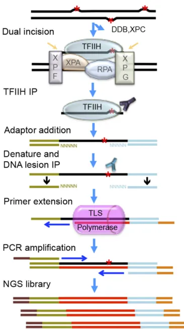
The basic features of sequencing library generation for tXR-seq are shown in Fig. 1. In brief, the excised oligomers are immuno-precipitated with anti-TFIIH (anti-XPB and anti-p62) antibodies, because the excision product is released in a relatively stable complex with TFIIH, which also protects the excision product from degradation by nonspecific nucleases. This is followed by extraction of the excised oligomers and ligation to adaptors and a second immunoprecipitation with either anti-CPD antibody or
Significance
Benzo[a]pyrene (BaP) is a widespread potent carcinogen found in food, coal tar, cigarette smoke, and industrial smoke. Ciga-rette smoking is the leading cause of lung cancer, and the mutagenesis in smoking-associated lung cancer is determined by multiple factors, including nucleotide excision repair. We have developed a general method for genome-wide mapping of nucleotide excision repair at single-nucleotide resolution and applied it to generate repair maps of UV- and BaP-induced DNA damage in human. Results show a novel sequence speci-ficity of BaP diol epoxide-deoxyguanosine repair. This general method can be used to study repair of all types of DNA dam-ages that undergo nucleotide excision repair.
Author contributions: W.L., J.H., and A.S. designed research; W.L. performed research; W.L., J.H., O.A., S.A., Y.Y., Y.-Y.C., and A.S. analyzed data; and W.L. and A.S. wrote the paper.
Reviewers: N.L.-B., Institute for Research in Biomedicine Barcelona; and G.P.P., Van Andel Research Institute of the City of Hope.
The authors declare no conflict of interest.
Freely available online through the PNAS open access option.
Data deposition: The data reported in this paper have been deposited in the Gene Ex-pression Omnibus (GEO) database,https://www.ncbi.nlm.nih.gov/geo/(accession no.
GSE97675).
1To whom correspondence should be addressed. Email: [email protected].
anti-BPDE antibody. Human DNA polymerasesηandκare used for primer extension in the presence of purified excised oligonu-cleotides containing either CPD or BPDE-dG, because polymerases ηandκcan bypass CPD and BPDE-dG damage, respectively, in an error-free manner (15–17). This is followed by PCR amplification with index primers and NGS on the Illumina HiSeq 2500 platform.
Length Distribution and Nucleotide Frequency Analysis of Excision Products. The PCR products, approximately 145 bp long, can be seen only in the presence of polymerasesηor κ, indicating that the resulting dsDNA libraries exclusively contain the se-quence information from the previously excised oligonucleotides attributed to efficient lesion bypass (15–17) (Fig. 2A). Fig. 2B shows the length distributions of the oligomers obtained by tXR-seq and conventional CPD XR-tXR-seq in which the CPD is repaired
by CPD photolyase. In both CPD seq and BPDE-dG tXR-seq cases, oligomers in the size range of 24–28 nt predominate, with a median size of 26 nt, in agreement with the well-established dual incision mode of the human nucleotide exci-sion repair system (18–20). Importantly, the length distribution of the oligomers obtained by tXR-seq for CPD is in remarkable agreement with that obtained by conventional CPD XR-seq, affirming the validity of tXR-seq.
We further analyzed the base sequence distributions along the excised CPD oligomers obtained by tXR-seq and conventional XR-seq (Fig. 2C). As expected, for CPD oligomers isolated by both conventional XR-seq and tXR-seq methods, positions 19– 21 from the 5′end were enriched for thymines (Ts), in agreement with the dual incision pattern of the human excision nuclease system (21, 22). In contrast, for the BPDE-dG oligomers, guanines (Gs) are enriched at positions 19–22, consistent with BPDE-dG monoadducts at one of these positions (Fig. 2D). As a control, when randomly chosen oligomers (10 million) of 26 nt from the human genome were analyzed for sequence composition, all four nucleotides were uniformly distributed throughout the length of the oligomers, with ∼60% AT and 40% GC at all positions, consistent with Chargaff’s rules (23) (Fig. 2D). We also analyzed the frequencies of di-pyrimidines (TT, TC, CT, and CC) at each position along the excised oligomers of 26–29 nt from both tXR-seq and conventional XR-tXR-seq for CPD (Fig. S1AandB). In both experiments, there was strong TT enrichment at the fixed position of 6 nt from the 3′end, further confirming the validity of tXR-seq.
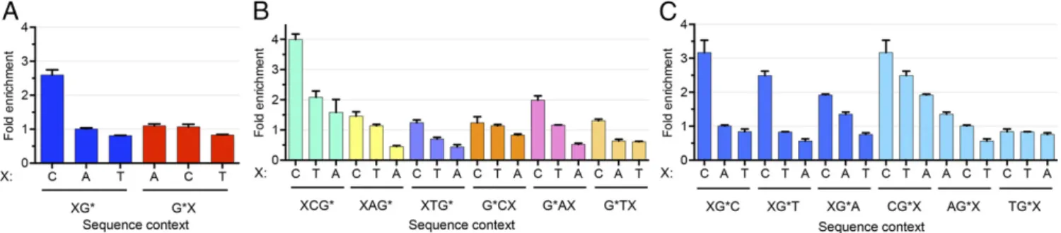
Sequence Specificity of BPDE-dG Repair.Several studies have been conducted on the effects of nearest-neighbor and distant-neighbor sequences on the repair rate of BPDE-dG. The data have been interpreted within the context of structural deformity and duplex backbone flexibility caused by the adduct in various sequence contexts, and some general rules have been derived (24–27). Although these pioneering studies have been quite useful, some of the rules were derived from the excision rates of the BPDE-dG adduct from in vitro experiments with human cell-free extracts and oligonucleotide duplexes 135 bp in length (28). The tXR-seq method, which produces at least∼20 million reads in each biological replicate, provides a model-independent means for analyzing the effects of nearest and distant (one base flanking the nearest bases) sequence neighbors on BPDE-dG excision ef-ficiencies. To secure the vast majority of the excised oligonucle-otides used in this analysis containing BPDE-dG at the same position, we classified the sequencing reads from tXR-seq based on read lengths. An analysis of single-nucleotide frequencies along reads 26–29 nt in length showed Gs enriched at two fixed positions (Fig. S2). For 26, 27, and 28 oligomers, we selected those with nucleotide is G at position 20, 21, and 22, respectively. We further filtered out those reads containing G within three bases upstream and four bases downstream of the specific G position to eliminate other possible BPDE-dG sites that could possibly interfere with our analysis. We applied the same criteria to the randomly se-lected 26, 27, and 28 oligomers from human genomic sequences and then calculated the frequencies of dinucleotides and trinu-cleotides for both excised oligomers and randomly selected olig-omers. Those frequencies for excised oligomers were normalized to their respective frequencies for randomly selected oligomers. We plotted the average fold enrichment of nearest- and distant-neighbor base sequence context frequencies for the damaged G base that had been excised in 26–28 oligomers (Fig. 3).
Based on the results, we can draw the following conclusions: First, the fold enrichment of CG context was∼2.5, whereas there was no apparent enrichment of the other dinucleotide contexts (Fig. 3A). Second, there was a significant effect of the nearest and distant neighbors on the frequency of BPDE-dG excision. In tri-nucleotide frequency analysis, the excision frequency generally followed the“C>T>A”rule at both the 5′and 3′distant-neighbor Fig. 1. Schematic of sequencing library construction strategy for tXR-seq.
The red asterisk represents a DNA lesion processed by nucleotide excision repair. Excised oligomers are precipitated with TFIIH antibodies, extracted, and ligated to adaptors. Then, the adaptor-containing oligomers are pre-cipitated with anti-lesion antibody, the primer with index is annealed and extended by an appropriate TLS DNA polymerase (pink), and the extension product is amplified by PCR to obtain the dsDNA library for NGS.
BIOCHEMISTR
positions (Fig. 3BandCand Table 1). The 3′nearest-neighbor base also followed the same rule, except for AG*, which had an A>C>T order. For G*A and G*T, the 5′nearest-neighbor bases with the second-highest excision frequency were A and T, re-spectively. We also selected the specific G at positions 19, 20, and 21 for 26, 27, and 28 oligomers, respectively, and analyzed the sequence context effect in the same way (Fig. S3andTable S1). The results suggest that the excision frequency was similar and also generally followed the C>T>A rule. Finally, these conclu-sions must be qualified, because they are based on the assumption of no sequence effect on adduct formation. In fact, BPDE pref-erentially binds at methylated CpG sites over any other NpG sites (29). In view of these reported sequence preferences for damage formation, genome-wide BPDE-dG damage distribution must be experimentally demonstrated by Damage-seq (10) for a definitive generalization of the sequence effect on repair.
Effect of Transcription on BPDE-dG Repair.Transcription stimulates repair on the transcribed strand (TS) (30). As a general rule, this effect is more apparent in highly transcribed genes and for le-sions that are poorly recognized by the core nucleotide excision repair machinery. To explore the effect of transcription on
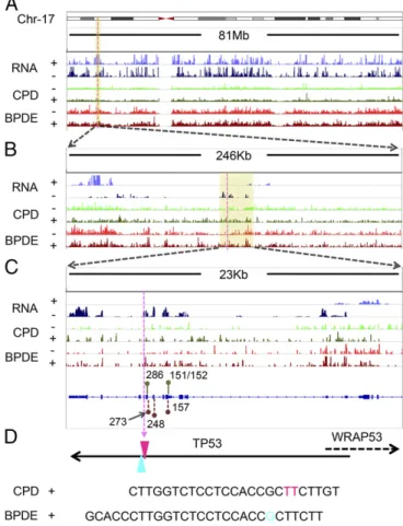
BPDE-dG repair, we first generated both CPD and BPDE-dG repair maps and integrated the total RNA-seq signal across the entire human genome using tXR-seq reads and ENCODE RNA-seq data from GM12878 cells (Fig. S4). We chose theTP53gene to analyze the effect of transcription on BPDE-dG repair for several reasons. First, it is mutated as a driver both in UV-induced and cigarette smoke-UV-induced cancers. Second, it is transcribed at moderately high levels in many cell lines, including the GM12878 cell line used in this study. Finally, previous high-resolution repair studies by ligation-mediated PCR (LM-PCR) on short fragments ofTP53may be used as references for tXR-seq data (31). Fig. 4 shows the transcription and repair maps of human chromosome 17, which carries theTP53gene, at resolu-tions ranging from megabase to single base or dinucleotide in TP53. This figure also shows tXR-seq data for both CPD and BPDE-dG repair in GM12878 cells, as well as two excision products that can be unambiguously assigned to specific TT and G damages, respectively (Fig. 4). As is apparent from the repair data at all resolutions, the TS for bothTP53and the adjacentWRAP53 gene transcribed in opposite direction were repaired at higher efficiency than the nontranscribed strand (NTS). In addition, for Fig. 2. Analyses of length distribution and single-nucleotide frequencies for the excised oligomers obtained from tXR-seq and conventional XR-seq. (A) dsDNA library for tXR-seq analyzed by 10% na-tive polyacrylamide gel electrophoresis. Primer ex-tension products (1%) were amplified by PCR using the indicated cycles. NC, nontemplate control. (B) Length distribution of excised oligomers from CPD and BPDE-dG tXR-seq (∼20 million each) and CPD XR-seq 6 (∼10 million) after removal of adaptors and duplicate reads. (C) Single-nucleotide frequen-cies for 26-mers obtained by CPD tXR-seq (∼13 million) and conventional CPD XR-seq 6 (∼10 million). (D) Single-nucleotide frequencies for the 26-mers from BPDE-dG tXR-seq (∼12 million) along with single-nucleotide distribution of randomly selected 26-mers from the human genome (10 million).
both damage types, there appeared to be hotspots for repair, al-though deeper sequencing is needed to be able to comment on the cause and significance of these repair hotspots.
In addition, we analyzed CPD and BPDE-dG repair around the transcription start sites (TSS) and transcription end sites (TES) in NHF1 and GM12878 cell lines, respectively (Fig. 5). The trend was in general agreement with the data obtained from the NHF1 cell line for the repair of CPD and (6-4)PP as measured by the conventional XR-seq method. As expected, all of the repair profiles had repair peaks around TSS, in agreement with the documented high RNA polymerase II density and nascent RNA levels in this region (13, 32–34). In addition, as we reported pre-viously (13), a low level of enriched excision repair near TSS was also seen in CSB cell line, which can be linked to the open chromatin structure close to TSS. In fact, the repair ratio at 1 h for BPDE-dG fell somewhere between that for CPD and (6-4)PP, indicating that BPDE-dG is recognized by the core excision repair complex at an affinity between that of (6-4)PP and CPD (Fig. 5A).
Effect of Chromatin States on Repair.Based on DNA sequence ele-ments and histone posttranslational modifications, 15 chromatin states have been defined (35). In previous studies, we found that although these states did not affect cisplatin-induced damage for-mation, they did affect repair efficiencies, particularly in active pro-moters, enhancers, and transcribed gene bodies (10, 13). We analyzed all 15 chromatin states for CPD, BPDE-dG, and (6-4)PP repair (Fig. 5B). Again, the general pattern observed in tXR-seq was similar to the patterns seen in CPD and (6-4)PP repair obtained by conven-tional XR-seq. Interestingly, for BPDE-dG and CPD repair at 1 h after UV treatment, both exhibited high repair levels over active chromatin states but had distinct repair levels over TS and NTS around TSS and TES (Fig. 5AandB). For BPDE-dG repair, there was only a minor increase on TS compared with NTS, whereas there was much higher repair on TS than on NTS in CPD repair. This finding further confirms the unique BPDE-dG repair characteristics. The foregoing findings not only show that BPDE-dG as a bulky adduct is processed by the cell in a manner similar to other bulky DNA lesions, but also show that tXR-seq, as a simpler and more versatile method, can be used for damages not suitable for analysis by the XR-seq method.
Discussion
The recently developed XR-seq method has been quite useful for mapping nucleotide excision repair of UV and cisplatin and oxaliplatin damage. However, the method in its original form is applicable only to damages that can be either enzymatically or chemically reversed. Here we have used TLS polymerases to circumvent this obstacle and made the method more widely applicable. In doing so, we have also generated a human genome repair map for one of the major carcinogens, BaP. Interestingly, we found that transcription-coupled repair has only a moderate effect on BPDE-dG adduct, because this lesion, like the UV-induced (6-4)PP, is relatively efficiently recognized by the core human nucleotide excision repair machinery. We also note that LM-PCR analysis ofTP53hotspots for cigarette smoke-related mutagenesis has led to the conclusion that BPDE adducts form preferentially at G residues in codons 157, 248, and 273 ofTP53,
and that these cancer-causing mutagenic adducts are in the nontranscribed strand and are repaired at slower rates than other BPDE-dG adducts in the same strand in TP53 gene, in the context of G*TC (157), CG*G (248), and CG*T (273) (31). The sequencing depth in this tXR-seq was insufficient to detect re-pair at these sites and thus make a meaningful comparison with repair at other sites to confirm these findings. With new im-provements in NGS technology, we expect that deeper se-quencing will be affordable and will allow us to address the issue of whether carcinogenic mutations at these sites are isolated with high damage frequency combined with poor repair efficiency to give rise to lung cancers.
We note that although the DNA polymerasesηandκthat we used in this study are not applicable to all lesions processed by nucleotide excision repair. However, many TLS polymerases have been identified and characterized in recent years (36, 37); thus, for virtually any bulky DNA lesion, it is possible to use an appropriate TLS polymerase or a combination of two TLS polymerases to accomplish translesion synthesis and construct genomic DNA repair maps. In conclusion, the tXR-seq method
Fig. 4. Screenshots of CPD and BPDE-dG tXR-seq signals for chromosome 17 along with the RNA-seq profile in GM12878 cells. (A–C) Representative CPD and BPDE-dG repair map at increasing resolution, zooming in on theTP53and WRAP53genes. The yellow shaded boxes indicate the zoomed-in regions. For the RNA signal, the“+”indicates genes transcribed from left to right, and the“−”
indicates genes transcribed from right to left. For the tXR-seq signal,“+”indicates plus-strand DNA (5′to 3′direction) and“−”represents minus-strand DNA (3′to 5′
direction). Green and dark-red lollipops indicate the CPD hotspot codons (286 and 151/152) (41) and BPDE-dG hotspot codons (273, 248, and 157) (31), respectively. (D) Two representative oligomers in CPD tXR-seq and BPDE-dG tXR-seq excised from theTP53. The purple dashed line indicates the excision positions inTP53at different scales. Pink TT (chr17: 7,576,999–7,577,000) and blue G (chr17: 7,576,997) indicate the positions of the respective damages in theTP53.
Table 1. Sequence specificity of BPDE-dG repair
5′distant XG* 3′nearest 5′nearest G*X 3′distant
C>T>A CG* C>T>A C>A>T G*C C>T>A
C>T>A AG* A>C>T C>A>T G*A C>T>A
C>T>A TG* C>T>A C>T>A G*T C>A>T
G* represents the BPDE-dG lesion, and X represents the possible A, C, and T bases.
BIOCHEMISTR
opens the door to genome-wide mapping repair of all types of DNA damages processed by nucleotide excision repair at single-nucleotide resolution.
Nonetheless, we note that although tXR-seq is a significant improvement over XR-seq, further technical and computational optimizations are needed for rigorous and quantitative analyses of all damages processed by nucleotide excision repair. The trans-lesion synthesis efficiency and accuracy of the TLS polymerases used need to be taken into consideration, as does the neighboring sequence effect on TLS. In the analysis of BPDE-dG adducts, the elimination of sequences of reads containing Gs three bases up-stream and four bases downup-stream of the adducted G residue precludes the analysis of neighboring Gs on excision efficiency of the adducted G. Although these limitations are important, we believe that we will be able to address them in the near future. The translesion synthesis efficiency and neighboring sequence effects for most TLS polymerases are known, and we expect to determine these factors for the TLS polymerases that have not yet been analyzed. Regarding the neighboring G effects on BPDE-dG ad-ducts, we are developing computational tools to predict with precision the adducted G even when there are neighboring Gs, and thus we will be able to analyze the effect of all sequence contexts on the repair of BPDE-dG damage and the damage in-duced by other agents, such asN-acetoxy-2-acetylaminofluorene, that specifically attack G residues.
Materials and Methods
Antibodies and TLS DNA Polymerases.The following antibodies were used in
this study: anti-mouse IgG 2025), anti-rabbit IgG 2027), anti-XPB (sc-293), and p62 (sc-292) from Santa Cruz Biotechnology; rabbit anti-mouse IgG (ab46540) from Abcam; anti-CPD from Kamiya Biomedical; and anti-BPDE (clone 8E11) from Trevigen. TLS DNA polymerasesηandκwere obtained from Enzymax.
Cell Line and Culture Conditions.Human GM12878 cells were cultured in RPMI
medium 1640 (no phenol red) with 15% FBS and 2 mM glutamine at 37 °C in a 5% CO2humidified chamber.
UV Irradiation and BPDE Treatment. GM12878 cells were grown to∼80%
confluence before UV irradiation or (±)-anti-BPDE (MRIGlobal) treatment. The UV (20 J/m2at 254 nm) irradiation was performed as described pre-viously (6), except that 20 mL of GM12878 cells were irradiated in RPMI medium 1640 (no phenol red) in a 150-mm Petri dish. BPDE stock solution (4 mM), which was freshly made by dissolving BPDE in DMSO, was added to the GM12878 cell suspension at a final concentration of 2μM, and cells were incubated for 1 h at 37 °C in a 5% CO2humidified chamber.
tXR-seq Library Preparation and Sequencing.After UV or BPDE treatment,
GM12878 cells were incubated for 4 h and 1 h respectively. At appropriate time points, cells were collected by centrifugation and lysed in ice-cold Buffer A (25 mM Hepes pH 7.9, 100 mM KCl, 12 mM MgCl2, 0.5 mM EDTA, 2 mM DTT, 12.5% glycerol, 0.5% Nonidet P-40) for 10 min, followed by 40 strokes in a glass hand homogenizer. The chromatin fraction was pelleted by centrifuga-tion at 16,800×gfor 30 min at 4 °C, and the supernatant was subjected to TFIIH immunoprecipitation by adding 2μg of anti-XPB, 1μg of anti-p62, and 200μg of RNase A (Sigma-Aldrich; R4642) per 20 mL of original cell culture. After 3–5 h rotation at 4 °C, 15μL of recombinant protein A/G Plus-agarose (Santa Cruz Biotechnology; sc2003) per 20 mL of original cell culture was added, and the mixture was gently rotated overnight at 4 °C. The excised oligonucleotides were eluted from the recombinant protein A/G Plus-agarose beads by 100μL of Buffer C (10 mM Tris·Cl pH 7.5, 1 mM EDTA, and 1% SDS) for 15 min at 65 °C after two washes with Buffer A and Buffer B (25 mM Hepes pH 7.9, 100 mM KCl, 12 mM MgCl2, 0.5 mM EDTA, 2 mM DTT, 12.5% glycerol, and 1% Nonidet P-40), respectively. The eluted excised oligonucleotides were extracted by phenol-chloroform, and the ethanol-precipitated excised oliogos were incubated with 5μL of RNase A/T1 mixture (Thermo Fisher Scientific; EN0551) for 1 h at 37 °C. Before adaptor ligation, another round of phenol-chloroform extraction was applied, and a G50 filtration column (GE Health-care) was used to further purify the excised oligonucleotides.
For one adaptor ligation reaction, 1μL of 5′adaptor (20 pmol), 2μL of 3′
adaptor (40 pmol), and 1.2μL of 10×hybridization buffer (20 mM Tris·HCl, pH 7.5, 200 mM NaCl, and 0.2 mM EDTA) were added into the 5.8μL of purified excised oligonucleotides, and the reaction mixture was incubated at 60 °C for 10 min, followed by a 5-min incubation at 16 °C. Then 4μL of 5×ligase buffer, 1μL of T4 DNA ligase HC (Thermo Fisher Scientific; 15224-041), 1μL of 50% PEG8000 (New England BioLabs), and 4μL of ddH2O were added into the mixture, and the ligation reaction mixture was incubated overnight at 16 °C. The 5′adaptor and 3′adaptor were the same as described previously (6).
After phenol-chloroform extraction and ethanol precipitation, the excised oligonucleotides with adaptors were boiled for 5 min and then immediately placed in ice water. Either 2μL of anti-CPD or 4μL of anti-BPDE was pre-incubated with 5 μL of Protein G Dynabeads (Thermo Fisher Scientific; 1004D), 5μL of anti-rabbit Dynabeads (Thermo Fisher Scientific; 11203D) and 4μL of 50% rabbit anti-mouse IgG (Abcam; ab46540) for 2–3 h, followed by mixing with 100μL of Reaction Buffer (20 mm Tris·Cl at pH 8.0, 2 mm EDTA, 150 mm NaCl, 1% Triton X-100, and 0.5% sodium deoxycholate) containing the denatured excised oligonucleotides with adaptors. The mixtures were rotated at 4 °C overnight. The subsequent washing, elution, phenol-chloroform extraction, and ethanol precipitation steps were performed as described previously (4).
addition of either 2μL of polymeraseηor 1μL of polymeraseκinto the reaction with mixing; and incubation at 37 °C for 30 min. Then the primer extension product was purified by phenol-chloroform extraction and ethanol precipitation.
The dsDNA library was constructed by PCR amplification using Kapa HotStart ReadyMix with RP1 and RPIn primers, and purified from 10% native polyacrylamide gel as described previously (6). In CPD tXR-seq and BPDE-dG tXR-seq, the primer extension products were amplified for 11 and 14 PCR cycles, respectively. All tXR-seq sequencing libraries were sequenced on the Illumina HiSeq 2500 platform.
Read Processing and Genome Alignment.At least 22 million reads were obtained in tXR-seq. All adaptors in reads were trimmed using Trimmomatic, and duplicate reads were removed by the FASTX-Toolkit (38). Reads>50 mer are filtered before further analysis. The processed reads were aligned to the hg19 human female genome using bowtie with the following command options: -x -q–nomaqround–
phred33-quals -m 4 -n 2 -e 70 -l 20–best -p 4–seed=123–S (39).
Data Visualization.All sequencing data were obtained from two biological
replicates of tXR-seq dsDNA libraries. To compare the repair signal, we nor-malized all count data by the sequencing depth and visualized them in the Integrative Genomics Viewer (40). Sequencing data for both CPD XR-seq and (6-4)PP XR-seq at 1 h in NHF1 cells were obtained from a previously published dataset (6) [Gene Expression Omnibus (GEO) accession no. GSE67941]. The raw data and bigwig tracks for tXR-seq in this study were deposited in the GEO database,https://www.ncbi.nlm.nih.gov/geo/(accession no. GSE97675).
Encode Data.GM12878 long nonpolyA RNA-seq [University of California
Santa Cruz (UCSC) genome browser accession no. wgEncodeEH000148],
chromatin state datasets for GM12878 (accession no. wgEncodeEH000784), and NHLF (accession no. wgEncodeEH000792) were downloaded from the ENCODE portal (genome.ucsc.edu/ENCODE/).
Length Distribution and Nucleotide Frequencies. We obtained the length
distribution and nucleotide frequencies using custom scripts after adaptor trimming and removal of duplicate reads. To normalize nucleotide fre-quencies in excised oligonucleotides to human whole genome nucleotide frequencies, we used our published undamaged human genomic sequencing dataset (10) (GEO accession no. GSE82213) and selected size-specific genomic sequences at random for further analysis.
Repair Profiles of Strands and Chromatin State Analysis.The transcript
coordi-nates from hg19 reference genome were downloaded from the UCSC genome browser. The transcripts with an expression score of≥300 were taken into ac-count. Among those transcripts, those with another transcript in the 6 kb up-downstream vicinity and those<15 kb were filtered out. Regions 5 kb upstream and 15 kb downstream from the transcription start and end sites were binned into 100-bp windows.
The dataset of chromatin states for GM12878 mapped on the hg19 reference genome was downloaded from ENCODE. Reads per kilobase per mapped million reads values were computed and plotted. Two biological replicate sets were combined to generate the boxplots. Bedtools software was used to count the reads for each analysis. All quantitative data were analyzed and plotted using R or GraphPad Prism 6 software.
ACKNOWLEDGMENTS.This work was supported by NIH Grants GM118102 and ES027255.
1. Alexandrov LB, et al. (2016) Mutational signatures associated with tobacco smoking in human cancer.Science354:618–622.
2. Conney AH (1982) Induction of microsomal enzymes by foreign chemicals and carci-nogenesis by polycyclic aromatic hydrocarbons: G. H. A. Clowes Memorial Lecture.
Cancer Res42:4875–4917.
3. Seeberg E, Steinum AL, Nordenskjöld M, Söderhäll S, Jernström B (1983) Strand-break formation in DNA modified by benzo[alpha]pyrene diolepoxide: Quantitative cleav-age byEscherichia coliuvrABC endonuclease.Mutat Res112:139–145.
4. Hu J, et al. (2013) Nucleotide excision repair in human cells: Fate of the excised oli-gonucleotide carrying DNA damage in vivo.J Biol Chem288:20918–20926. 5. Choi JH, et al. (2014) Highly specific and sensitive method for measuring nucleotide excision
repair kinetics of ultraviolet photoproducts in human cells.Nucleic Acids Res42:e29. 6. Hu J, Adar S, Selby CP, Lieb JD, Sancar A (2015) Genome-wide analysis of human
global and transcription-coupled excision repair of UV damage at single-nucleotide resolution.Genes Dev29:948–960.
7. Choi JH, Kim SY, Kim SK, Kemp MG, Sancar A (2015) An integrated approach for analysis of the DNA damage response in mammalian cells: Nucleotide excision repair, DNA damage checkpoint, and apoptosis.J Biol Chem290:28812–28821.
8. Zavala AG, Morris RT, Wyrick JJ, Smerdon MJ (2014) High-resolution characterization of CPD hotspot formation in human fibroblasts.Nucleic Acids Res42:893–905. 9. Mao P, Smerdon MJ, Roberts SA, Wyrick JJ (2016) Chromosomal landscape of UV
damage formation and repair at single-nucleotide resolution.Proc Natl Acad Sci USA
113:9057–9062.
10. Hu J, Lieb JD, Sancar A, Adar S (2016) Cisplatin DNA damage and repair maps of the human genome at single-nucleotide resolution.Proc Natl Acad Sci USA113:11507–11512. 11. Canturk F, et al. (2016) Nucleotide excision repair by dual incisions in plants.Proc Natl
Acad Sci USA113:4706–4710.
12. Adebali O, Chiou YY, Hu J, Sancar A, Selby CP (2017) Genome-wide transcription-coupled repair inEscherichia coliis mediated by the Mfd translocase.Proc Natl Acad Sci USA114:E2116–E2125.
13. Adar S, Hu J, Lieb JD, Sancar A (2016) Genome-wide kinetics of DNA excision repair in relation to chromatin state and mutagenesis. Proc Natl Acad Sci USA 113: E2124–E2133.
14. Consortium EP; ENCODE Project Consortium (2012) An integrated encyclopedia of DNA elements in the human genome.Nature489:57–74.
15. Johnson RE, Prakash S, Prakash L (1999) Efficient bypass of a thymine-thymine dimer by yeast DNA polymerase, Poleta.Science283:1001–1004.
16. Masutani C, et al. (1999)Xeroderma pigmentosumvariant (XP-V) correcting protein from HeLa cells has a thymine dimer bypass DNA polymerase activity.EMBO J18:3491–3501. 17. Zhang Y, Wu X, Guo D, Rechkoblit O, Wang Z (2002) Activities of human DNA
poly-merase kappa in response to the major benzo[a]pyrene DNA adduct: Error-free lesion bypass and extension synthesis from opposite the lesion.DNA Repair (Amst)1:559–569. 18. Wood RD (1997) Nucleotide excision repair in mammalian cells.J Biol Chem272:
23465–23468.
19. Reardon JT, Sancar A (2005) Nucleotide excision repair.Prog Nucleic Acid Res Mol Biol
79:183–235.
20. Huang JC, Svoboda DL, Reardon JT, Sancar A (1992) Human nucleotide excision nuclease removes thymine dimers from DNA by incising the 22nd phosphodiester bond 5′and the 6th phosphodiester bond 3′to the photodimer.Proc Natl Acad Sci USA89:3664–3668.
21. Sancar A, Lindsey-Boltz LA, Unsal-Kaçmaz K, Linn S (2004) Molecular mechanisms of mammalian DNA repair and the DNA damage checkpoints.Annu Rev Biochem73: 39–85.
22. Sancar A (2016) Mechanisms of DNA repair by photolyase and excision nuclease (Nobel Lecture).Angew Chem Int Ed Engl55:8502–8527.
23. Chargaff E, Zamenhof S, Green C (1950) Composition of human desoxypentose nu-cleic acid.Nature165:756–757.
24. Cai Y, Patel DJ, Geacintov NE, Broyde S (2007) Dynamics of a benzo[a]pyrene-derived guanine DNA lesion in TGT and CGC sequence contexts: Enhanced mobility in TGT explains conformational heterogeneity, flexible bending, and greater susceptibility to nucleotide excision repair.J Mol Biol374:292–305.
25. Cai Y, Patel DJ, Geacintov NE, Broyde S (2009) Differential nucleotide excision repair susceptibility of bulky DNA adducts in different sequence contexts: Hierarchies of recognition signals.J Mol Biol385:30–44.
26. Cai Y, et al. (2010) Distant neighbor base sequence context effects in human nucle-otide excision repair of a benzo[a]pyrene-derived DNA lesion.J Mol Biol399:397–409. 27. Menzies GE, Reed SH, Brancale A, Lewis PD (2015) Base damage, local sequence context and TP53 mutation hotspots: A molecular dynamics study of benzo[a]pyrene-induced DNA distortion and mutability.Nucleic Acids Res43:9133–9146.
28. Kropachev K, et al. (2009) The sequence dependence of human nucleotide excision repair efficiencies of benzo[a]pyrene-derived DNA lesions: Insights into the structural factors that favor dual incisions.J Mol Biol386:1193–1203.
29. Chen JX, Zheng Y, West M, Tang MS (1998) Carcinogens preferentially bind at methylated CpG in the p53 mutational hot spots.Cancer Res58:2070–2075. 30. Mellon I, Spivak G, Hanawalt PC (1987) Selective removal of transcription-blocking
DNA damage from the transcribed strand of the mammalian DHFR gene.Cell51: 241–249.
31. Denissenko MF, Pao A, Tang M, Pfeifer GP (1996) Preferential formation of benzo[a] pyrene adducts at lung cancer mutational hotspots in P53.Science274:430–432. 32. Gyenis A, et al. (2014) UVB induces a genome-wide acting negative regulatory
mechanism that operates at the level of transcription initiation in human cells.PLoS Genet10:e1004483.
33. Nojima T, et al. (2015) Mammalian NET-seq reveals genome-wide nascent transcrip-tion coupled to RNA processing.Cell161:526–540.
34. Guenther MG, Levine SS, Boyer LA, Jaenisch R, Young RA (2007) A chromatin land-mark and transcription initiation at most promoters in human cells.Cell130:77–88. 35. Ernst J, et al. (2011) Mapping and analysis of chromatin state dynamics in nine human
cell types.Nature473:43–49.
36. Prakash S, Johnson RE, Prakash L (2005) Eukaryotic translesion synthesis DNA poly-merases: Specificity of structure and function.Annu Rev Biochem74:317–353. 37. Goodman MF, Woodgate R (2013) Translesion DNA polymerases.Cold Spring Harb
Perspect Biol5:a010363.
38. Bolger AM, Lohse M, Usadel B (2014) Trimmomatic: A flexible trimmer for Illumina sequence data.Bioinformatics30:2114–2120.
39. Langmead B, Trapnell C, Pop M, Salzberg SL (2009) Ultrafast and memory-efficient alignment of short DNA sequences to the human genome.Genome Biol10:R25. 40. Robinson JT, et al. (2011) Integrative genomics viewer.Nat Biotechnol29:24–26. 41. Tornaletti S, Rozek D, Pfeifer GP (1994) Mapping of UV photoproducts along the
human P53 gene.Ann N Y Acad Sci726:324–326.
BIOCHEMISTR