ACUTE EFFECTS OF POSTURE SHIRT USE ON SCAPULAR KINEMATICS AND ROUNDED SHOULDER POSTURE IN COLLEGE STUDENTS
John Peter Manor
A thesis submitted to the faculty of the University of North Carolina at Chapel Hill in partial fulfillment of the requirements for the degree of Master of Arts in the Department of
Exercise & Sport Science in the College of Arts & Sciences.
Chapel Hill 2013
Approved by:
Joseph B. Myers
Meredith Petschauer
Posture shirts are used as an adjunct to traditional therapies to correct rounded shoulder posture, however there is no current literature to indicate their use. The purpose of this study was to evaluate the acute effects of posture shirt use on scapular kinematics and rounded shoulder posture in college students. Participants with rounded shoulder posture were recruited and put through a postural assessment and an electromagnetic assessment of scapular kinematics under three test conditions. No significant differences were found between the posture shirt and the control conditions with posture or kinematic assessment. A significant decrease in forward shoulder angle was found between the sham shirt and the control and posture shirt conditions. Additionally, a significant increase in protraction was found between the sham condition and the control and posture shirt conditions at high levels of humeral elevation.
ABSTRACT
John Peter Manor: Acute Effects of Posture Shirt Use on Scapular Kinematics and Rounded Shoulder Posture in College Students
(Under the direction of Joseph B. Myers)
PREFACE
TABLE OF CONTENTS
LIST OF FIGURES ... VIII LIST OF TABLES ... IX
CHAPTER I ... 1
INTRODUCTION ... 1
PURPOSE ... 4
RESEARCH QUESTIONS ... 5
INDEPENDENT VARIABLES ... 5
DEPENDENT VARIABLES ... 6
HYPOTHESES ... 6
NULL HYPOTHESES ... 7
OPERATIONAL DEFINITIONS ... 8
ASSUMPTIONS ... 8
DELIMITATIONS: ... 8
LIMITATIONS ... 8
CHAPTER II ... 10
REVIEW OF THE LITERATURE ... 10
SHOULDER INJURIES ... 12
Impingement Syndrome ... 12
Myofascial Pain ... 13
Thoracic Outlet Syndrome ... 14
CAUSES OF SHOULDER PAIN ... 14
Muscle Imbalances ... 14
Kinematics... 16
Posture ... 18
CORRECTIVE TECHNIQUES ... 21
Strengthening ... 22
Manual Therapy ... 22
Bracing ... 23
POSTURE SHIRTS ... 24
INSTRUMENTATION ... 25
Clinical Assessment ... 25
Laboratory Assessment ... 26
CLINICAL RELEVANCE ... 27
CHAPTER III ... 28
PARTICIPANTS ... 28
INSTRUMENTATION ... 28
DESIGN ... 29
PROCEDURES ... 29
Kinematics... 30
Photographic Posture Assessment ... 32
vii
Photographic Data Reduction ... 35
STATISTICAL ANALYSIS ... 35
CHAPTER IV: MANUSCRIPT ... 36
INTRODUCTION ... 36
METHODS ... 38
Design ... 38
Participants ... 38
Procedures ... 39
Statistical Analyses ... 43
RESULTS ... 44
DISCUSSION ... 44
CONCLUSION ... 48
FIGURES ... 49
TABLES ... 54
LIST OF FIGURES
FIGURE 1: RECEIVER AND REFLECTIVE MARKER PLACEMENT FOR THE HUMERUS, SCAPULA, TRAGUS, AND
SPINOUS PROCESS OF C7 ...49 FIGURE 2: PARTICIPANT PERFORMING ELEVATION TASK
WHILE EQUIPPED WITH ELECTROMAGNETIC
MOTION TRACKING SYSTEM ...50 FIGURE 3: FORWARD SHOULDER ANGLE ASSESSMENT USING
IMAGEJ SOFTWARE ...50 FIGURE 4: COORDINATE SYSTEMS OF THE THORAX, SCAPULA,
HUMERUS, AND CLAVICLE ...51 FIGURE 5: SCAPULAR POSITION AND ORIENTATION DEPENDENT
VARIABLES ...51 FIGURE 6: SIGNIFICANT FINDINGS IN FORWARD SHOULDER ANGLE ...52 FIGURE 7: SIGNIFICANT FINDINGS IN SCAPULAR PROTRACTION
AT 90° OF HUMERAL ELEVATION ...52 FIGURE 8: SIGNIFICANT FINDINGS IN SCAPULAR PROTRACTION
ix
LIST OF TABLES
TABLE 1: POSTURE SHIRT SIZING CHART ...54
TABLE 2: DIGITIZED LANDMARKS OF THE THORAX, SCAPULA, CLAVICLE AND HUMERUS ...55
TABLE 3: COORDINATE SYSTEMS OF THE THORAX - XTYTZT ...56
TABLE 4: COORDINATE SYSTEM OF THE SCAPULA - XSYSZS ...56
TABLE 5: COORDINATE SYSTEM OF THE CLAVICLE - XCYCZC ...56
TABLE 6: COORDINATE SYSTEM OF THE HUMERUS - XHYHZH ...57
TABLE 7: INTRASESSION AND INTERSESSION RELIABILITY OF FORWARD SHOULDER ANGLE (FSA) MEASUREMENT ...57
TABLE 8: INTRASESSION AND INTERTESTER RELIABILITY AND PRECISION OF ELECTROMAGNETIC TRACKING OF SCAPULAR KINEMATICS ...57
TABLE 9: PARTICIPANT DEMOGRAPHIC INFORMATION ...58
TABLE 10: MEANS AND STANDARD DEVIATIONS OF FORWARD SHOULDER ANGLE ...58
TABLE 11: CHANGE SCORE MEANS AND STANDARD DEVIATIONS FOR SCAPULAR KINEMATICS AT EACH LEVEL OF HUMERAL ELEVATION ...59
CHAPTER I INTRODUCTION
Shoulder injuries are common throughout the student population with some estimates ranging from 26 to 35% (Katz, Amick et al. 2000; Schlossberg, Morrow et al. 2004; Bruls, Bastiaenen et al. 2013). Additionally, shoulder injuries account for 8-20% of all sport related injuries (Powell and Barber-Foss 1999). These estimates were further supported by the 2007 Injury Surveillance System (ISS) employed by the National Collegiate Athletic Association, which found that between 6% and 20% of all injuries were shoulder injuries in sports such as football, men’s lacrosse, baseball, wrestling, volleyball, and softball (Agel, Palmieri-Smith et al. 2007; Agel, Ransone et al. 2007; Dick, Ferrara et al. 2007; Dick, Romani et al. 2007; Dick, Sauers et al. 2007; Marshall, Hamstra-Wright et al. 2007).
2
decreased sensation and decreased blood flow to the extremities (Ferrante 2004). All of these injuries have been attributed in the literature to rounded shoulder posture, muscular
imbalances, and altered kinematics (Greenfield, Catlin et al. 1995; Brossmann, Preidler et al. 1996).
Rounded shoulder posture is often described in tandem with shoulder pathology as it can be identified clinically when the tip of the acromion process presents protracted relative to other body landmarks (Griegel-Morris, Larson et al. 1992). Rounded shoulder posture is thought to be caused by an increase in scapular anterior tipping, internal rotation and
protraction as well as a decrease in upward rotation (Ludewig and Cook 2000; Borstad 2006; Thigpen, Padua et al. 2010). These changes in shoulder posture have been associated with shoulder pathology such as impingement syndrome (Lukasiewicz, McClure et al. 1999); and persons with rounded shoulder posture have been shown to exhibit an increased incidence of interscapular pain (Griegel-Morris, Larson et al. 1992). This is important to athletes, as it has been shown that even healthy athletes may present with increased shoulder protraction and anterior tipping on their dominant side (Oyama, Myers et al. 2008). Rounded shoulder posture may be the result of a combination of altered scapular kinematics or muscular imbalance (Finley and Lee 2003; Thigpen, Padua et al. 2010).
Altered scapular kinematics refers to changes in scapular internal/external rotation, upward/downward rotation, and anterior/posterior tipping, as well changes in scapular elevation/depression and protraction/retraction during shoulder motion (Lukasiewicz,
McClure et al. 1999; Ludewig and Cook 2000). These changes in kinematics may be an attempt to minimize pain in a symptomatic shoulder and may be the result of muscular imbalances (Kebaetse, McClure et al. 1999; Ludewig and Cook 2000; Lewis, Green et al. 2005).
Muscle imbalances such as tightness, weakness, over activity, or under activity can play an important role in shoulder motion, position, and may predispose someone to pathology (Kebaetse, McClure et al. 1999; Lewis, Green et al. 2005). Tightness or over activity of the anterior muscles combined with weakness or under activity of the posterior musculature of the shoulder girdle may lead to a relative protraction, anterior tipping or downward rotation of the shoulder (Ludewig and Cook 2000; Thigpen, Padua et al. 2010). These muscle imbalances may result in either static changes in shoulder position, i.e. rounded shoulder posture, or they may result in kinematic changes to the shoulder (Langford 1994; Finley and Lee 2003).
With rounded shoulder posture and altered scapular kinematics being linked to shoulder pathology in the literature many clinicians have sought to correct these
abnormalities (Wang, McClure et al. 1999; Lynch, Thigpen et al. 2010; Wong, Coleman et al. 2010). Many different means of posture correction have been examined in the literature, including stretching of tight structures, strengthening of weak and lengthened structures, and the use of manual therapy (Wang, McClure et al. 1999; Kluemper, Uhl et al. 2006; Lynch, Thigpen et al. 2010; Wong, Coleman et al. 2010).
4
Scapular Stabilizing System (S3) brace (Alignmed, Santa Ana, CA) which manually retracts the scapula using non-elastic bands embedded into the shirt (Apparel 2012). While there has been limited research on the use of posture braces, evidence has suggested that the S3 brace acutely decreased forward shoulder angle (rounded shoulder posture) (Cole, Prentice et al. 2008).
The S3 brace, designed to be manipulative, is only recommended to be worn 2-4 hours each day (Apparel 2012). Recently released, a corrective Posture Shirt (Alignmed, Santa Ana, CA), may provide longer lasting relief as it can be worn for extended periods of time (Apparel 2012). However, there is no current literature that confirms this assumption. If poor scapular kinematics and rounded shoulder posture can be acutely corrected through the use of posture shirts, it may be possible to reduce the incidence of shoulder pain.
Additionally, posture shirts may provide a useful adjunct to current therapies by providing temporary relief for sufferers of shoulder pain while other rehabilitative techniques take longer to be effective.
PURPOSE
Posture is shown in the literature to be a contributor to shoulder pain. This is
continuation of daily activities in a pain free manor; or for the correction of posture and kinematics or relief of symptoms while performing traditional rehabilitation exercises.
RESEARCH QUESTIONS
RQ1: What are the acute effects of posture shirt use on forward shoulder angle (FSA) in college students?
RQ2: What are the effects of posture shirt use on scapular kinematics during an elevation task in the scapular plane in college students with rounded shoulder posture?
RQ2.1: What is the effect on anterior/posterior tipping?
RQ2.2: What is the effect on upward/downward rotation?
RQ2.3: What is the effect on internal/external rotation?
RQ2.4: What is the effect on elevation/depression?
RQ2.5: What is the effect on protraction/retraction?
INDEPENDENT VARIABLES
1. Condition (treatment, sham, control) a. Treatment – posture shirt
6 DEPENDENT VARIABLES
1. Forward shoulder angle as measured by a lateral photograph.
2. Scapular anterior and posterior tipping during an elevation task.
3. Scapular upward and downward rotation during an elevation task.
4. Scapular internal and external rotation during an elevation task.
5. Scapular elevation and depression during an elevation task.
6. Scapular protraction and retraction during an elevation task.
HYPOTHESES
RH1: Forward shoulder angle (FSA) will be decreased at rest when wearing the posture shirt as compared to the sham and control conditions.
HO: μFSA Control = μFSA Sham = μFSA Treatment
HA: μFSA Control = μFSA Sham < μFSA Treatment
RH2: Scapular kinematics will be different when wearing the posture shirt as compared to the sham and control conditions.
RH2.1: Posture shirt use will result in increased posterior tipping (PT) of the scapula.
HA: μ PT Control = μ PT Sham < μ PTTreatment
RH2.2: Posture shirt use will result in increased upward rotation (UR) of the scapula.
HO: μ URControl = μ URSham = μ URTreatment
HA: μ URControl = μ URSham < μ URTreatment
RH2.3: Posture shirt use will result in increased external rotation (ER) of the scapula.
HO: μ ERControl = μ ERSham = μ ERTreatment
HA: μ ERControl = μ ERSham < μ ERTreatment
RH2.4: Posture shirt use will result in increased depression (SD) of the scapula.
HO: μ SDControl = μ SDSham = μ SDTreatment
HA: μ SDControl = μ SDSham < μ SDTreatment
RH2.5: Posture shirt use will result in increased retraction (SR) of the scapula.
HO: μ SRControl = μ SRSham = μ SRTreatment
HA: μ SRControl = μ SRSham < μ SRTreatment
NULL HYPOTHESES
1. Posture shirt use will not result in acute improvements in Forward Shoulder Angle.
8 OPERATIONAL DEFINITIONS
1. Rounded shoulder posture: A person will be defined as having rounded shoulder posture if they present with a Forward Shoulder Angle (FSA) greater than or equal to 52°, as described by Thigpen et al (2010).
2. Corrective posture shirt: A cotton/polyester blend shirt, with non-elastic bands of fabric sewn in, which is designed to provide mechanical and neurological feedback to the wearer and encourages a change in posture.
3. Sham treatment: The use of a non-corrective shirt that is similar in appearance and feel to the corrective shirt, without the inclusion of the corrective bands.
ASSUMPTIONS
1. Participant had no prior experience with posture corrective devices.
2. Participant did not know the difference between the corrective and non-corrective shirts.
DELIMITATIONS:
1. Participants were truthful about their injury history.
2. Analysis was only performed on the participant’s dominant arm. 3. Participants had pre-existing rounded shoulder posture.
LIMITATIONS
1. College students between the ages of 18 and 25 may not represent all people between the ages of 18 and 25
3. Participants were attached to electromagnetic tracking system. a. May have affected how each participant performs the task.
4. Counterbalanced research design may have allowed participants to glean information from each external device placed on them.
CHAPTER II REVIEW OF THE LITERATURE
Shoulder injuries are a common occurrence with some estimates ranging from 20 to 30% of injuries (van der Windt, Koes et al. 1995; Katz, Amick et al. 2000; Agel, Palmieri-Smith et al. 2007; Agel, Ransone et al. 2007; Dick, Ferrara et al. 2007; Dick, Romani et al. 2007; Dick, Sauers et al. 2007; Bruls, Bastiaenen et al. 2013). Many times these injuries are classified as being overuse injuries, including impingement, which has been described as one of the most common contributors to shoulder pain (Griegel-Morris, Larson et al. 1992; Bigliani and Levine 1997; Feleus, Bierma-Zeinstra et al. 2008). Impingement syndrome can be extremely debilitating due to the constant sensation of pain (Corso 1995). Other
McClure et al. 1999; Ludewig and Cook 2000; Hibberd, Oyama et al. 2012). These kinematic differences may present themselves clinically as rounded shoulder posture (RSP) (Kibler, Sciascia et al. 2008; Thigpen, Padua et al. 2010), which, along with thoracic kyphosis and forward head posture (FHP), have been shown to be associated with an increased incidence of interscapular pain which can be debilitating, resulting in loss of work or removal from activity (Griegel-Morris, Larson et al. 1992).
With rounded shoulder posture being identifiable clinically, it has also been suggested that it could be corrected through the use of modalities such as strengthening, stretching, and manual therapy (Wang, McClure et al. 1999; Kluemper, Uhl et al. 2006; Lynch, Thigpen et al. 2010; Wong, Coleman et al. 2010). It has also been examined if external means of correction are effective, such as corrective braces. One such device is the Scapular
Stabilizing System (S3) brace (Alignmed, Santa Ana, CA) which has been found effective in acutely reducing rounded shoulder posture (Cole, Prentice et al. 2008). This brace is designed to manually retract the scapula through the use of non-elastic straps (Apparel 2012) If
rounded shoulder posture is corrective through the physical act of repositioning the scapula, then improving posture may reduce the incidence of shoulder pain in a population that experiences a large number of shoulder injuries. Recently a new form of corrective garment has been released, a Posture Shirt (Apparel 2012); but there has been no research to
12 SHOULDER INJURIES
Impingement Syndrome
Shoulder impingement is the most commonly reported cause of shoulder pain among an active population (Griegel-Morris, Larson et al. 1992; van der Windt, Koes et al. 1995; Bigliani and Levine 1997). Shoulder impingement is a catch all term that may include pathologies such as rotator cuff tendonitis or bursitis and is thought to be caused by decreased subacromial space between the coracoacromial arch and the underlying tissues including the subacromial bursa, the rotator cuff tendons and the long head of the biceps brachii (Neer 1972; Neer 1983; Graichen, Bonel et al. 1999). Impingement syndrome has many causes which may include anatomical variations in the acromion process, fatigue in the rotator cuff muscles, or poor scapular mechanics (Hawkins, Misamore et al. 1985; Hardy, Vogler et al. 1986; Graichen, Bonel et al. 1999; Neer 2005). The acromion may present in one of three shapes. Type 1 acromion may present as flat and may be associated with low incidence of impingement, a type 2 acromion may appear curved and associated with increased incidence of impingement, while a type 3, or hooked acromion may be associated with the greatest incidence of shoulder impingement syndrome (Bigliani and Levine 1997; Worland, Lee et al. 2003). Decreased upward rotation of the scapula or uncontrolled humeral motion, caused by weak or underactive scapular stabilizing or rotator cuff musculature, has been suggested to decrease the subacromial space and lead to impingement (Hawkins, Misamore et al. 1985; Graichen, Bonel et al. 1999; Reddy, Mohr et al. 2000). During shoulder elevation uncontrolled humeral motion may lead to humeral head superior
rotator cuff tendons begins at approximately 30° of abduction and reach its maximum at 90° of abduction (Brossmann, Preidler et al. 1996) These altered kinematics may be present in someone who also exhibits rounded shoulder posture (Lukasiewicz, McClure et al. 1999; Borstad 2006; Thigpen, Padua et al. 2010).. The alterations in posture or scapular position may play a role in the incidence of impingement. If these biomechanical factors could be restored to their normal state it may be possible to reduce the incidence of impingement related pain.
Myofascial Pain
Myofascial pain is a very common pathology throughout the body, but can be
14 Thoracic Outlet Syndrome
Thoracic outlet syndrome (TOS) is another pathology that has been associated with rounded shoulder posture. Thoracic outlet syndrome can be broken into three classifications; neurogenic TOS in which there is compression of the brachial plexus, vascular TOS which includes compression of the subclavian blood vessels, and nonspecific-type, or common, TOS which may present as chronic pain or features suggestive of brachial plexus
involvement (Wilbourn 1990; Ferrante 2004). Symptoms of TOS may include decreased blood flow, distal upper extremity edema, heaviness of the arms, and possibly numbness or tingling in the extremities (Urschel 1972; Huang and Zager 2004). Two common sites of compression are the anterior scalene muscles which may be tightened in someone who
exhibits forward head posture, or in the subcoracoid space where the neurovascular structures pass between the coracoid process and pectoralis minor muscle, and the rib cage (Huang and Zager 2004). If the pectoralis minor muscle is overactive or tight, as in someone who exhibits rounded shoulder posture (Lewis and Valentine 2007), the scapula may present as being protracted. This may cause predisposition to thoracic outlet syndrome, as the neurovascular structures that pass beneath the subcoracoid space will be at increased risk of compression (Ferrante 2004). If rounded shoulder posture were to be corrected it may relieve some of the tension placed on these neurovascular structures and could possibly alleviate symptoms of thoracic outlet syndrome.
CAUSES OF SHOULDER PAIN Muscle Imbalances
muscles being equal and opposite in strength in flexibility (Paine and Voight 1993; Inman, Saunders et al. 1996). However, changes in normal function may result in changes to shoulder kinematics (Wang, McClure et al. 1999; Finley and Lee 2003; Lucas, Rich et al. 2010). The anterior musculature of the shoulder, the pectoralis major, pectoralis minor and anterior deltoid, have a tendency to become over-active or tightened (Sahrman 2002; Wong, Coleman et al. 2010). Conversely, the stabilizers of the shoulder girdle, the rhomboids, serratus anterior, and the rotator cuff, are more prone to weakness and under-activity (Wang, McClure et al. 1999; Sahrman 2002). Changes to length tension relationships have been explored in the literature as fatigue, trauma, and/or painful conditions can result in inhibition of the muscles surrounding the shoulder girdle and changes to the kinematics of the shoulder (Fleisig, Barrentine et al. 1996; McQuade, Dawson et al. 1998). One study found that
scapular kinematics were altered following an external rotator fatigue protocol. After the fatigue protocol the participants demonstrated increased upward scapular rotation and decreased lower trapezius activity which altered the force couples of the shoulder girdle (Joshi, Thigpen et al. 2011). Another study has shown that decreased pectoralis minor length led to changes in the resting position of the scapula including increased scapular protraction, increased internal rotation, and a decreased distance between the coracoid process and sternal notch (Borstad 2006). Positional alterations, such as scapular protraction, may result
16 Kinematics
To increase total motion of the shoulder girdle the scapula has motions in three dimensions including intern/external rotation, upward/downward rotation, anterior/posterior tipping, protraction/retraction, and elevation/depression (McClure, Michener et al. 2001; Myers, Laudner et al. 2005; Wu, van der Helm et al. 2005). These motions, in conjunction with glenohumeral motion, allow the shoulder to have extensive range of motion. Scapular and humeral kinematics are frequently measured using an electromagnetic or optoelectric motion capture system (Lukasiewicz, McClure et al. 1999; Karduna, McClure et al. 2001; Finley and Lee 2003; McClure, Michener et al. 2006; Joshi, Thigpen et al. 2011; Hibberd, Oyama et al. 2012). Electromagnetic motion capture systems use electromagnetic receivers, tethered to the participant and attached to significant bodily landmarks, to collect kinematic data in real time inside an electromagnetic field generated by a transmitter (McClure,
recreation of body segments (Brochard, Lempereur et al. 2011). For this study the use of an electromagnetic motion capture system is the most appropriate due to the application of an external appliance (posture or sham shirt) to the participant. The use of an optoelectric motion capture system would require the repeated removal and reapplication of the reflective markers and recalibration of the system, which may invalidate the data collected (Bourne, Choo et al. 2011; Chu, Akins et al. 2012).
With extensive motion available at the shoulder girdle, the scapula must have proper orientation relative to the trunk and humerus in order to facilitate efficient movement.
Normal scapular resting orientation has been defined as having approximately 40° of internal rotation, 11° of upward rotation, and 10° of posterior tipping (Ludewig and Cook 2000). When moved into abduction, at 90° degrees abduction the orientation of the scapula was 22° of posterior tipping, 28° of upward rotation, and 41° of internal rotation. At maximal
abduction the orientation of the scapula changed to 34° of posterior tipping, 40° of upward rotation, and 39° of internal rotation (Lukasiewicz, McClure et al. 1999). However, with injury, normal kinematics may be altered (Lukasiewicz, McClure et al. 1999; Ludewig and Cook 2000; Lewis, Green et al. 2005). In one study of participants with and without signs of shoulder impingement it was found that participants with symptoms of impingement
18
position can be generally defined as rounded shoulder posture, usually represented by excessive scapular protraction, anterior tipping, and internal rotation (Borstad 2006; Oyama, Myers et al. 2008; Thigpen, Padua et al. 2010).
Posture
Many ways of measuring posture have been cited in the literature. For overall
posture, the use of a plumb line has been described (Griegel-Morris, Larson et al. 1992; Ono, Bastrom et al. 2012). The plumb line is aligned with various landmarks on the body and abnormal posture is identified by finding landmarks that fall outside the plumb line. Forward head posture has been defined as the tragus of the ear lying in front of the plumb line, and rounded shoulder posture is defined as the acromion process lying in front of the plumb line (Griegel-Morris, Larson et al. 1992). Other methods include the use of lateral radiographs, a double square, a Baylor square, and a measure of scapular position using a measuring tape (Peterson, Blankenship et al. 1997). The Baylor square involves the participant standing against a wall while the physical distance is measured from the spinous process of C7 to the anterior border of the acromion process (Peterson, Blankenship et al. 1997). The double square uses a modified carpenter’s square, similar to the Baylor square, to measure the distance between the anterior border of the acromion process to the wall (Peterson,
20
static position. The largest downside to each system as a tool for posture assessment is that they are not portable and can only be used in a laboratory setting.
athletic populations that participate in overhead sports such as baseball, volleyball,
swimming, and tennis (Beach, Whitney et al. 1992; Myers, Laudner et al. 2005; Kluemper, Uhl et al. 2006; Oyama, Myers et al. 2008; Laudner, Moline et al. 2010; Lynch, Thigpen et al. 2010; Hibberd, Oyama et al. 2012). Oyama et al. found that the dominant scapula for select baseball, tennis, and volleyball athletes was more internally rotated and anteriorly tipped. They also found that in tennis players, specifically, the scapula of the dominant arm was more protracted than the non-dominant one (Oyama, Myers et al. 2008). In baseball athletes it has been found that the dominant arm, the throwing arm, displays increased forward shoulder posture than the non-dominant arm (Laudner, Moline et al. 2010). Postural changes are not just common in throwing sports. Swimmers are also commonly affected by changes in posture due to the repetitive pushing and pulling nature of their sport (Lynch, Thigpen et al. 2010). Swimming places increased demand on the anterior musculature of the shoulder, causing them to become overactive and tight which can sometimes result in a forward head and rounded shoulder posture (Kluemper, Uhl et al. 2006). This postural change in swimmers, like that in other athletes, can result in changes in muscle function, decreased subacromial space, or increased incidence of shoulder pain.
CORRECTIVE TECHNIQUES
22 Strengthening
Strengthening exercises targeting posterior shoulder musculature such as the trapezius and rhomboids have been shown to be effective at reducing rounded shoulder posture (Wang, McClure et al. 1999; Kluemper, Uhl et al. 2006; Lynch, Thigpen et al. 2010). These studies used exercises such as scapular retraction, shoulder shrugs, and shoulder external rotation in an attempt to strengthen the posterior musculature. These exercises have been shown to activate the serratus anterior, upper trapezius, and lower trapezius at levels that have been shown to increase strength (Kibler, Sciascia et al. 2008). Kluemper et al. who employed a 6 week intervention program found that, between 2 groups of swimmers, the experimental group that strengthened as well as stretched showed significant improvements in rounded shoulder posture as compared to a control group that received no intervention (Kluemper, Uhl et al. 2006). Another study by Hibberd et al. found that, while not statistically
significant, following a 6 week intervention period swimmers that performed scapular stabilizing exercises such as I’s T’s and W’s displayed slightly increased shoulder flexion and abduction strength (Hibberd, Oyama et al. 2012). They posit that an increase in the intervention period may result in clinical significance. An increase in strength may result in increased neural activity of the muscle, altering kinematics and resulting in increased posterior tipping and scapular retraction (Wang, McClure et al. 1999). Also, it may result in increased stiffness or tightness that would create a posterior pull on the shoulder girdle, thereby reducing forward shoulder posture (Kluemper, Uhl et al. 2006).
Manual Therapy
1999; Roddey, Olson et al. 2001; Kluemper, Uhl et al. 2006; Wong, Coleman et al. 2010). Stretching of anterior musculature such as the pectoralis minor muscle may increase the static length of the muscles and may decrease its stiffness; resulting in a relative laxity in the musculature. In a study by Wong et al. the combination of soft tissue massage and static stretching directed at the pectoralis minor muscle resulted in a decrease in rounded shoulder posture that persisted for two weeks following a sing treatment session (Wong, Coleman et al. 2010). Kluemper et al. found that the combination of static stretching of the pectoralis major and minor and strengthening using rubber tubing resulted in a decrease in forward shoulder posture (Kluemper, Uhl et al. 2006). This relative laxity may result in a decreased anterior pull on the shoulder girdle and may decrease rounded shoulder posture and may create less protraction during shoulder motion (Borstad 2006). However, static posture changes may not be the only effects of an intervention protocol. Wang et al. found that following a 6-week intervention protocol there were no significant changes to resting scapular position (rounded shoulder posture), but participants demonstrated an increase in scapular stability and altered scapulohumeral rhythm (Wang, McClure et al. 1999). Bracing
24
shoulder posture by stretching the anterior shoulder musculature such as the pectoralis minor. It is also thought that the mechanical pressure of the shirt and its straps will “correct
improper posture by re-educating and re-engineering the musculoskeletal system surrounding the shoulder and spine (Apparel 2012).” Although limited research has been performed to determine the effects of bracing, one study of 38 college athletes found that the use of the S3 braces resulted in acute reductions in forward shoulder angle and increased muscle activation of the trapezius muscle when compared to the control group without a brace, while
performing select rehabilitation exercises (Cole, Prentice et al. 2008). It is also interesting to note that, in this study, there was minimal difference between the treatment group that wore the shirt as it was designed to and the sham group that wore the shirt incorrectly. Cole et al. postulated that this discrepancy was the result of the shirts’ construction, meaning that the neural bands built into the shirt portion of the brace may still be effective even without the use of the straps that mechanically pull the shoulders posteriorly (Cole, Prentice et al. 2008). The posture braces, however, are not a perfect tool. The braces are recommended by the manufacturer to be worn only 2-4 hours each day. This limited amount of exposure to the brace may make it an ineffective tool in the correction of rounded shoulder posture. POSTURE SHIRTS
shirt (Apparel 2012). The posture shirt and the sewn in bands are theorized to work in a very similar fashion to the S3 brace by creating “a mechanical pull on the muscles, ligaments and tendons” and through “neurological signals to the brain” (Apparel 2012). These two
mechanisms are thought to encourage the wearer to self-correct their posture, and “create a downward tilting of the scapula that creates more space in your shoulder joint” (Apparel 2012). The mechanical pull and increased neurological signals are theorized to provide a posterior force through the shoulder girdle and increase muscle activity of the posterior musculature of the shoulder girdle which might alter the wearer’s posture.
Although posture shirts are used by clinicians there have been no published studies on the effect of posture shirts as a therapeutic modality in the correction of rounded shoulder posture or scapular kinematics. Should studies confirm that posture shirts are effective at altering forward shoulder posture or scapular kinematics, it would legitimize the use of posture shirts as a simple, inexpensive, and valuable adjunct to current modalities used in treating poor posture.
INSTRUMENTATION Clinical Assessment
26
determine forward shoulder angle. The backdrop allows for a reference to true vertical so that angle measurements can be standardized between participants. The measured distances and fixed orientation of the camera and backdrop allow for more consistent measures as a rotation of the participant or a misplacement of the backdrop or camera may result in an apparent change in the participants forward shoulder angle. This method of postural
assessment is preferable to other methods as it has been shown to be equally reliable or more reliable than other methods such as the Baylor square or double square (Peterson,
Blankenship et al. 1997; Thigpen, Padua et al. 2010), and can be assessed without moving the participant or without the removal of electromagnetic motion tracking hardware as would be required for methods such as the pectoralis length test measurement (Lewis and Valentine 2007).
Laboratory Assessment
Electromagnetic motion capture system is used for laboratory assessment of kinematic upper extremity motion. Receivers are placed on bodily landmarks and other significant points are digitized to generate a three-dimensional representation of the
participant. These points are analyzed for changes in position in both a world axis system and segment axis systems and are analyzed to obtain changes in position and orientation which allow the clinician to assess motion of the participant. As compared to video-based motion tracking systems, this method is preferable for this study, as the application of an external appliance to the body (posture or sham shirt) would require the removal and replacement of the reflective markers which could invalidate the data (Chu, Akins et al. 2012).
CLINICAL RELEVANCE
CHAPTER III PARTICIPANTS
Forty nine participants were screened for this study with twenty four participants qualifying for participation. Participants were recruited from the general student population of the University of North Carolina at Chapel Hill. Participants were both males and females between the ages of 18 and 25 years old. Participants in this study also presented with a pre-existing rounded shoulder posture. This abnormality was identified during a pre-screening of the participant.
Participants were excluded from this study if they had a history of shoulder, neck, or back surgery, a history of scoliosis, if they were actively seeking regular treatment for shoulder pain, or if they had experienced shoulder pain or injury that resulted in the loss of practice or play time greater than three consecutive days in the previous three months.
INSTRUMENTATION
Postural assessments were made using a lateral photograph taken with a Canon PowerShot SD1000 digital camera (Canon USA Inc., Lake Success, NY). Images were then downloaded into ImageJ. ImageJ is a photo-analysis program, developed by the National Institutes of Health (Bethesda, MD), which allows for identification of forward shoulder angle using an angle measurement tool.
DESIGN
The design of this study was a repeated measures intervention study with
counterbalanced conditions, where each participant was tested four times under three unique conditions; control (no shirt), treatment and sham (test conditions). First every participant completed posture and kinematic measurement (testing) under the control condition,
followed by testing under one of the test conditions (treatment or sham). The participant then completed testing under a second control condition followed by the other test condition, counterbalanced to the first (treatment or sham). Each participant had their kinematics and posture assessed at each phase of testing.
PROCEDURES
30
repeated three times resulting in three photographs per participant. Using ImageJ the forward shoulder angle (FSA) was measured from the acromion process marker relative to a vertical line placed at C7 in each photograph (Figure 2). The measurements were averaged and a FSA for the participant was recorded. Participants with a FSA ≥ 52° were defined as having rounded shoulder posture (Thigpen, Padua et al. 2010).
If the participant qualified as having a FSA ≥ 52° they were called back to the SMRL for a second, testing, session and the collection of scapular kinematic and posture data. Each participant completed a shoulder elevation task in the scapular plane to assess scapular kinematics and posture under the three test conditions, performed in a counterbalanced order chosen at random; control, sham, and treatment. The control condition involved the
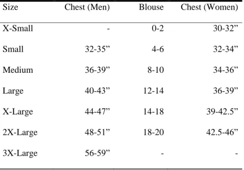
participant wearing no shirt; males were bare skinned, females were in either a tank top shirt or sports bra. The sham condition involved the participant wearing a non-corrective shirt. The treatment condition involved the wearing of a posture shirt. The appropriate shirt was selected by a circumferential measurement of the participant’s chest per the manufacturer’s recommendations (Table 1).
Kinematics
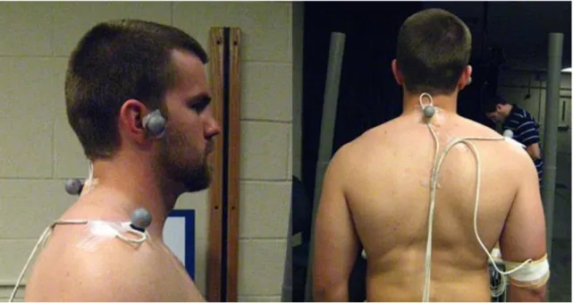
Participants were fitted with electromagnetic tracking receivers that are used with the electromagnetic motion capture system to collect scapulothoracic and humerothoracic data which was analyzed to obtain scapular kinematics during a shoulder elevation task. The male participants removed their shirt, and female participants wore a tank top or sports bra to make receiver placement more accurate and secure. A total of 4 receivers were used for the
and the humeral shaft of the dominant arm (Figure 1) (Oyama, Myers et al. 2008). The receiver on C7 was placed over the spinous process, defined as the point with the least amount of soft tissue covering it. The receiver placed on the acromion processes was placed over the lateral one third of the process where the soft tissue is of least thickness (the
acromial angle). The receiver placed on the humerus was placed on the posterior humerus, distal to the muscle belly of the triceps brachii. All receivers were secured by means of hypoallergenic tape. The receiver on the humeral shaft was also wrapped with foam under-wrap and then covered with athletic tape. The cables attached to each receiver were taped to the participant in such a way that the participant could don a shirt without disrupting the cables (Figure 1).
32
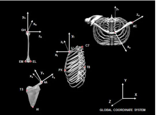
The local coordinate systems for the thorax, scapula, clavicle and humerus were constructed based on the digitization of these anatomical landmarks. The position and orientation of the scapula relative to the thorax were calculated based on these local coordinate systems (Wu, van der Helm et al. 2005; Myers and Jolly 2006).
After digitization, the participants were then instructed on a humeral elevation task in the scapular plane. Guidance for the task was provided by vertical posts positioned in front of the participant; that they used as a guide to their motion. The scapular plane was defined as 30 degrees anterior to the frontal plane for this study. Shoulder rotation was in the neutral position and the participant was instructed to maintain this position by keeping their thumbs pointing towards the ceiling (Figure 2). Each participant performed 15 elevations at a rate of 4 seconds per repetition, 2 seconds raising and 2 seconds lowering (Karduna, McClure et al. 2000; Hibberd, Oyama et al. 2012).
Photographic Posture Assessment
reapplication of the reflective marker on the acromion process. To ensure accurate placement during each condition the marker was aligned with the most anterior prominence of the acromion process, identified through palpation of the area. Additionally, this marker was placed by the same clinician in an effort to standardize placement.
DATA REDUCTION Kinematic Data Reduction
Raw kinematic data was filtered with a 10Hz Butterworth filter (Challis and Kitney 1983). Receiver position and orientation data of the thoracic, scapular, and humeral receivers were transformed into a local coordinate system for each of the respective segments. The coordinate systems used were in accordance with recommendations from the International Society of Biomechanics (Tables 3-6) (Wu, van der Helm et al. 2005). With the participant standing in anatomical position, the coordinate system for each segment was: vertical (y-axis), horizontal to the right (z-axis) and anterior (x-axis) (Figure 3). Euler angles (Y-X-Y order) were used to determine the position of the humerus relative to the thorax. The rotation sequence of the Euler angle was chosen from the recommendations of the International Shoulder Group (Wu, van der Helm et al. 2005). Humeral orientation was determined as rotation about the y-axis of the humerus (plane of elevation), rotation about the z-axis (elevation), and rotation about the y-axis (axial rotation). Using Matlab software (The
MathWorks Inc., Natick, Massachusetts) humeral elevation angles of 30°, 60°, 90°, and 120° were identified relative to the thorax during the ascending phase of the elevation task. The time points at said humeral elevation angles were used for the assessment of scapular
34
Euler angles (Y-X-Z order) were used to determine the scapular orientation, with respect to the thorax, at the time points identified for a given humeral elevation angle. Orientation of the scapula relative to the thorax was determined as rotation about the y-axis of the scapula (internal/external rotation), rotation about the z-axis of the scapula
(upward/downward rotation) and rotation about the x-axis of the scapula (anterior/posterior tipping) (Figure 4). Scapular position (protraction/retraction, elevation/depression) was represented by clavicular kinematic measurements; not actual clavicular motions. Scapular protraction/retraction was calculated as the angle formed between the vector extending from the SC joint to the AC joint, projected onto the transverse plane of the thorax, relative to the frontal plane of the thorax. Scapular elevation/depression was calculated as the angle formed between the vector extending from the SC joint to the AC joint, projected onto the frontal plane of the thorax, relative to the transverse plane of the thorax.
Based on the recommendations from the ISB, scapular movements in internal rotation, downward rotation and posterior tipping directions were indicated by the positive numbers (Wu, van der Helm et al. 2005). Scapular upward rotation values were multiplied by -1 to make upward rotation a positive movement. This was done to make clinical
difference between the sham condition and preceding control condition, and the difference between the treatment condition and the preceding control condition. These data were used in statistical analysis.
Photographic Data Reduction
Three photographs were taken of each participant under each test condition. The Forward Shoulder Angle was measured in each photograph. These data for each participant and condition were averaged and the means were used for statistical analysis of FSA. STATISTICAL ANALYSIS
36
CHAPTER IV: MANUSCRIPT INTRODUCTION
Shoulder injuries are common throughout the student population with some estimates as high as 35% (Katz, Amick et al. 2000; Schlossberg, Morrow et al. 2004). Additionally, shoulder injuries account for 8-20% of all sport related injuries (Agel, Ransone et al. 2007; Dick, Ferrara et al. 2007). Many shoulder injuries are chronic in nature and may be classified as overuse injuries such as shoulder impingement syndrome, myofascial pain or thoracic outlet syndrome (Simons and Travell 1981; Griegel-Morris, Larson et al. 1992; Ferrante 2004). These injuries have been partially attributed in the literature to rounded shoulder posture, muscle imbalance, and altered scapular kinematics (Greenfield, Catlin et al. 1995).
and abnormal scapular position (elevation/depression and protraction/retraction) during shoulder motion (Lukasiewicz, McClure et al. 1999; Ludewig and Cook 2000; Thigpen, Padua et al. 2010). Often, persons with symptoms of shoulder impingement present clinically with changes in shoulder motion; specifically increased anterior tipping and greater upward rotation
(Lukasiewicz, McClure et al. 1999; Ludewig and Cook 2000). These abnormal kinematics may be an attempt to minimize pain in a symptomatic shoulder and may result from muscular
imbalances (Kebaetse, McClure et al. 1999; Ludewig and Cook 2000). Tightness or over activity of the anterior muscles combined with weakness or under activity of the posterior musculature of the shoulder girdle may lead to a relative protraction, anterior tipping or downward rotation of the shoulder (Ludewig and Cook 2000; Thigpen, Padua et al. 2010). These muscle imbalances may result in either static changes in shoulder position, i.e. rounded shoulder posture, or may result in kinematic changes to the shoulder (Langford 1994; Finley and Lee 2003).
With rounded shoulder posture and altered scapular kinematics being linked to shoulder pathology, clinicians have sought ways to correct these abnormalities (Wang, McClure et al. 1999; Lynch, Thigpen et al. 2010; Wong, Coleman et al. 2010). Many different means of posture correction have been examined in the literature, including stretching of tight structures,
38
rounded shoulder posture through the use of specialized neural bands that extend across the superior shoulder girdle from pectorals to the spine. The neural bands are designed to improve posture through increased proprioception and manipulation of the scapula and muscles of the shoulder girdle (Apparel 2012), however there is little published evidence in favor of or against their efficacy.
The purpose of this study was to determine if application of the corrective posture shirt, as compared to a sham shirt and control (no shirt) condition, has an acute effect on scapular kinematics or posture in college students with rounded shoulder posture. Clinically, if excessive rounded shoulder posture and abnormal scapular kinematics can be acutely corrected through the use of posture shirts, it may be possible to reduce the incidence of shoulder pain and may provide a useful adjunct to current therapies by providing temporary relief for sufferers of shoulder pain while other rehabilitative techniques take longer to be effective. Additionally, if effective, posture shirts could be worn in conjunction with traditional therapies such as rehabilitation exercises as an additive benefit.
METHODS Design
A repeated measures intervention study with counterbalanced conditions was used to determine how a posture shirt changes excessive forward shoulder posture and scapular movement patterns compared to both control and sham conditions.
Participants
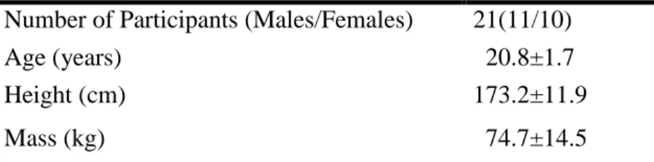
student body population at a large university and were between 18 and 25 years old. Three participants (2 females, 1 male) were excluded from final analysis for being clear outliers relative to the other participants. Final analysis included twenty-one participants (11 males, 10 females; age 20.8±1.7 y, height 173.2±11.9 cm, weight 74.7±14.5 kg) (Table 9). Participants were excluded from the study if they had a history of upper extremity surgery, history of
scoliosis, active shoulder pain, or shoulder pain in the last three months that restricted activity for greater than three consecutive days.
Procedures
Lateral photographs were taken with a Canon PowerShot SD1000 digital camera (Canon USA Inc., Lake Success, NY) and the images were downloaded to ImageJ software (National Institutes of Health, Bethesday, MD) for analysis.
Kinematic assessment was performed using the MotionStar model 800 wide-range transmitter and miniaturized birds (Ascension Technologies Inc., Milton, VT). The device used an electromagnetic transmitter and 4 electromagnetic receivers, 3 of which were attached to the participant with the fourth being used for digitization of landmarks as described in previous literature (Meskers, Fraterman et al. 1999). The tracking device recorded position and orientation data of the receivers relative to the transmitter. All kinematic data were collected at 100 Hz and analysis was done using Motion Monitor version 8.64 electromagnetic tracking software (Innovative Sports Training Inc., Chicago, IL).
40
distance away from a camera. Participants were equipped with three reflective markers, on the spinous process of the seventh cervical vertebra (C7), the most anterior border of the acromion process and on the tragus of the dominant side of the participant’s body (Figures 2&3).
Participants were instructed to touch their toes and reach overhead three times and then stand in their normal posture, at which time a high definition lateral photograph was taken. This
procedure was repeated three times with three photographs used for calculation of FSA.
Photographs were analyzed using the angle calculation function of the ImageJ software. Forward shoulder angle, defined as the angle formed between the reflective marker on the acromion process and a vertical line through the marker on the spinous process at C7 (Thigpen, Padua et al. 2010), was calculated for each photograph and averaged to determine participant eligibility (Figure 2). This measurement of posture, as described by Thigpen et al. (Thigpen, Padua et al. 2010), was used for identification of FSA and was shown to be reliable during pilot testing for this study (Intraclass Correlation Coefficient (ICC)(3,1)=0.99, Standard Error of the Mean
(SEM)=0.23°, Minimum Detectable Difference (MDD)=0.78°). This same manner of data collection was used during testing with those data used in statistical analysis of FSA.
Upon qualifying for continuation in the study with a forward shoulder angle greater than 52°, participants were called back to the research laboratory for additional posture and kinematic testing. Posture testing procedures were identical to the screening process. Kinematic assessment was done through electromagnetic motion tracking using 4 electromagnetic receivers. This method has been shown a precise, valid and reliable method of collection of scapular kinematics (McClure, Michener et al. 2001; Myers and Jolly 2006), Receivers were placed on the
of the posterior humerus. All receivers were secured with double-sided adhesive tape, as well as pre-wrap and athletic tap on the humeral receiver, to minimize receiver movement. The fourth receiver was used for digitization of anatomical landmarks on the thorax, scapula, clavicle, and upper arm. Digitized landmarks included the spinous process of the seventh cervical vertebra, spinous process of the eighth thoracic vertebra, suprasternal notch, xiphoid process, root of the spine of the scapula, scapular inferior angle, scapular acromial angle, sternoclavicular joint, acromioclavicular joint, and the medial and lateral epicondyles of the humerus. The
glenohumeral joint center, defined as the point that moves the least with respect to the scapula as the humerus is moved through short arcs of motion (Veeger 2000), was estimated through a least squares algorithm. Construction of local coordinate systems were done using the digitized landmarks for each body segment; thorax, scapula, and humerus. Local coordinate systems were used for calculation of position and orientation of the scapula.
Participants completed an elevation task consisting of 15 repetitions of continuous bilateral full-shoulder elevation in the scapular plane, defined as 30 degrees anterior to the frontal plane verified with a goniometer. A guide was placed in front of the participant to ensure that their arms remained in the scapular plane (Figure 3) (Karduna, McClure et al. 2000;
Hibberd, Oyama et al. 2012). The participant completed each cycle to the beat of a metronome with elevation taking two seconds and lowering taking two seconds.
42
posture. The sham shirt was chosen as an analog to the posture shirt that provided no posture correction to the wearer. Both the sham shirt and posture shirt were fitted according to
manufacturer recommendations using a circumferential measurement of the participant’s chest which was compared to sizing charts from the respective manufacturer. The control conditions involved the participant wearing no shirt (males), or a tank top or sports bra (females). No
electromagnetic receivers were removed during the testing process and only the reflective marker on the acromion process was removed to accommodate for the shirt conditions. To ensure proper replacement of the reflective marker on the acromion process all placements were done by the same clinician, and by palpating the most anterior border of the acromion process.
For kinematic data reduction the middle 5 repetitions (of 15 total repetitions) of the elevation task were used to account for a learning effect and fatigue. Raw kinematic data were filtered using a low-pass, 10Hz Butterworth filter; and reduced using Matlab software (The MathWorks Inc., Natick, Massachusetts). Position and orientation data from each receiver were used to construct local coordinate systems for the thorax, scapula and humerus; in accordance with International Society of Biomechanics (ISB) recommendations (Wu, van der Helm et al. 2005). With the participant standing in anatomic position each segment was defined as vertical (y-axis), horizontal to the right (z-axis) and anterior (x-axis). Scapular orientation was
formed between the vector extending from the sternoclavicular joint to the acromioclavicular joint projected onto the transverse plane of the thorax, relative to the frontal plane of the thorax. Scapular elevation/depression was calculated as the angle formed between the vector extending from the sternoclavicular joint to the acromioclavicular joint, projected onto the frontal plane of the thorax, relative to the transverse plane of the thorax.
For each kinematic variable, mean position and orientation data were identified at 30°, 60°, 90°, and 120° of humeral elevation during the ascension phase of the elevation task. These data were further reduced by calculating change scores at each level of humeral elevation, for each kinematic variable, for the treatment condition (posture shirt) minus the preceding control condition, the sham condition minus the preceding control condition, and the difference between the two control conditions. These data were used in statistical analysis.
Statistical Analyses
A one-way, repeated measures ANOVA was run with one within-subjects factor (condition) to compare forward shoulder angles between conditions (posture shirt , sham shirt, control condition). A post-hoc analysis with a Bonferroni correction was used to assess pair-wise comparisons of test conditions. Using change scores between the sham shirt, the posture shirt, and the two control conditions, two-way repeated measures ANOVAs were run with two within-subjects factors (condition and angle) to assess each kinematic variable including scapular upward rotation, internal rotation, posterior tipping, protraction, and elevation. Post-hoc analyses with Bonferroni corrections of each ANOVA were used to assess main effects as well as
44
Chicago, IL). Bonferonni corrections were performed, adjusted for multiple comparisons and giving an adjusted alpha level of 0.0027 for all post-hoc testing.
RESULTS
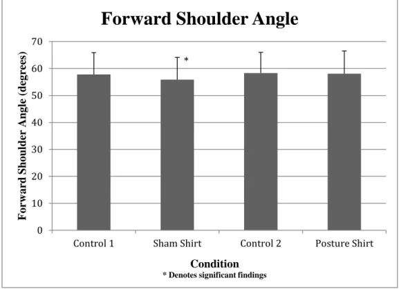
A significant difference was found between postural analysis conditions for the forward shoulder angle (F(4,92)=3.726, p=0.007)(Table 10). Post-hoc testing revealed that participants
wearing the sham shirts demonstrated a significant decrease of 2.40° in FSA between the sham shirt and its preceding control condition as compared to the posture shirt and its preceding control condition and between the two control conditions (p=0.048)(Figure 6). No other significant differences were found between postural assessment conditions.
A significant interaction effect (F(6,120)=3.776, p=0.002) was found for scapular
protraction (Table 11). Post-hoc testing revealed a significant increase in protraction at 90° of elevation (Figure 7) between the sham shirt and the control (t(20)=5.035, p<0.001), and at 120° of
elevation (Figure 8), a significant increase in protraction between the sham shirt and both the control (t(20)= - 4.070, p<0.001) and the posture shirt change scores (t(20)=8.113, p=0.001) (Table
11). Finally, while there was a significant main effect (F(3,120)=8.955, p<0.001)and significant
interaction effect (F(6,120)=3.479, p=0.003) for scapular internal rotation as well as a significant
main effect (F(3,120)=6.782, p=0.001) and interaction effect (F(6,120)=4.504, p<0.001) for posterior
tipping, post-hoc testing revealed no statistically significant changes in either kinematic variable. There were no significant findings for scapular upward rotation or elevation.
DISCUSSION
There were no statistically significant findings for the posture shirts with regard to
no current literature on posture shirts, a comparison to literature on posture braces is the closest analogue available. One particular study assessed posture via forward shoulder angle and muscle activation through electromyographic analysis (EMG) while wearing a posture brace under various conditions (Cole, Prentice et al. 2008). The results of Cole et al,’s study demonstrated a significant decrease in forward shoulder angle (FSA) and a change in EMG activity for select muscles during rehabilitation exercises while wearing both a sham brace and treatment brace as compared to a control (no brace) condition. The decreases in FSA and increases in EMG activity that were seen in the sham condition, where the bracing straps were not placed correctly, suggest that the strapping procedures may not be necessary for changes in muscle activation or posture to occur. A parallel could be drawn between the sham condition employed by Cole et al. and the treatment condition (posture shirt) used in this study, in that the posture shirts do not employ bracing straps and are designed to obtain the same result as the posture braces. However, no change was found in FSA with posture shirt use in this study; and without EMG analysis in this study it is not possible to compare the EMG results found by Cole et al. The lack of significant change in FSA or kinematics in this study may be due to the differences in construction between the posture brace and the posture shirt, or the nature of the posture brace which is designed to be a tighter fitting and manipulative appliance. The posture shirt is designed for extended wear and may be looser fitting and more flexible than a brace. This may result in decreased acute
46
anecdotal evidence with regard to improving posture and scapular kinematics and remain in use in clinics that specialize in rehabilitation, but that evidence could not be replicated by this study and may require further research to confirm.
The sham shirt, contrary to the posture shirt, was found to significantly decrease forward shoulder angle as compared to the treatment and control conditions (Figure 6); however the sham shirt also increased scapular protraction at higher angles of humeral elevation relative to both the control and treatment conditions (Figures 7&8). This suggests that while the posture shirt does not acutely alter rounded shoulder posture or scapular kinematics, the sham shirt may decrease rounded shoulder posture while simultaneously altering scapular kinematics in a sub-optimal fashion. With decreased rounded shoulder posture, the sham shirts may decrease a contributing factor to shoulder pain (Griegel-Morris, Larson et al. 1992; Borstad 2006). Conversely, the increased protraction at higher levels of humeral elevation could be detrimental to the patient as increased protraction has been shown to be a contributing factor to shoulder pathology
(Lukasiewicz, McClure et al. 1999; Ludewig and Cook 2000). Finally, despite being statistically significant, these changes may not be clinically significant as they show a change of only a few degrees.
fitted by a clinician trained in the application of external devices such as braces and by fitting both shirts according to manufacturer’s recommendations.
Another thing to note about this study is that participants may have self-corrected their posture by simply being aware that they were participating in a posture related study, participants were given no verbal cueing as to how to improve their posture while standing or during the kinematic task. This is of particular importance to clinicians as instruction and verbal cueing are consistently used for training a patient to perform rehabilitative exercises correctly. The lack of verbal cueing in this study may provide additional insight as to why no significant results were found.
48
population that was free of shoulder pain. As the shirts are commonly used in a symptomatic population this study may not be generalizable to other populations.
This study is a preliminary assessment of the effects of posture shirt use on scapular kinematics and rounded shoulder posture. Additional studies may focus on the use of posture shirts during functional tasks, specifically concentrating on their effect on muscle activation and movement patterns. Those studies may focus on the use of a posture shirt as an adjunct to traditional therapies as a means to improve muscle function during rehabilitation exercises, in combination with verbal cueing, or as a palliative treatment for people with active shoulder pain, none of which were the focus of this study.
CONCLUSION
FIGURES
50
Figure 2 – Participant performing elevation task while equipped with electromagnetic motion tracking system
52
Figure 6 – Significant findings for forward shoulder angle
Figure 7 – Significant findings of scapular protraction at 90 degrees of elevation.
0 10 20 30 40 50 60 70
Control 1 Sham Shirt Control 2 Posture Shirt
F or w ar d Sho ul der Angl e (deg re es ) Condition * Denotes significant findings
Forward Shoulder Angle
* -1 0 1 2 3 4 5 6 7 8
Posture Shirt-Control Sham-Control Control 1-Control 2
D if fe renc e Sco re (deg rees ) Conditions Assessed
*Denotes significant increase in prtraction relative to control condition comparisons.
Protraction Difference Scores at 90°
Humeral Elevation
Figure 8 – Significant findings of scapular protraction at 120 degrees of elevation.
0 1 2 3 4 5 6 7 8 9
Posture Shirt-Control Sham-Control Control 1-Control 2
Dif
fe
re
nc
e
Sco
re
s
(deg
re
es
)
Conditions Assessed
*Denotes significant increase in protraction relative to Control 1-Control 2 and Shirt-Control assessments.
Protraction Difference Scores at 120° Humeral
Elevation
54 TABLES
Table 1: Posture Shirt Sizing Chart
Size Chest (Men) Blouse Chest (Women)
X-Small - 0-2 30-32”
Small 32-35” 4-6 32-34”
Medium 36-39” 8-10 34-36”
Large 40-43” 12-14 36-39”
X-Large 44-47” 14-18 39-42.5”
2X-Large 48-51” 18-20 42.5-46”
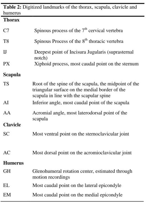
Table 2: Digitized landmarks of the thorax, scapula, clavicle and humerus
Thorax
C7 Spinous process of the 7th cervical vertebra T8 Spinous Process of the 8th thoracic vertebra IJ Deepest point of Incisura Jugularis (suprasternal
notch)
PX Xiphoid process, most caudal point on the sternum Scapula
TS Root of the spine of the scapula, the midpoint of the triangular surface on the medial border of the scapula in line with the scapular spine
AI Inferior angle, most caudal point of the scapula AA Acromial angle, most laterodorsal point of the
scapula Clavicle
SC Most ventral point on the sternoclavicular joint
AC Most dorsal point on the acromioclavicular joint Humerus
GH Glenohumeral rotation center, estimated through motion recordings
56 Table 3: Coordinate Systems of the Thorax XtYtZt
Ot The origin coincident with IJ.
Yt The line parallel to the line connecting the midpoint between
PX and T8 and the midpoint between IJ and C7, pointing upward.
Zt The line perpendicular to the plane formed by IJ, C7, and the
midpoint between PX and T8, pointing to the right. Xt The common line perpendicular to the Zt- and Yt-axis,
pointing forwards.
Table 4: Coordinate System of the Scapula - XsYsZs
Os The origin coincident with AA.
Zs The line connecting TS and AA, pointing to AA.
Xs The line perpendicular to the plane formed by AI, AA, and
TS, pointing forward. Note that because of the use of AA instead of AC, this plane is not the same as the visual plane of the scapula bone.
Ys The common line perpendicular to the Xs- and Zs-axis,
pointing upward.
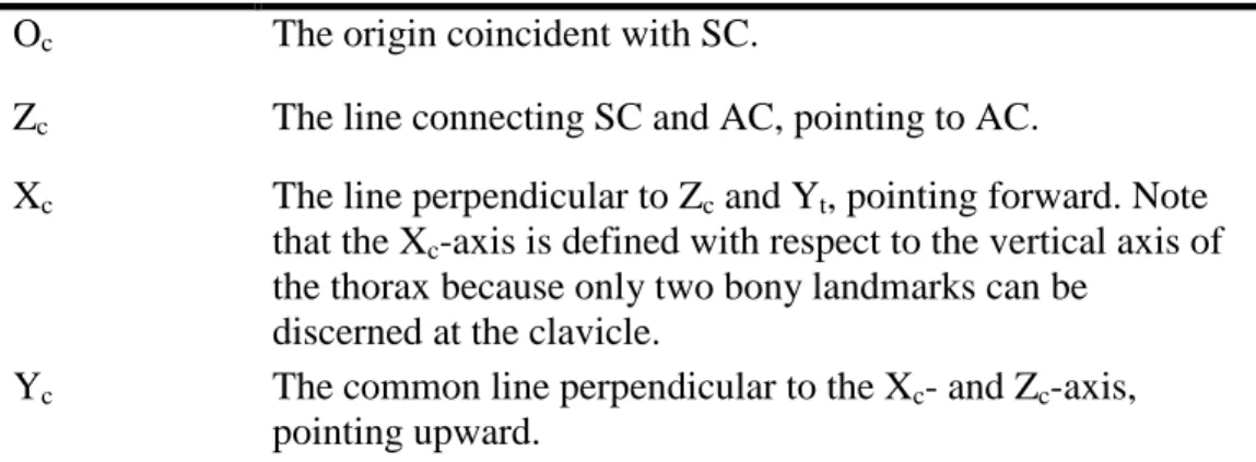
Table 5: Coordinate System of the Clavicle – XcYcZc
Oc The origin coincident with SC.
Zc The line connecting SC and AC, pointing to AC.
Xc The line perpendicular to Zc and Yt, pointing forward. Note
that the Xc-axis is defined with respect to the vertical axis of
the thorax because only two bony landmarks can be discerned at the clavicle.
Yc The common line perpendicular to the Xc- and Zc-axis,
Table 6: Coordinate System of the Humerus - XhYhZh
Oh The origin coincident with GH.
Yh The line connecting GH and the midpoint of EL and
EM, pointing to GH.
Xh The line perpendicular to the plane formed by EL,
EM, and GH, pointing forward.
Zh The common line perpendicular to the Yh- and Zh
-axis, pointing to the right.
Table 7: Intrasession and Intersession Reliability of Forward Shoulder Angle (FSA) Measurement (Myers and Jolly 2006)
Posture (°) Intrasession ICC Intrasession SEM Intersession ICC Intersession SEM Forward Shoulder Angle
0.89 5° 0.72 7°
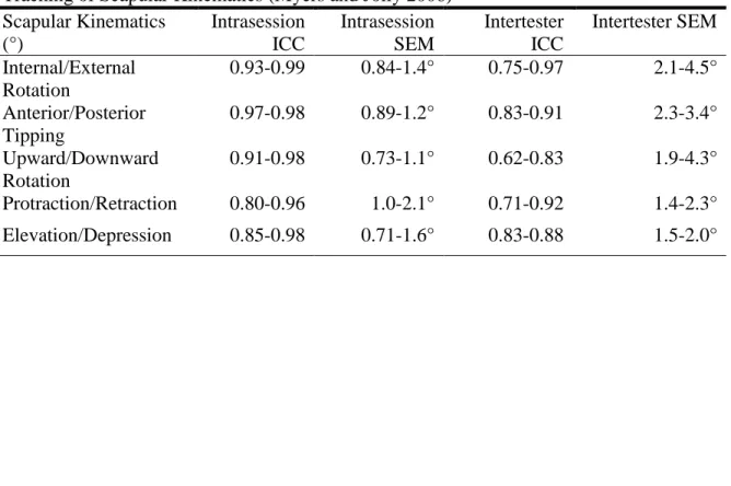
Table 8: Intrasession and Intertester Reliability and Precision of Electromagnetic Tracking of Scapular Kinematics (Myers and Jolly 2006)
Scapular Kinematics (°) Intrasession ICC Intrasession SEM Intertester ICC Intertester SEM Internal/External Rotation
0.93-0.99 0.84-1.4° 0.75-0.97 2.1-4.5° Anterior/Posterior
Tipping
0.97-0.98 0.89-1.2° 0.83-0.91 2.3-3.4° Upward/Downward
Rotation
58 Table 9: Participant demographic information
Number of Participants (Males/Females) 21(11/10)
Age (years) 20.8±1.7
Height (cm) 173.2±11.9
Mass (kg) 74.7±14.5
Table 10: Means and standard deviations (SD) of forward shoulder angle in degrees. Pre-Sham
Control (1)
Sham Pre-Shirt
Control (2)
Posture Shirt
Forward Shoulder Angle (FSA)
57.8±8.1 55.8±8.2* 58.3±7.7 58.1±8.4