2-[(4-Methylbenzoyl)hydrazono]-propionic acid monohydrate
Hon Wee Wong, Kong Mun Lo and Seik Weng Ng*
Department of Chemistry, University of Malaya, 50603 Kuala Lumpur, Malaysia Correspondence e-mail: [email protected]
Received 21 January 2009; accepted 24 January 2009
Key indicators: single-crystal X-ray study;T= 100 K; mean(C–C) = 0.003 A˚;
Rfactor = 0.030;wRfactor = 0.083; data-to-parameter ratio = 7.8.
In the title compound, C11H12N2O3H2O, the water molecule is a hydrogen-bond donor to the double-bond amide and the carbonyl O atoms of two acid molecules; it is also a hydrogen-bond acceptor to the acid –OH and amide –NH– groups. These hydrogen-bonding interactions give rise to a layer structure, with the layers parallel to theabplane.
Related literature
The deprotonated anion of 2-aroylhydrazonopropionic acid furnishes a number of metal complexes; see, for example: Wu, Chenet al.(2006); Liuet al.(2007); Wu & Zeng (2007); Wuet al.(2006a,b); Yanget al.(2004); Yin & Chen (2006); Zhaiet al.
(2007).
Experimental
Crystal data
C11H12N2O3H2O
Mr= 238.24
Monoclinic, P21
a= 6.8464 (1) A˚ b= 11.9753 (2) A˚ c= 7.0005 (1) A˚
= 102.169 (1)
V= 561.06 (2) A˚3
Z= 2
MoKradiation
= 0.11 mm1
T= 100 (2) K 0.200.100.10 mm
Data collection
Bruker SMART APEX diffractometer
Absorption correction: none 5272 measured reflections
1335 independent reflections 1211 reflections withI> 2(I) Rint= 0.029
Refinement
R[F2> 2(F2)] = 0.030 wR(F2) = 0.083
S= 1.02 1335 reflections 172 parameters 5 restraints
H atoms treated by a mixture of independent and constrained refinement
max= 0.19 e A˚ 3
min=0.16 e A˚ 3
Absolute structure: 1126 Friedel pairs were merged
Table 1
Hydrogen-bond geometry (A˚ ,).
D—H A D—H H A D A D—H A
O1—H1 O1W 0.83 (2) 2.03 (2) 2.777 (2) 149 (3) O1W—H11 O3 0.84 (2) 1.97 (2) 2.794 (2) 165 (4) O1W—H12 O2i
0.84 (2) 2.00 (1) 2.829 (2) 168 (3) N2—H2 O1Wii 0.87 (2) 2.35 (1) 3.210 (2) 168 (3)
Symmetry codes: (i)xþ3;y1
2;zþ2; (ii)x1;y;z.
Data collection:APEX2(Bruker, 2007); cell refinement:APEX2; data reduction: SAINT (Bruker, 2007); program(s) used to solve structure:SHELXS97(Sheldrick, 2008); program(s) used to refine structure: SHELXL97 (Sheldrick, 2008); molecular graphics:
X-SEED (Barbour, 2001); software used to prepare material for publication:publCIF(Westrip, 2009).
The autors thank the University of Malaya (grant Nos. FS339/2008A and PS206/2008A) for supporting this study.
Supplementary data and figures for this paper are available from the IUCr electronic archives (Reference: CV2513).
References
Barbour, L. J. (2001).J. Supramol. Chem.1, 189–191.
Bruker (2007).APEX2andSAINT. Bruker AXS Inc., Madison, Wisconsin, USA.
Liu, F., Wu, W.-T., Zhang, W.-P., Chen, F.-Y. & He, S.-Y. (2007).Acta Cryst. E63, m2450–m2451.
Sheldrick, G. M. (2008).Acta Cryst.A64, 112–122. Westrip, S. P. (2009).publCIF. In preparation.
Wu, W.-T., Chen, F.-Y., He, S.-Y., Hu, H.-M., Yang, M.-L. & Wang, Y.-Y. (2006). Chin. J. Chem.24, 711–713.
Wu, W.-T., He, S.-Y., Hu, H.-M., Yang, M.-L., Wang, Y.-Y. & Shi, Q.-Z. (2006a). J. Coord. Chem.59, 1785–1791.
Wu, W.-T., He, S.-Y., Hu, H.-M., Yang, M.-L., Wang, Y.-Y. & Shi, Q.-Z. (2006b). J. Coord. Chem.60, 125–130.
Wu, W.-P. & Zeng, F.-C. (2007).Acta Cryst.E63, m2664.
Yang, R., He, S.-Y., Wu, W.-T., Wen, Z.-Y., Shi, Q.-Z. & Wang, D.-Q. (2004). Acta Chim. Sin.62, 2040–2044.
Yin, H.-D. & Chen, S.-W. (2006).Inorg. Chim. Acta,359, 3330–3338. Zhai, J., Yin, H., Li, F. & Wang, D. (2007).Acta Cryst.E63, m3066. Acta Crystallographica Section E
Structure Reports Online
supporting information
Acta Cryst. (2009). E65, o419 [doi:10.1107/S1600536809003067]
2-[(4-Methylbenzoyl)hydrazono]propionic acid monohydrate
Hon Wee Wong, Kong Mun Lo and Seik Weng Ng
S1. Experimental
4-Toluihydrazide (1 g, 0.007 mol) and pyruvic acid (0.6 g, 0.007 mol) were dissolved in methanol (30 ml). The solution
was heated for 3 h; slow evaporation of the solvent gave colorless crystals.
S2. Refinement
Carbon-bound H atoms were placed in calculated positions (C—H 0.93–0.99 Å) and were included in the refinement in
the riding model approximation, with U(H) set to 1.2 to 1.5U(C). The methyl H atoms were rotated to fit the electron
density.
The oxygen- and nitrogen-bound H atoms were located in a difference Fourier map, and were refined with distance
[image:2.610.117.489.361.601.2]restraints [N—H 0.88 (2) and O—H 0.84 (2) Å]; their temperature factors were freely refined.
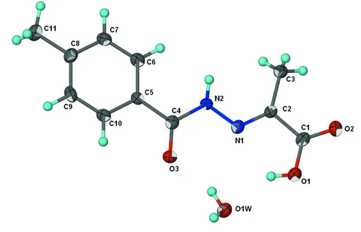
Figure 1
Displacement ellipsoids plot (Barbour, 2001) of the title compound at the 70% probability level. Hydrogen atoms are
2-[(4-Methylbenzoyl)hydrazono]propionic acid monohydrate
Crystal data
C11H12N2O3·H2O
Mr = 238.24
Monoclinic, P21
Hall symbol: P 2yb
a = 6.8464 (1) Å
b = 11.9753 (2) Å
c = 7.0005 (1) Å
β = 102.169 (1)°
V = 561.06 (2) Å3
Z = 2
F(000) = 252
Dx = 1.410 Mg m−3
Mo Kα radiation, λ = 0.71073 Å
Cell parameters from 1743 reflections
θ = 3.0–26.9°
µ = 0.11 mm−1
T = 100 K
Irregular block, colourless 0.20 × 0.10 × 0.10 mm
Data collection
Bruker SMART APEX diffractometer
Radiation source: fine-focus sealed tube Graphite monochromator
ω scans
5272 measured reflections 1335 independent reflections
1211 reflections with I > 2σ(I)
Rint = 0.029
θmax = 27.5°, θmin = 3.0°
h = −8→8
k = −14→15
l = −9→9
Refinement
Refinement on F2
Least-squares matrix: full
R[F2 > 2σ(F2)] = 0.030
wR(F2) = 0.083
S = 1.02
1335 reflections 172 parameters 5 restraints
Primary atom site location: structure-invariant direct methods
Secondary atom site location: difference Fourier map
Hydrogen site location: inferred from neighbouring sites
H atoms treated by a mixture of independent and constrained refinement
w = 1/[σ2(Fo2) + (0.0559P)2 + 0.0248P]
where P = (Fo2 + 2Fc2)/3
(Δ/σ)max = 0.001
Δρmax = 0.19 e Å−3
Δρmin = −0.16 e Å−3
Absolute structure: 1126 Friedel pairs were merged
Special details
Geometry. All e.s.d.'s (except the e.s.d. in the dihedral angle between two l.s. planes) are estimated using the full covariance matrix. The cell e.s.d.'s are taken into account individually in the estimation of e.s.d.'s in distances, angles and torsion angles; correlations between e.s.d.'s in cell parameters are only used when they are defined by crystal symmetry. An approximate (isotropic) treatment of cell e.s.d.'s is used for estimating e.s.d.'s involving l.s. planes.
Fractional atomic coordinates and isotropic or equivalent isotropic displacement parameters (Å2)
x y z Uiso*/Ueq
O1 1.4450 (2) 0.99988 (13) 0.8798 (3) 0.0255 (4)
O2 1.2160 (2) 1.13156 (13) 0.8463 (2) 0.0269 (4)
O3 1.2831 (2) 0.64501 (13) 0.7200 (2) 0.0260 (4)
C2 1.0961 (3) 0.94692 (18) 0.7861 (3) 0.0183 (4)
C3 0.8847 (3) 0.98625 (18) 0.7447 (3) 0.0232 (5)
H3A 0.8131 0.9412 0.6686 0.035*
H3B 0.8400 0.9951 0.8651 0.035*
H3C 0.8754 1.0568 0.6777 0.035*
C4 1.1067 (3) 0.65752 (18) 0.7236 (3) 0.0198 (4)
C5 0.9684 (3) 0.56054 (18) 0.7054 (3) 0.0178 (4)
C6 0.7608 (3) 0.5709 (2) 0.6510 (3) 0.0218 (4)
H6 0.7018 0.6422 0.6192 0.026*
C7 0.6408 (3) 0.47675 (19) 0.6437 (3) 0.0231 (5)
H7 0.4998 0.4845 0.6067 0.028*
C8 0.7226 (3) 0.37199 (18) 0.6891 (3) 0.0214 (5)
C9 0.9315 (3) 0.3617 (2) 0.7379 (3) 0.0221 (5)
H9A 0.9904 0.2901 0.7659 0.026*
C10 1.0524 (3) 0.45480 (19) 0.7457 (3) 0.0202 (4)
H10 1.1936 0.4467 0.7787 0.024*
C11 0.5927 (3) 0.27036 (19) 0.6870 (4) 0.0288 (5)
H11A 0.6157 0.2411 0.7985 0.043*
H11B 0.4542 0.2924 0.6689 0.043*
H11C 0.6071 0.2211 0.5815 0.043*
H11 1.496 (3) 0.755 (3) 0.813 (5) 0.059 (10)*
H12 1.653 (4) 0.748 (2) 0.963 (3) 0.052 (10)*
H1 1.445 (5) 0.9307 (9) 0.867 (5) 0.056 (11)*
H2 0.906 (2) 0.773 (2) 0.761 (4) 0.031 (7)*
Atomic displacement parameters (Å2)
U11 U22 U33 U12 U13 U23
O1 0.0193 (8) 0.0198 (9) 0.0371 (10) −0.0029 (6) 0.0049 (7) −0.0031 (7)
O2 0.0234 (7) 0.0182 (8) 0.0384 (9) −0.0001 (6) 0.0047 (7) −0.0028 (7)
O3 0.0171 (7) 0.0206 (8) 0.0416 (9) −0.0005 (6) 0.0094 (6) −0.0039 (7)
O1W 0.0205 (7) 0.0208 (8) 0.0331 (10) −0.0018 (6) 0.0036 (7) 0.0028 (7)
N1 0.0186 (9) 0.0171 (9) 0.0218 (9) −0.0017 (7) 0.0042 (7) 0.0008 (7)
N2 0.0139 (8) 0.0165 (9) 0.0301 (10) −0.0001 (7) 0.0048 (7) −0.0012 (7)
C1 0.0198 (10) 0.0212 (11) 0.0202 (11) −0.0033 (8) 0.0043 (8) −0.0013 (8)
C2 0.0167 (9) 0.0179 (10) 0.0209 (10) −0.0012 (8) 0.0054 (8) −0.0004 (8)
C3 0.0174 (10) 0.0189 (11) 0.0329 (13) 0.0007 (8) 0.0044 (9) 0.0033 (9)
C4 0.0200 (10) 0.0186 (10) 0.0207 (10) −0.0004 (8) 0.0041 (8) 0.0004 (9)
C5 0.0170 (10) 0.0169 (10) 0.0201 (10) 0.0010 (8) 0.0049 (8) −0.0013 (8)
C6 0.0205 (10) 0.0204 (10) 0.0240 (11) 0.0024 (9) 0.0036 (8) −0.0003 (9)
C7 0.0159 (10) 0.0238 (11) 0.0289 (12) 0.0017 (9) 0.0034 (8) −0.0049 (9)
C8 0.0225 (11) 0.0216 (11) 0.0211 (11) −0.0046 (9) 0.0068 (8) −0.0057 (9)
C9 0.0260 (11) 0.0154 (10) 0.0254 (11) 0.0042 (9) 0.0067 (9) 0.0005 (8)
C10 0.0160 (9) 0.0214 (11) 0.0226 (11) 0.0021 (8) 0.0030 (8) −0.0004 (9)
Geometric parameters (Å, º)
O1—C1 1.312 (3) C4—C5 1.487 (3)
O1—H1 0.83 (2) C5—C10 1.395 (3)
O2—C1 1.213 (3) C5—C6 1.397 (3)
O3—C4 1.222 (2) C6—C7 1.390 (3)
O1W—H11 0.84 (2) C6—H6 0.9500
O1W—H12 0.82 (2) C7—C8 1.383 (3)
N1—C2 1.277 (3) C7—H7 0.9500
N1—N2 1.377 (2) C8—C9 1.404 (3)
N2—C4 1.369 (3) C8—C11 1.506 (3)
N2—H2 0.87 (2) C9—C10 1.383 (3)
C1—C2 1.508 (3) C9—H9A 0.9500
C2—C3 1.491 (3) C10—H10 0.9500
C3—H3A 0.8400 C11—H11A 0.8400
C3—H3B 0.9620 C11—H11B 0.9663
C3—H3C 0.9620 C11—H11C 0.9662
C1—O1—H1 108 (2) C6—C5—C4 123.14 (19)
H11—O1W—H12 106 (3) C7—C6—C5 119.8 (2)
C2—N1—N2 118.80 (17) C7—C6—H6 120.1
C4—N2—N1 116.00 (17) C5—C6—H6 120.1
C4—N2—H2 124.9 (19) C8—C7—C6 121.34 (19)
N1—N2—H2 118.1 (19) C8—C7—H7 119.3
O2—C1—O1 121.2 (2) C6—C7—H7 119.3
O2—C1—C2 120.35 (19) C7—C8—C9 118.5 (2)
O1—C1—C2 118.40 (18) C7—C8—C11 121.41 (19)
N1—C2—C3 128.72 (19) C9—C8—C11 120.1 (2)
N1—C2—C1 113.55 (17) C10—C9—C8 120.6 (2)
C3—C2—C1 117.73 (19) C10—C9—H9A 119.7
C2—C3—H3A 109.5 C8—C9—H9A 119.7
C2—C3—H3B 109.9 C9—C10—C5 120.40 (18)
H3A—C3—H3B 112.0 C9—C10—H10 119.8
C2—C3—H3C 109.5 C5—C10—H10 119.8
H3A—C3—H3C 106.6 C8—C11—H11A 109.5
H3B—C3—H3C 109.3 C8—C11—H11B 110.0
O3—C4—N2 121.82 (19) H11A—C11—H11B 102.8
O3—C4—C5 121.12 (19) C8—C11—H11C 110.2
N2—C4—C5 117.06 (17) H11A—C11—H11C 115.2
C10—C5—C6 119.3 (2) H11B—C11—H11C 108.8
C10—C5—C4 117.61 (17)
C2—N1—N2—C4 173.5 (2) N2—C4—C5—C6 20.0 (3)
N2—N1—C2—C3 −3.9 (3) C10—C5—C6—C7 2.0 (3)
N2—N1—C2—C1 176.43 (17) C4—C5—C6—C7 −177.78 (19)
O2—C1—C2—N1 179.1 (2) C5—C6—C7—C8 −0.1 (3)
O1—C1—C2—C3 178.78 (19) C7—C8—C9—C10 1.8 (3)
N1—N2—C4—O3 −5.9 (3) C11—C8—C9—C10 −178.2 (2)
N1—N2—C4—C5 173.67 (17) C8—C9—C10—C5 0.1 (3)
O3—C4—C5—C10 19.8 (3) C6—C5—C10—C9 −2.0 (3)
N2—C4—C5—C10 −159.73 (19) C4—C5—C10—C9 177.77 (19)
O3—C4—C5—C6 −160.4 (2)
Hydrogen-bond geometry (Å, º)
D—H···A D—H H···A D···A D—H···A
O1—H1···O1W 0.83 (2) 2.03 (2) 2.777 (2) 149 (3)
O1W—H11···O3 0.84 (2) 1.97 (2) 2.794 (2) 165 (4)
O1W—H12···O2i 0.84 (2) 2.00 (1) 2.829 (2) 168 (3)
N2—H2···O1Wii 0.87 (2) 2.35 (1) 3.210 (2) 168 (3)