organic papers
Acta Cryst.(2005). E61, o1631–o1633 doi:10.1107/S1600536805012031 Rodrigueset al. C
3H7NO2NH4+NO3
o1631
Acta Crystallographica Section EStructure Reports
Online
ISSN 1600-5368
Sarcosine ammonium nitrate
V. H. Rodrigues, M. Ramos Silva,* A. Matos Beja, J. A. Paixa˜o and M.M.R.R. Costa
CEMDRX, Departamento de Fı´sica, Faculdade de Cieˆncias e Tecnologia, Universidade de Coimbra, P-3004-516 Coimbra, Portugal
Correspondence e-mail: [email protected]
Key indicators
Single-crystal X-ray study
T= 293 K
Mean(C–C) = 0.003 A˚
Rfactor = 0.041
wRfactor = 0.130
Data-to-parameter ratio = 11.8
For details of how these key indicators were automatically derived from the article, see http://journals.iucr.org/e.
#2005 International Union of Crystallography
Printed in Great Britain – all rights reserved
In the title compound, C3H7NO2NH4 +
NO3
, the sarcosine zwitterions have an almost perfectly planar N—C—COO main chain and assemble in chains viahydrogen bonds. The ammonium and nitrate ions are arranged in sheets that alternate with sheets of sarcosine chains. The whole structure is stabilized by an extensive three-dimensional network of hydrogen bonds.
Comment
Sarcosine (N-methylglycine, CH3NHþ2CH2COO
) is an -amino acid found in many biological materials (Mostad & Natarajan, 1989). Several structural studies of sarcosine with inorganic acids and metallic ions have been published, span-ning a variety of applications in which the sarcosine molecule plays a role. We report here the crystal structure of this amino acid cocrystallized with ammonium nitrate, viz. the title compound, (I), which could be particularly relevant given the importance of hydrogen-bonded interactions in chemistry and biology. This is the first reported crystal structure of an amino acid cocrystallized with ammonium nitrate.
The ionization state of the sarcosine molecule inferred from the C—O distances in the carboxyl group was found to be a zwitterionic form with a protonated amino group and a deprotonated carboxylate group. The sarcosine skeleton including atoms O1, O2, C1 and C2 is planar within 0.003 (2) A˚ . The N atom deviates 0.090 (3) A˚ from this plane. The title compound is slightly less distorted from planarity than pure sarcosine (Mostad & Natarajan, 1989), the corre-sponding values of the torsion angles O1—C1—C2—N1 and O2—C1—C2—N1 being 173.7 (2) and 6.8 (2). The methyl group breaks the almost perfect planarity of the whole main chain, and atom C3 is displaced by 1.435 (4) A˚ from the previously mentioned least-squares plane, in the same direc-tion as the N atom. The nitrate anion, which is planar within 0.0016 (8) A˚ , deviates slightly from the usual (non-crystal-lographic) D3h symmetry; one of the N—O bonds is
0.010 (3) A˚ shorter than the other two. In addition, two of the O—N—O angles deviate by about 2 from the ideal 120 value. The three-dimensional hydrogen-bonded network can be described as the stacking of linear chains of head-to-tail hydrogen-bonded sarcosine zwitterions, running along the a
axis between sheets of nitrates and ammonium ions. Further-more, each sarcosine molecule is hydrogen-bonded to another sarcosine in a neighbouring chain related by an inversion centre. The ammonium and nitrate ions are arranged in a two-dimensional network in which each nitrate is anchored to four ammonium cations via bifurcated hydrogen bonds and, conversely, each ammonium is anchored to four nitrate anions viathe same bifurcated hydrogen bonds.
Experimental
Colourless block-shaped crystals were obtained by recrystallization of the solution resulting from pouring an excess of nitric acid directly over sarcosine crystals (0.5 g) as purchased from Aldrich (98%), followed by neutralization with ammonia solution (25%).
Crystal data
C3H7NO2NH4+NO3
Mr= 169.15 Monoclinic,P21=c a= 5.7208 (9) A˚
b= 7.9144 (8) A˚
c= 17.447 (2) A˚
= 101.682 (13)
V= 773.58 (17) A˚3 Z= 4
Dx= 1.452 Mg m
3
MoKradiation Cell parameters from 25
reflections
= 6.6–17.3
= 0.14 mm1 T= 293 (2) K Block, colourless 0.430.400.39 mm
Data collection
Enraf–Nonius CAD-4 diffractometer
!–2scans
Absorption correction: none 2819 measured reflections 1423 independent reflections 901 reflections withI> 2(I)
Rint= 0.037
max= 25.5
h=6!6
k=9!9
l=21!6 3 standard reflections
frequency: 7200 min intensity decay: 0.3%
Refinement
Refinement onF2 R[F2> 2(F2)] = 0.041
wR(F2) = 0.130 S= 0.98 1423 reflections 121 parameters
H atoms treated by a mixture of independent and constrained refinement
w= 1/[2(F
o2) + (0.0885P)2]
whereP= (Fo 2
+ 2Fc 2
)/3 (/)max< 0.001
max= 0.19 e A˚
3
min=0.26 e A˚
3
Table 1
Selected geometric parameters (A˚ ,).
O1—C1 1.261 (3) O2—C1 1.234 (3) O3—N2 1.241 (3)
O4—N2 1.243 (3) O5—N2 1.231 (2)
C3—N1—C2—C1 75.5 (2) O2—C1—C2—N1 4.4 (3)
[image:2.610.311.564.71.350.2]O1—C1—C2—N1 176.4 (2)
Table 2
Hydrogen-bond geometry (A˚ ,).
D—H A D—H H A D A D—H A
N1—H1A O1i
0.89 1.97 2.822 (2) 158 N1—H1A O2i
0.89 2.49 3.160 (2) 132 N1—H1B O2ii
0.89 1.95 2.826 (2) 167 N3—H4 O1iii
0.94 (2) 1.92 (2) 2.839 (2) 164 (2) N3—H5 O1i
0.95 (2) 2.20 (2) 2.942 (2) 135 (2) N3—H5 O5iii
0.95 (2) 2.45 (3) 2.961 (3) 114 (2) N3—H5 O3iv 0.95 (2) 2.59 (3) 3.072 (3) 112 (2) N3—H6 O4i
0.92 (2) 2.05 (2) 2.946 (3) 164 (3) N3—H6 O5i
0.92 (2) 2.42 (3) 3.171 (3) 139 (3) N3—H6 N2i
0.92 (2) 2.55 (2) 3.443 (3) 164 (3) N3—H7 O3 0.94 (2) 2.11 (2) 2.977 (3) 153 (3) N3—H7 O4 0.94 (2) 2.31 (2) 3.151 (3) 149 (3) N3—H7 N2 0.94 (2) 2.56 (2) 3.494 (3) 175 (3)
Symmetry codes: (i) x1;y;z; (ii) xþ1;y;z; (iii)xþ1;y1 2;zþ
1 2; (iv) x;y1
2;zþ 1 2.
The ammonium H atoms were located in a difference Fourier map and refined isotropically subject to appropriate restraints to ensure an average tetrahedral geometry; all N—H distances were restrained to be equal within 0.02 A˚ , with a corresponding restraint on all H H distances. All other H atoms were positioned geometrically and
organic papers
o1632
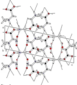
Rodrigueset al. C3H7NO2NH4+NO3 Acta Cryst.(2005). E61, o1631–o1633 Figure 2
[image:2.610.56.281.72.246.2]The packing, showing the layers of sarcosine chains alternating with the ammonium nitrate layers. Dashed lines represent the extensive hydrogen-bond network that stabilizes the structure
Figure 1
[image:2.610.314.564.534.664.2]subsequently refined as riding, with the bond length also allowed to refine (N—H = 0.90 A˚ and C—H = 0.94–0.97 A˚).Uiso(H) = 1.2Ueq(N)
for NH2, Uiso(H) = 1.2Ueq(C) for CH2 and 1.5Ueq(C) for CH3.
Examination of the crystal structure with PLATON(Spek, 2003) showed that there are no solvent-accessible voids.
Data collection: CAD-4 Software (Enraf–Nonius, 1989); cell refinement:CAD-4 Software; data reduction:PLATON(Spek, 2003); program(s) used to solve structure: SHELXS97 (Sheldrick, 1997); program(s) used to refine structure:SHELXL97(Sheldrick, 1997); molecular graphics: ORTEPII (Johnson, 1976); software used to prepare material for publication:SHELXL97.
This work was supported by Fundac¸a˜o para a Cieˆncia e a Tecnologia (FCT).
References
Enraf–Nonius (1989).CAD-4 Software. Version 5.0. Enraf–Nonius, Delft, The Netherlands.
Johnson, C. K. (1976).ORTEPII. Report ORNL-5138. Oak Ridge National Laboratory, Tennessee, USA.
Mostad, A. & Natarajan, S. (1989).Acta Chem. Scand.43, 1004–1006. Sheldrick, G. M. (1997). SHELXS97 and SHELXL97. University of
Go¨ttingen, Germany.
Spek, A. L. (2003).J. Appl. Cryst.36, 7–13.
organic papers
Acta Cryst.(2005). E61, o1631–o1633 Rodrigueset al. C
supporting information
sup-1 Acta Cryst. (2005). E61, o1631–o1633
supporting information
Acta Cryst. (2005). E61, o1631–o1633 [https://doi.org/10.1107/S1600536805012031]
Sarcosine ammonium nitrate
V. H. Rodrigues, M. Ramos Silva, A. Matos Beja, J. A. Paix
ã
o and M.M.R.R. Costa
′N-methylglycine ammonium nitrate′
Crystal data
C3H7NO2·H4N+·NO3−
Mr = 169.15 Monoclinic, P21/c
Hall symbol: -P 2ybc
a = 5.7208 (9) Å
b = 7.9144 (8) Å
c = 17.447 (2) Å
β = 101.682 (13)°
V = 773.58 (17) Å3
Z = 4
F(000) = 360
Dx = 1.452 Mg m−3
Mo Kα radiation, λ = 0.71074 Å Cell parameters from 25 reflections
θ = 6.6–17.3°
µ = 0.14 mm−1
T = 293 K Block, colourless 0.43 × 0.40 × 0.39 mm
Data collection
Enraf–Nonius CAD-4 diffractometer
Radiation source: fine-focus sealed tube Graphite monochromator
profile data from ω–2θ scans 2819 measured reflections 1423 independent reflections 901 reflections with I > 2σ(I)
Rint = 0.037
θmax = 25.5°, θmin = 2.8°
h = −6→6
k = −9→9
l = −21→6
3 standard reflections every 7200 min intensity decay: 0.3%
Refinement
Refinement on F2
Least-squares matrix: full
R[F2 > 2σ(F2)] = 0.041
wR(F2) = 0.130
S = 0.98 1423 reflections 121 parameters 10 restraints
Primary atom site location: structure-invariant direct methods
Secondary atom site location: difference Fourier map
Hydrogen site location: inferred from neighbouring sites
H atoms treated by a mixture of independent and constrained refinement
w = 1/[σ2(F
o2) + (0.0885P)2]
where P = (Fo2 + 2Fc2)/3
(Δ/σ)max < 0.001
Δρmax = 0.19 e Å−3
Δρmin = −0.26 e Å−3
Special details
supporting information
sup-2 Acta Cryst. (2005). E61, o1631–o1633
Refinement. Refinement of F2 against ALL reflections. The weighted R-factor wR and goodness of fit S are based on F2,
conventional R-factors R are based on F, with F set to zero for negative F2. The threshold expression of F2 > σ(F2) is used
only for calculating R-factors(gt) etc. and is not relevant to the choice of reflections for refinement. R-factors based on F2
are statistically about twice as large as those based on F, and R- factors based on ALL data will be even larger.
Fractional atomic coordinates and isotropic or equivalent isotropic displacement parameters (Å2)
x y z Uiso*/Ueq
O1 0.9102 (3) 0.23951 (19) 0.13575 (9) 0.0392 (4) O2 0.7208 (3) 0.1325 (2) 0.02244 (9) 0.0442 (5) C1 0.7251 (4) 0.1963 (2) 0.08725 (13) 0.0306 (5) N1 0.2800 (3) 0.1830 (2) 0.05347 (10) 0.0332 (5) H1A 0.154 (2) 0.1719 (3) 0.0763 (4) 0.040* H1B 0.3046 (5) 0.0830 (19) 0.0326 (4) 0.040* C2 0.4902 (3) 0.2253 (3) 0.11365 (13) 0.0325 (5) H2A 0.4887 (3) 0.1597 (11) 0.1582 (8) 0.039* H2B 0.4808 (4) 0.3387 (19) 0.1277 (3) 0.039* C3 0.2213 (4) 0.3082 (4) −0.01002 (16) 0.0563 (8) H3A 0.195 (4) 0.418 (2) 0.0119 (4) 0.084* H3B 0.078 (3) 0.2733 (14) −0.0464 (9) 0.084* H3C 0.353 (3) 0.3163 (19) −0.0374 (9) 0.084* N3 0.0319 (3) 0.0545 (2) 0.28475 (11) 0.0389 (5) H4 0.066 (4) −0.055 (3) 0.3039 (15) 0.083 (10)* H5 −0.008 (5) 0.051 (4) 0.2295 (11) 0.082 (10)* H6 −0.097 (5) 0.098 (4) 0.3025 (17) 0.109 (14)* H7 0.173 (4) 0.118 (4) 0.2998 (19) 0.128 (16)* O3 0.3624 (3) 0.3477 (2) 0.31995 (13) 0.0661 (6) O4 0.5828 (3) 0.1282 (2) 0.33614 (14) 0.0686 (7) O5 0.7443 (3) 0.3697 (2) 0.32930 (13) 0.0610 (6) N2 0.5642 (3) 0.2840 (2) 0.32817 (11) 0.0399 (5)
Atomic displacement parameters (Å2)
U11 U22 U33 U12 U13 U23
supporting information
sup-3 Acta Cryst. (2005). E61, o1631–o1633
Geometric parameters (Å, º)
O1—C1 1.261 (3) C3—H3B 0.9710
O2—C1 1.234 (3) C3—H3C 0.9710
C1—C2 1.524 (3) N3—H4 0.94 (2)
N1—C2 1.466 (3) N3—H5 0.945 (19)
N1—C3 1.474 (3) N3—H6 0.92 (2)
N1—H1A 0.8942 N3—H7 0.94 (2)
N1—H1B 0.8942 O3—N2 1.241 (3)
C2—H2A 0.9351 O4—N2 1.243 (3)
C2—H2B 0.9351 O5—N2 1.231 (2)
C3—H3A 0.9710
O2—C1—O1 125.72 (19) N1—C3—H3A 109.5 O2—C1—C2 118.90 (19) N1—C3—H3B 109.5 O1—C1—C2 115.37 (19) H3A—C3—H3B 109.5 C2—N1—C3 114.45 (19) N1—C3—H3C 109.5
C2—N1—H1A 108.6 H3A—C3—H3C 109.5
C3—N1—H1A 108.6 H3B—C3—H3C 109.5
C2—N1—H1B 108.6 H4—N3—H5 108.8 (19)
C3—N1—H1B 108.6 H4—N3—H6 110.5 (19)
H1A—N1—H1B 107.6 H5—N3—H6 108.1 (19) N1—C2—C1 113.26 (18) H4—N3—H7 106.9 (18)
N1—C2—H2A 108.9 H5—N3—H7 109 (2)
C1—C2—H2A 108.9 H6—N3—H7 114 (2)
N1—C2—H2B 108.9 O5—N2—O3 122.30 (19)
C1—C2—H2B 108.9 O5—N2—O4 119.5 (2)
H2A—C2—H2B 107.7 O3—N2—O4 118.2 (2)
C3—N1—C2—C1 −75.5 (2) O1—C1—C2—N1 176.4 (2) O2—C1—C2—N1 −4.4 (3)
Hydrogen-bond geometry (Å, º)
D—H···A D—H H···A D···A D—H···A
N1—H1A···O1i 0.89 1.97 2.822 (2) 158
N1—H1A···O2i 0.89 2.49 3.160 (2) 132
N1—H1B···O2ii 0.89 1.95 2.826 (2) 167
N3—H4···O1iii 0.94 (2) 1.92 (2) 2.839 (2) 164 (2)
N3—H5···O1i 0.95 (2) 2.20 (2) 2.942 (2) 135 (2)
N3—H5···O5iii 0.95 (2) 2.45 (3) 2.961 (3) 114 (2)
N3—H5···O3iv 0.95 (2) 2.59 (3) 3.072 (3) 112 (2)
N3—H6···O4i 0.92 (2) 2.05 (2) 2.946 (3) 164 (3)
N3—H6···O5i 0.92 (2) 2.42 (3) 3.171 (3) 139 (3)
N3—H6···N2i 0.92 (2) 2.55 (2) 3.443 (3) 164 (3)
supporting information
sup-4 Acta Cryst. (2005). E61, o1631–o1633
N3—H7···O4 0.94 (2) 2.31 (2) 3.151 (3) 149 (3) N3—H7···N2 0.94 (2) 2.56 (2) 3.494 (3) 175 (3)