Crystal structure of (5
000S
,8
000S
)-3-(2,5-di-
methylphenyl)-8-methoxy-3-nitro-1-aza-spiro[4.5]decane-2,4-dione
Gao-Bo Hu, Da-Wei Jiang, Jiang-Yan Li, Yan Rao and Li-Yuan Jiang*
Medical College, Quzhou College of Technology, Quzhou 324000, People’s Republic of China. *Correspondence e-mail: [email protected]
Received 15 December 2014; accepted 7 March 2015
Edited by W. T. A. Harrison, University of Aberdeen, Scotland
The title compound, C18H22N2O5, was synthesized by nitrification of its enol precursor. The pyrrolidine ring plane adopts a twisted conformation about the C—C bond linking the spiro centre and the C O group remote from the N atom. It makes dihedral angles of 71.69 (9) and 88.92 (9),
respectively, with the benzene ring plane and the plane defined by the four C atoms that form the seat of the of the cyclohexane chair. At the spiro centre, the NH group is axial and the C O group is equatorial with respect to the cyclohexane ring. In the crystal, inversion dimers linked by pairs of N—H O hydrogen bonds generateR2
2
(8) loops. The dimers are linked by C—H O interactions, generating a three-dimensional network.
Keywords:crystal structure; 1-azaspiro[4.5]decane-2,4-dione; hydrogen bonding; pesticide; spirotetramat.
CCDC reference:1052631
1. Related literature
For the pesticide spirotetramat, the central unit of the title compound, see: Fischer & Weiss (2008); Maus (2008); Brucket al.(2009); Campbellet al.(1985); Schobert & Schlenk (2008). For structures of spirotetramat derivatives, see: Fischeret al.
(2010).
2. Experimental
2.1. Crystal data
C18H22N2O5 Mr= 346.38
Monoclinic,P21=c a= 9.5707 (9) A˚ b= 8.4181 (7) A˚ c= 22.8720 (19) A˚
= 100.703 (8)
V= 1810.7 (3) A˚3 Z= 4
MoKradiation
= 0.09 mm1 T= 170 K
0.360.320.23 mm
2.2. Data collection
Agilent Xcalibur (Atlas, Gemini ultra) diffractometer
Absorption correction: multi-scan (CrysAlis PRO; Agilent, 2011) Tmin= 0.954,Tmax= 1.000
6891 measured reflections 3308 independent reflections 2600 reflections withI> 2(I) Rint= 0.034
2.3. Refinement
R[F2> 2(F2)] = 0.050 wR(F2) = 0.139 S= 1.04 3308 reflections
229 parameters
H-atom parameters constrained
max= 0.60 e A˚
3
min=0.31 e A˚
3
Table 1
Hydrogen-bond geometry (A˚ ,).
D—H A D—H H A D A D—H A
N2—H2 O3i
0.88 2.02 2.8853 (19) 167
C4—H4 O5ii 0.95 2.57 3.287 (3) 132
C7—H7B O1iii
0.98 2.49 3.454 (3) 168
C14—H14B O2iv
0.99 2.54 3.265 (3) 130
Symmetry codes: (i)xþ1;yþ2;zþ1; (ii)x;yþ3 2;z
1
2; (iii)x;y 1 2;zþ
1 2;
(iv)x;yþ2;zþ1.
Data collection: CrysAlis PRO (Agilent, 2011); cell refinement: CrysAlis PRO; data reduction: CrysAlis PRO; program(s) used to solve structure: SHELXS97(Sheldrick, 2008); program(s) used to refine structure:SHELXL97(Sheldrick, 2008); molecular graphics: OLEX2(Dolomanovet al., 2009); software used to prepare material for publication:OLEX2.
Supporting information for this paper is available from the IUCr electronic archives (Reference: HB7342).
data reports
o238
Huet al. doi:10.1107/S2056989015004715 Acta Cryst.(2015).E71, o238–o239References
Bru¨ck, E., Elbert, A., Fischer, R., Krueger, S., Ku¨hnhold, J., Klueken, A. M., Nauen, R., Niebes, J. F., Reckmann, U., Schnorbach, H. J., Steffens, R. & van Waetermeulen, X. (2009).Crop Prot.28, 838–844.
Campbell, A. C., Maidment, M. S., Pick, J. H. & Stevenson, D. F. M. (1985).J. Chem. Soc. Perkin Trans. 1, p. 1567.
Dolomanov, O. V., Bourhis, L. J., Gildea, R. J., Howard, J. A. K. & Puschmann, H. (2009).J. Appl. Cryst.42, 339–341.
Fischer, R., Bretschneider, T., Lehr, S., Arnold, C., Dittgen, J., Feucht, D., Kehne, H., Malsam, O., Rosinger, C. H., Franken, E. M. & Goergens, U. (2010). US Patent No. 20100279873A1.
Fischer, R. & Weiss, H. C. (2008).Bayer CropSci. J.61(2), 127–140. Maus, C. (2008).Bayer CropSci. J.61, 159–180.
supporting information
sup-1
Acta Cryst. (2015). E71, o238–o239
supporting information
Acta Cryst. (2015). E71, o238–o239 [doi:10.1107/S2056989015004715]
Crystal structure of (5
′
S
,8
′
S
)-3-(2,5-dimethylphenyl)-8-methoxy-3-nitro-1-aza-spiro[4.5]decane-2,4-dione
Gao-Bo Hu, Da-Wei Jiang, Jiang-Yan Li, Yan Rao and Li-Yuan Jiang
S1. Comment
Spirotetramat is a new systemic insecticide which chemically belongs to the class of spirocyclic tetramic acid derivatives and be developed by Bayer CropScience AG (Fischer et al., 2008; Maus, 2008). A unique mode of action coupled with a high degree of activity on targeted pests and low toxicity to nontarget organisms make spirocyclic tetronic acid
compounds as a new tool for integrated pest management (Bruck et al., 2009; Campbell et al.,1985; Schobert et al., 2008) In order to study the influence of new substituents on the activity of the Spirotetramat derivative, the title
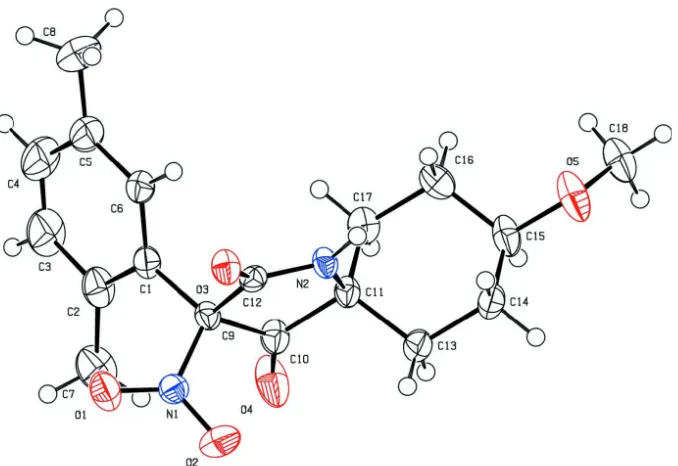
compound, has been synthesized and its structure has been determined (Fig. 1). The molecule contains one benzene ring, one six membered ring, and one five membered ring. The cyclohexane ring adopts a chair conformation;the C13, C14, C16 and C17 atoms are on one plane with C15 and C11 deviating by 0.658 (5)and -0.676 (9) Å, respectively. There are three planes in the molecule: atoms of C10, C11, C12 and N2 generate the pyrrolidine plane (I), C1—C6 yield the benzene plane (II) and C13—C14 and C16—C17 form the cyclohexane plane(III). The angle between planes I and II is 71.69 (9) °, and that between planes I and III is 88.92 (9) °. The space arrangement might result from the space factor between groups.
S2. Experimental
A solution of fuming nitric acid (0.92 g, 16mmoL) in anhydrous chloroform (10 ml) was added dropwise to a solution of compound 2 (1.78 g, 5.9mmoL) in anhydrous chloroform (20 ml) at 0 degree and stirred for 2 h. The reaction mixture was then washed with ice water (15 ml), and saturated sodium chloride solution and dried over anhydrous Na2SO4. The
solvent was evaporated, and the residual solid was crystallized from ethanol to afford 1.84 g compound 3 as a pale yellow solid: yield 90%. The 1H NMR, 13 C-NMR and ESI-MS data testified the title compound's structure. ESI-MS: 347 (M+H)+ (100%); 1H NMR (500 MHz, CDCl3): 7.48 (s, 1H, –NH–),7.55 (s, 1H, Ph—H), 7.14 (d, 1H, Ph—H), 7.07 (d, 1H, Ph—H), 3.54 (s, 3H, –OCH3), 3.34–3.33 (m, 1H, –CH—O—C–), 2.36 (s, 3H, Ph—Me), 2.27 (s, 3H, Ph—Me), 2.12–2.07 (m, 4H, Cyclohexane-H4), 1.99–1.56 (m, 4H, Cyclohexane-H4); 13 C-NMR (100 MHz, CDCl3): 199.9, 165.1, 136.7, 136.0, 133.2, 131.5, 129.7, 127.4, 95.8, 76.8, 75.6, 66.1, 55.7, 32.5, 31.5, 26.4, 26.3, 21.0, 20.3.
S3. Refinement
Figure 1
The molecular structure of title molecule, showing 50% displacement ellipsoids.
Figure 2
Reaction scheme.
(5′S,8′S)-3-(2,5-Dimethylphenyl)-8-methoxy-3-nitro-1-azaspiro[4.5]decane-2,4-dione
Crystal data
C18H22N2O5
Mr = 346.38
Monoclinic, P21/c
a = 9.5707 (9) Å
b = 8.4181 (7) Å
c = 22.8720 (19) Å
β = 100.703 (8)°
V = 1810.7 (3) Å3
Z = 4
F(000) = 736
Dx = 1.271 Mg m−3
Mo Kα radiation, λ = 0.71073 Å Cell parameters from 2205 reflections
θ = 3.2–29.5°
µ = 0.09 mm−1
T = 170 K Block, colourless 0.36 × 0.32 × 0.23 mm
Data collection
Agilent Xcalibur (Atlas, Gemini ultra) diffractometer
Radiation source: Enhance (Mo) X-ray Source Graphite monochromator
Detector resolution: 10.3592 pixels mm-1
ω scans
Absorption correction: multi-scan (CrysAlis PRO; Agilent, 2011)
Tmin = 0.954, Tmax = 1.000
supporting information
sup-3
Acta Cryst. (2015). E71, o238–o239
3308 independent reflections 2600 reflections with I > 2σ(I)
Rint = 0.034
θmax = 25.4°, θmin = 3.3°
h = −11→7
k = −8→10
l = −22→27
Refinement
Refinement on F2
Least-squares matrix: full
R[F2 > 2σ(F2)] = 0.050
wR(F2) = 0.139
S = 1.04 3308 reflections 229 parameters 0 restraints
Primary atom site location: structure-invariant direct methods
Secondary atom site location: difference Fourier map
Hydrogen site location: inferred from neighbouring sites
H-atom parameters constrained
w = 1/[σ2(F
o2) + (0.0602P)2 + 0.8521P]
where P = (Fo2 + 2Fc2)/3
(Δ/σ)max < 0.001
Δρmax = 0.60 e Å−3
Δρmin = −0.31 e Å−3
Special details
Experimental. Absorption correction: CrysAlisPro, Agilent Technologies, Version 1.171.35.11 (release 16-05-2011 CrysAlis171 .NET) (compiled May 16 2011,17:55:39) Empirical absorption correction using spherical harmonics, implemented in SCALE3 ABSPACK scaling algorithm.
Geometry. All e.s.d.'s (except the e.s.d. in the dihedral angle between two l.s. planes) are estimated using the full covariance matrix. The cell e.s.d.'s are taken into account individually in the estimation of e.s.d.'s in distances, angles and torsion angles; correlations between e.s.d.'s in cell parameters are only used when they are defined by crystal symmetry. An approximate (isotropic) treatment of cell e.s.d.'s is used for estimating e.s.d.'s involving l.s. planes.
Refinement. Refinement of F2 against ALL reflections. The weighted R-factor wR and goodness of fit S are based on F2,
conventional R-factors R are based on F, with F set to zero for negative F2. The threshold expression of F2 > σ(F2) is used
only for calculating R-factors(gt) etc. and is not relevant to the choice of reflections for refinement. R-factors based on F2
are statistically about twice as large as those based on F, and R- factors based on ALL data will be even larger.
Fractional atomic coordinates and isotropic or equivalent isotropic displacement parameters (Å2)
x y z Uiso*/Ueq
O1 0.18294 (17) 1.31285 (17) 0.34437 (6) 0.0406 (4) O2 0.09810 (17) 1.2327 (2) 0.42014 (7) 0.0500 (5) O3 0.44250 (14) 1.14145 (16) 0.43813 (5) 0.0295 (3) O4 −0.00240 (16) 0.9051 (2) 0.37430 (8) 0.0579 (5) O5 0.32239 (19) 0.4533 (2) 0.56929 (7) 0.0558 (5) N1 0.16126 (17) 1.2114 (2) 0.37880 (7) 0.0281 (4) N2 0.33749 (16) 0.90936 (19) 0.45886 (6) 0.0245 (4)
H2 0.4057 0.8790 0.4880 0.029*
C1 0.2650 (2) 1.0141 (2) 0.31529 (8) 0.0277 (4) C2 0.1642 (2) 1.0202 (3) 0.26206 (9) 0.0381 (5) C3 0.2143 (3) 0.9836 (3) 0.21029 (10) 0.0558 (7)
H3 0.1491 0.9859 0.1735 0.067*
C4 0.3526 (3) 0.9447 (3) 0.20978 (11) 0.0612 (8)
H4 0.3801 0.9187 0.1731 0.073*
C5 0.4534 (3) 0.9422 (3) 0.26167 (10) 0.0483 (6) C6 0.4058 (2) 0.9761 (2) 0.31421 (9) 0.0342 (5)
H6 0.4721 0.9732 0.3507 0.041*
H7A −0.0125 1.0761 0.2968 0.076*
H7B −0.0505 0.9924 0.2330 0.076*
H7C −0.0024 1.1747 0.2379 0.076*
C8 0.6086 (3) 0.9084 (4) 0.26190 (13) 0.0732 (9)
H8A 0.6263 0.7941 0.2671 0.110*
H8B 0.6674 0.9663 0.2947 0.110*
H8C 0.6327 0.9428 0.2241 0.110*
C9 0.22283 (19) 1.0470 (2) 0.37501 (8) 0.0237 (4) C10 0.1216 (2) 0.9211 (2) 0.39383 (8) 0.0305 (5) C11 0.20633 (19) 0.8179 (2) 0.44258 (8) 0.0258 (4) C12 0.34921 (19) 1.0401 (2) 0.42782 (8) 0.0228 (4) C13 0.1293 (2) 0.8034 (3) 0.49537 (9) 0.0346 (5)
H13A 0.1177 0.9104 0.5118 0.042*
H13B 0.0334 0.7582 0.4815 0.042*
C14 0.2111 (2) 0.6980 (3) 0.54417 (9) 0.0374 (5)
H14A 0.3025 0.7494 0.5613 0.045*
H14B 0.1555 0.6857 0.5763 0.045*
C15 0.2394 (2) 0.5373 (3) 0.52057 (9) 0.0384 (5)
H15 0.1470 0.4806 0.5074 0.046*
C16 0.3164 (2) 0.5505 (2) 0.46846 (9) 0.0349 (5)
H16A 0.3296 0.4430 0.4527 0.042*
H16B 0.4117 0.5974 0.4823 0.042*
C17 0.2336 (2) 0.6532 (2) 0.41893 (9) 0.0328 (5)
H17A 0.1417 0.6015 0.4026 0.039*
H17B 0.2882 0.6635 0.3864 0.039*
C18 0.3060 (3) 0.2894 (3) 0.56796 (12) 0.0524 (7)
H18A 0.3302 0.2475 0.5311 0.079*
H18B 0.2071 0.2627 0.5696 0.079*
H18C 0.3691 0.2424 0.6023 0.079*
Atomic displacement parameters (Å2)
U11 U22 U33 U12 U13 U23
supporting information
sup-5
Acta Cryst. (2015). E71, o238–o239
C9 0.0233 (10) 0.0254 (10) 0.0212 (9) 0.0009 (8) 0.0009 (7) 0.0036 (8) C10 0.0258 (11) 0.0363 (11) 0.0275 (10) −0.0049 (9) 0.0003 (8) 0.0060 (9) C11 0.0215 (10) 0.0308 (10) 0.0237 (9) −0.0033 (8) 0.0011 (7) 0.0057 (8) C12 0.0207 (9) 0.0286 (10) 0.0192 (9) 0.0010 (8) 0.0039 (7) −0.0007 (8) C13 0.0297 (11) 0.0420 (12) 0.0340 (11) 0.0025 (9) 0.0106 (9) 0.0106 (10) C14 0.0338 (12) 0.0508 (14) 0.0294 (11) 0.0028 (10) 0.0108 (9) 0.0135 (10) C15 0.0332 (12) 0.0409 (13) 0.0367 (11) −0.0065 (10) −0.0047 (9) 0.0161 (10) C16 0.0354 (12) 0.0279 (11) 0.0381 (12) 0.0004 (9) −0.0021 (9) −0.0013 (9) C17 0.0348 (12) 0.0326 (11) 0.0289 (10) −0.0060 (9) 0.0000 (8) −0.0008 (9) C18 0.0645 (17) 0.0363 (13) 0.0544 (15) 0.0038 (12) 0.0059 (12) 0.0190 (12)
Geometric parameters (Å, º)
O1—N1 1.206 (2) C8—H8A 0.9800
O2—N1 1.226 (2) C8—H8B 0.9800
O3—C12 1.226 (2) C8—H8C 0.9800
O4—C10 1.195 (2) C9—C10 1.550 (3)
O5—C15 1.430 (2) C9—C12 1.544 (2)
O5—C18 1.388 (3) C10—C11 1.523 (3)
N1—C9 1.513 (2) C11—C13 1.532 (3)
N2—H2 0.8800 C11—C17 1.529 (3)
N2—C11 1.461 (2) C13—H13A 0.9900
N2—C12 1.325 (2) C13—H13B 0.9900
C1—C2 1.407 (3) C13—C14 1.523 (3)
C1—C6 1.390 (3) C14—H14A 0.9900
C1—C9 1.520 (3) C14—H14B 0.9900
C2—C3 1.392 (3) C14—C15 1.500 (3)
C2—C7 1.506 (3) C15—H15 1.0000
C3—H3 0.9500 C15—C16 1.518 (3)
C3—C4 1.366 (4) C16—H16A 0.9900
C4—H4 0.9500 C16—H16B 0.9900
C4—C5 1.384 (4) C16—C17 1.525 (3)
C5—C6 1.391 (3) C17—H17A 0.9900
C5—C8 1.511 (4) C17—H17B 0.9900
C6—H6 0.9500 C18—H18A 0.9800
C7—H7A 0.9800 C18—H18B 0.9800
C7—H7B 0.9800 C18—H18C 0.9800
C7—H7C 0.9800
C18—O5—C15 115.49 (19) N2—C11—C10 101.63 (15) O1—N1—O2 124.68 (18) N2—C11—C13 110.96 (15) O1—N1—C9 119.64 (15) N2—C11—C17 111.93 (15) O2—N1—C9 115.55 (16) C10—C11—C13 110.73 (16)
C11—N2—H2 121.5 C10—C11—C17 111.17 (15)
C12—N2—H2 121.5 C17—C11—C13 110.18 (16)
C12—N2—C11 117.08 (15) O3—C12—N2 127.30 (17)
C2—C1—C9 121.12 (18) O3—C12—C9 124.13 (16)
C6—C1—C9 118.57 (17) C11—C13—H13A 109.3
C1—C2—C7 125.23 (19) C11—C13—H13B 109.3
C3—C2—C1 116.0 (2) H13A—C13—H13B 108.0
C3—C2—C7 118.7 (2) C14—C13—C11 111.56 (16)
C2—C3—H3 118.4 C14—C13—H13A 109.3
C4—C3—C2 123.2 (2) C14—C13—H13B 109.3
C4—C3—H3 118.4 C13—C14—H14A 109.4
C3—C4—H4 119.4 C13—C14—H14B 109.4
C3—C4—C5 121.2 (2) H14A—C14—H14B 108.0
C5—C4—H4 119.4 C15—C14—C13 111.35 (17)
C4—C5—C6 116.8 (2) C15—C14—H14A 109.4
C4—C5—C8 122.3 (2) C15—C14—H14B 109.4
C6—C5—C8 121.0 (2) O5—C15—C14 106.08 (17)
C1—C6—C5 122.4 (2) O5—C15—H15 109.3
C1—C6—H6 118.8 O5—C15—C16 111.55 (18)
C5—C6—H6 118.8 C14—C15—H15 109.3
C2—C7—H7A 109.5 C14—C15—C16 111.32 (17)
C2—C7—H7B 109.5 C16—C15—H15 109.3
C2—C7—H7C 109.5 C15—C16—H16A 109.4
H7A—C7—H7B 109.5 C15—C16—H16B 109.4
H7A—C7—H7C 109.5 C15—C16—C17 111.29 (18)
H7B—C7—H7C 109.5 H16A—C16—H16B 108.0
C5—C8—H8A 109.5 C17—C16—H16A 109.4
C5—C8—H8B 109.5 C17—C16—H16B 109.4
C5—C8—H8C 109.5 C11—C17—H17A 109.5
H8A—C8—H8B 109.5 C11—C17—H17B 109.5
H8A—C8—H8C 109.5 C16—C17—C11 110.53 (16)
H8B—C8—H8C 109.5 C16—C17—H17A 109.5
N1—C9—C1 112.92 (15) C16—C17—H17B 109.5
N1—C9—C10 109.82 (15) H17A—C17—H17B 108.1
N1—C9—C12 104.28 (14) O5—C18—H18A 109.5
C1—C9—C10 114.16 (16) O5—C18—H18B 109.5
C1—C9—C12 113.32 (15) O5—C18—H18C 109.5
C12—C9—C10 101.33 (14) H18A—C18—H18B 109.5 O4—C10—C9 127.03 (18) H18A—C18—H18C 109.5 O4—C10—C11 124.44 (18) H18B—C18—H18C 109.5 C11—C10—C9 108.53 (15)
supporting information
sup-7
Acta Cryst. (2015). E71, o238–o239
N1—C9—C10—O4 53.2 (3) C9—C10—C11—C17 −106.48 (18) N1—C9—C10—C11 −126.63 (16) C10—C9—C12—O3 −164.71 (18) N1—C9—C12—O3 −50.6 (2) C10—C9—C12—N2 14.88 (19) N1—C9—C12—N2 128.95 (15) C10—C11—C13—C14 178.93 (18) N2—C11—C13—C14 −69.0 (2) C10—C11—C17—C16 −179.12 (16) N2—C11—C17—C16 68.0 (2) C11—N2—C12—O3 171.67 (18) C1—C2—C3—C4 −0.4 (4) C11—N2—C12—C9 −7.9 (2) C1—C9—C10—O4 −74.7 (3) C11—C13—C14—C15 −55.5 (2) C1—C9—C10—C11 105.40 (18) C12—N2—C11—C10 −3.1 (2) C1—C9—C12—O3 72.5 (2) C12—N2—C11—C13 −120.87 (18) C1—C9—C12—N2 −107.87 (17) C12—N2—C11—C17 115.59 (18) C2—C1—C6—C5 −0.3 (3) C12—C9—C10—O4 163.1 (2) C2—C1—C9—N1 −59.8 (2) C12—C9—C10—C11 −16.77 (19) C2—C1—C9—C10 66.6 (2) C13—C11—C17—C16 −56.0 (2) C2—C1—C9—C12 −178.06 (18) C13—C14—C15—O5 176.97 (17) C2—C3—C4—C5 −1.3 (4) C13—C14—C15—C16 55.5 (2) C3—C4—C5—C6 2.1 (4) C14—C15—C16—C17 −56.5 (2) C3—C4—C5—C8 −176.6 (3) C15—C16—C17—C11 56.7 (2) C4—C5—C6—C1 −1.3 (3) C17—C11—C13—C14 55.5 (2) C6—C1—C2—C3 1.1 (3) C18—O5—C15—C14 150.9 (2) C6—C1—C2—C7 −175.7 (2) C18—O5—C15—C16 −87.7 (2)
Hydrogen-bond geometry (Å, º)
D—H···A D—H H···A D···A D—H···A
N2—H2···O3i 0.88 2.02 2.8853 (19) 167
C4—H4···O5ii 0.95 2.57 3.287 (3) 132
C7—H7B···O1iii 0.98 2.49 3.454 (3) 168
C14—H14B···O2iv 0.99 2.54 3.265 (3) 130