A COMPARATIVE STUDY BETWEEN TITANIUM MINI PLATES AND 3 DIMENSIONAL TITANIUM
PLATES WITH LOCKING SCREWS USING BITE FORCE EVALUATION OF ANTERIOR
1
Denny George,
2Suresh, V.,
1
Post graduate, Department of Oral & Maxillofacial Surgery, Indira Gandhi institute of dental sciences,
Sri Balaji Vidyapeeth university (Deemed to this
2
Professor, Department of Oral & Maxillofacial Surgery, Indira Gandhi institute of dental sciences,
Sri Balaji Vidyapeeth university (Deemed to this university), Pondicherry
3
Professor and Head, Department of Oral & Maxillofacial
Sri Balaji Vidyapeeth university (Deemed to this university),
4
Post graduate, Department of Oral & Maxillofacial Surgery, Indira Gandhi institute of dental sciences,
Sri Balaji Vidyapeeth university (D
5
Post graduate, Department of Oral & Maxillofacial Surgery, Indira Gandhi institute of dental sciences,
Sri Balaji Vidyapeeth universit
ARTICLE INFO ABSTRACT
Purpose:
dimensional (3D) titanium plate with locking screws and standard titanium miniplates for mandibular anterior fractures treatment using bite force measurements.
randomized controlled clinical study for the treatment of mandibular fractures was conducted as a multicentre study at I
of 2012
underwent osteosynthesis with standard titanium miniplates and ten patients in group II underwent osteosynthesis with 3D titanium locking miniplates. The surgical technique was
clinical parameter assessed was the bite force measurement in relation to the incisor, right molar, and left molar regions. Follow up was done at seventh day, third week, sixth week and third month Results:
in each follow up in groups I and II. The difference in the incisor, right molar and left molar bite force was significantly higher for group II compared to group I. There were no statistical
changes in the clinical parameters such as infection, operating time and occlusion.
titanium miniplates with locking screws are better in fracture stabilization for anterior mandibular fractures when compared with titanium min
in both groups.
Copyright © 2018, Denny George et al. This is an open distribution, and reproduction in any medium, provided
INTRODUCTION
Understanding the anatomic and physiologic relationship of the mandible is very important in the management of mandibular fractures.
*Corresponding author: Rajkumar, R.,
Post graduate, Department of Oral & Maxillofacial Surgery, Indira Gandhi institute of dental sciences, Sri Balaji Vidyapeeth university (Deemed to this university), Pondicherry.
DOI: https://doi.org/10.24941/ijcr.30635.09.2018
ISSN: 0975-833X
Vol. 10, Issue, 09,
Article History:
Received 24th June, 2018
Received in revised form 17th July, 2018
Accepted 25th August, 2018
Published online 30th September, 2018
Citation:Denny George, Suresh, V., Sathyanarayanan, R., Rajkumar, R. and Harish, V.
3 dimensional titanium plates with locking screws using bite force evaluation of anterior mandibular fractures
Research, 10, (09), 73279-73285.
Key Words:
3D Locking plates. Miniplates
Biteforce Instrument. anterior Mandibular Fracture.
RESEARCH ARTICLE
A COMPARATIVE STUDY BETWEEN TITANIUM MINI PLATES AND 3 DIMENSIONAL TITANIUM
PLATES WITH LOCKING SCREWS USING BITE FORCE EVALUATION OF ANTERIOR
MANDIBULAR FRACTURES
Suresh, V.,
3Sathyanarayanan, R.,
4,*Rajkumar, R. and
Post graduate, Department of Oral & Maxillofacial Surgery, Indira Gandhi institute of dental sciences,
Sri Balaji Vidyapeeth university (Deemed to this university), Pondicherry
Professor, Department of Oral & Maxillofacial Surgery, Indira Gandhi institute of dental sciences,
Sri Balaji Vidyapeeth university (Deemed to this university), Pondicherry
Head, Department of Oral & Maxillofacial Surgery, Indira Gandhi institute of dental sciences,
Sri Balaji Vidyapeeth university (Deemed to this university),
Post graduate, Department of Oral & Maxillofacial Surgery, Indira Gandhi institute of dental sciences,
Sri Balaji Vidyapeeth university (Deemed to this university), Pondicherry
Post graduate, Department of Oral & Maxillofacial Surgery, Indira Gandhi institute of dental sciences,
Sri Balaji Vidyapeeth university (Deemed to this university), Pondicherry
ABSTRACT
Purpose: The purpose of this study is to compare the efficacy, stability and rigidity of three dimensional (3D) titanium plate with locking screws and standard titanium miniplates for mandibular anterior fractures treatment using bite force measurements. Patients and Methods:
randomized controlled clinical study for the treatment of mandibular fractures was conducted as a multicentre study at Indira Gandhi Institute of Dental Science, Puducherry, India, in the time period of 2012–2014. Twenty patients were randomly divided into two equal groups. Ten patients in group I underwent osteosynthesis with standard titanium miniplates and ten patients in group II underwent osteosynthesis with 3D titanium locking miniplates. The surgical technique was
clinical parameter assessed was the bite force measurement in relation to the incisor, right molar, and left molar regions. Follow up was done at seventh day, third week, sixth week and third month Results: In this study, a statistically significant change was present between the change in bite force in each follow up in groups I and II. The difference in the incisor, right molar and left molar bite force was significantly higher for group II compared to group I. There were no statistical
changes in the clinical parameters such as infection, operating time and occlusion.
titanium miniplates with locking screws are better in fracture stabilization for anterior mandibular fractures when compared with titanium miniplates. Other postoperative findings show similar changes in both groups.
access article distributed under the Creative Commons Attribution the original work is properly cited.
Understanding the anatomic and physiologic relationship of the mandible is very important in the management of
Post graduate, Department of Oral & Maxillofacial Surgery, Indira Gandhi Sri Balaji Vidyapeeth university (Deemed to this
The mandible can be regarded as a tubular long bone which is bent into a blunt “V” shape. As with all tubular bones, the strength of the mandible resid
The cortical bone is thicker anteriorly and at the lower border of the mandible, while posteriorly the lower border is relatively thin. The central cancellous bone of the body forms a loose network with frequent large, bone
Williams, 2009). The two important differences between the mandible and other long bones is that in the mandible:
International Journal of Current Research Vol. 10, Issue, 09, pp.73279-73285, September, 2018
Denny George, Suresh, V., Sathyanarayanan, R., Rajkumar, R. and Harish, V., 2018. “A comparative study between titanium mini plates and plates with locking screws using bite force evaluation of anterior mandibular fractures
A COMPARATIVE STUDY BETWEEN TITANIUM MINI PLATES AND 3 DIMENSIONAL TITANIUM
PLATES WITH LOCKING SCREWS USING BITE FORCE EVALUATION OF ANTERIOR
Rajkumar, R. and
5Harish, V.
Post graduate, Department of Oral & Maxillofacial Surgery, Indira Gandhi institute of dental sciences,
university), Pondicherry
Professor, Department of Oral & Maxillofacial Surgery, Indira Gandhi institute of dental sciences,
Sri Balaji Vidyapeeth university (Deemed to this university), Pondicherry
Surgery, Indira Gandhi institute of dental sciences,
Sri Balaji Vidyapeeth university (Deemed to this university),
Post graduate, Department of Oral & Maxillofacial Surgery, Indira Gandhi institute of dental sciences,
eemed to this university), Pondicherry
Post graduate, Department of Oral & Maxillofacial Surgery, Indira Gandhi institute of dental sciences,
Pondicherry
The purpose of this study is to compare the efficacy, stability and rigidity of three-dimensional (3D) titanium plate with locking screws and standard titanium miniplates for mandibular
Patients and Methods: A prospective randomized controlled clinical study for the treatment of mandibular fractures was conducted as a ndira Gandhi Institute of Dental Science, Puducherry, India, in the time period patients were randomly divided into two equal groups. Ten patients in group I underwent osteosynthesis with standard titanium miniplates and ten patients in group II underwent osteosynthesis with 3D titanium locking miniplates. The surgical technique was standardized. The clinical parameter assessed was the bite force measurement in relation to the incisor, right molar, and left molar regions. Follow up was done at seventh day, third week, sixth week and third month. significant change was present between the change in bite force in each follow up in groups I and II. The difference in the incisor, right molar and left molar bite force was significantly higher for group II compared to group I. There were no statistically significant changes in the clinical parameters such as infection, operating time and occlusion. Conclusion: 3D titanium miniplates with locking screws are better in fracture stabilization for anterior mandibular iplates. Other postoperative findings show similar changes
ribution License, which permits unrestricted use,
The mandible can be regarded as a tubular long bone which is bent into a blunt “V” shape. As with all tubular bones, the strength of the mandible resides in its dense cortical plates. The cortical bone is thicker anteriorly and at the lower border of the mandible, while posteriorly the lower border is relatively thin. The central cancellous bone of the body forms a loose network with frequent large, bone-free spaces (Rowe and The two important differences between the mandible and other long bones is that in the mandible:
OF CURRENT RESEARCH
Any movement causes both the condyles to move with respect to the skull base.
Anatomically, the condyles are the articular surfaces of
the mandible. Functionally the occlusal surface serves this role.
So, in a functional sense, the occlusal surface is
analogous to a joint space.
In the numerous investigations carried out by Huelke(1961)
and Hodgson (1967),they found that the bone fractures at sites
of tensile strain, as resistance to compressive forces is greater. Anterior forces applied to the symphysis menti, over one mental foramen or over the mandibular body, led to strain at the condylar necks and lingual plates over opposite molar. The mandible is a strong bone, the energy required to fracture being about 44.6–74.4 kg/m (Rowe and Williams, 2009). The mandible can be divided into the symphysis, parasymphysis, body, ramus, and condylar and coronoid process. The symphysis and parasymphysis form the anterior aspect of the
mandible. The muscles surrounding the symphyseal
parasymphysis area are an important consideration in the management of these fractures. The blood supply to the symphysis particularly on the lingual aspect is mainly from genioglossus and geniohyoid muscles. When evaluating mandibular fractures it is important to take a good history and do a proper physical exam. Knowledge of the mechanism of injury can help the physician in arriving at a proper treatment plan. Radiographic evaluation includes a panoromic view and the sub-mental view, which is good for evaluating symphysis
fractures (Heath Stacey et al., 2006). The occlusal radiograph
is indispensable for assessment of fractures involving the lingual cortex. Treatment mainly begins with a tetanus booster based on immunization schedule, followed by routine blood investigations to rule out any systemic disorders. A maxillomandibular fixation can be done to correct any occlusal derangement followed by open reduction and fixation. The most common approach used for the management of symphysis and parasymphysis is by the intraoral vestibular incision, or they can also be approached by existing lacerations.
Significant advances have been made in the management of mandibular fractures due to improved understanding of biomechanical principles and instrumentation. Over the years, two different schools of thoughts have evolved in the treatment of mandibular fractures with the use of plates and screws. In 1970, Spiessl advocated rigid fixation that prevented interfragmentary mobility even during active use of the mandible. The goal of treatment was to ensure primary bone union by compression osteosynthesis for which large bone plates and bicortical screws were used. The primary disadvantages of this technique included increased risk of nerve damage, stress shielding, scar formation due to extraoral approach and the bulky plates that made adaptation difficult. Champy (1978) advocated a modification of the technique
used by Michelet et al (1973). in 1973 for mandibular
osteosynthesis, in which there is use of monocortical juxta-alveolar and subapical osteosynthesis, without compression and without IMF. These plates were placed near the tension zone produced by physiological strain. Since their introduction, miniplates have been the preferred method of fixation in Craniomaxillofacial surgery. Their advantages include their relatively small size, adaptability, ease of
placement via intraoral approach (Cawood, 1985). The
disadvantages include controversies regarding their rigidity
and stability in case of angle fractures and comminuted fractures. To overcome these disadvantages Farmand
(Farmand, 1995) in 1995 developed three-dimensional (3D)
plates which are designed in a quadrangular shape that is formed by joining two miniplates with interconnecting crossbars. In this way, three-dimensional stability is achieved not because of the length or thickness but because of the configuration. There is also good resistance against torsional
forces. The internal Mini Locking System (Gutwald et al.,
2003) was developed in 2003 to avoid loosening of hardware and for a perfect adaptation. It is believed that 3D locking plates provide better stability and ease of plate adaptation for the open reduction and internal fixation of mandibular fractures. The biomechanical and technical advantages of 3D locking plates compared with titanium 2.0 mm miniplates has prompted us to perform the present study to compare 3D titanium locking plates with titanium 2.0 mm miniplates in the osteosynthesis of mandibular fractures according to the clinical parameters and the preoperative and postoperative bite force measurement.
Maximum voluntary bite force (MVBF) is related to the health
of the masticatory system. Maximum bite force is a useful
indicator of the functional state of the masticatory system and the loading of the teeth. The force results from the action of the jaw elevator muscles (in turn, determined by the central nervous system and feedback from muscle spindles,
mechanoreceptors and nociceptors) modified by the
craniomandibular biomechanics (Bakke, 1993).Maximum bite
force is highest in the molar region. Unilateral measurement of maximum bite force in the molar region averages between 300 and 600 Newtons (N) in healthy adults with natural teeth (Hagberg, 1987). Since the strength of 3D locking titanium plates appears to be in their rigidity and stability, bite force measurement can be a useful indicator to assess the jaw function and thereby the clinical efficiency of 3D locking titanium plates over conventional 2.0 mm titanium plates.
MATERIALS AND METHODS
Fig 1. Titanium plating
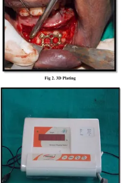
Fig 2. 3D Plating
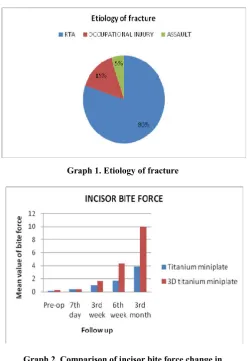
Fig 3. AXPERT Bite Force Instrument
The surgical technique was standardized. Follow up was done at 7th day, 3 and 6 weeks and 3 months and the clinical parameter assessed was bite force measurement in relation to the incisor, right molar and left molar regions. All bite force measurements were recorded using ‘AXPERT’ electronic weighing balance bite force recorder, made by AXPERT Enterprises in Ahmadabad, India. This device consists of an ABS mould cabinet, single mould stainless steel pan and battery backup of 18-20 hours. AXPERT electronic balances will have tactile membrance switches, LED display. AXPERT electronic balances are having auto Zero tracking, Auto display ON/OFF (Battery saving mode), extra zero and other software facilities Figure 3. Load changes in the steel pan produce a measurable digital value in kilogram force (kg). The measurements were made with subject comfortably seated facing forward. The patients were asked to maintain the same posture until the measurements were recorded and asked to bite on the pads of the bite force gauge using maximum voluntary bite force possible.
Statistics: A Prospective, randomized controlled clinical Study
was conducted in the Department of Oral and Maxillofacial Surgery, Indira Gandhi Institute of Dental Sciences and paired sample t test was used to analyse the collected data.
RESULTS
In this study, a total of 20 patients with isolated fractures of the symphysis or parasymphysis and that met the inclusion criteria were included. In group I, 10 patients underwent fixation with standard titanium miniplates and in group II, 10 patients underwent fixation with 3D titanium plates with locking screws. Most of the patients who reported at the emergency unit were victims of road traffic accident. In group I, the most common age group was from 22 to 45 and in group II, from 21 to 41 so that there was no bias in terms of the age of the patient in both the groups. Male patients were predominant in the study in that in the entire study only one female patient satisfied the inclusion criteria, probably due to the predominance of road traffic accidents among the male gender. The main cause for anterior mandibular fractures according to this study is road traffic accident (80%), occupational injury (15%) and assault (5%) Graph 1. The preoperative occlusion in all the patients was deranged. Postoperatively, functionally acceptable occlusion was obtained in almost all the patients except for two patients in group I and one patient in group II, for whom intermaxillary fixation had been given for an additional 1 week until functionally acceptable occlusion was attained. However, functionally acceptable occlusion was achieved postoperatively in all the patients. There was no significant difference in operating time between the two groups. The average operating time in both groups was 30–40 min. Almost all the patients were treated within 5–9 days of injury, and mean interval time duration between injury and surgery was 7 days. A significant difference in the bite force measurement was found between the two groups. The patients with 3D titanium plates with locking screws had progressive
improvement of bite force measurements after 7th day, 3, 6
[image:3.595.38.288.385.762.2]In group I, the bite force measurement at the incisor area had increased progressively at each of the postoperative follow-up visits like 7th day, 3rd week, 6th week and 3rd month compared with the previously recorded preoperative visits Table 1. In group II, the bite force measurement at the incisor area had also increased progressively at each of the postoperative follow-up visits like 7th day, 3rd week, 6th week, 3rd month compared with the previously recorded preoperative visits Table 2. At 7th day to 3 weeks, no notable change was seen in the difference in the incisor area bite force in groups II and I Table 3. From 7th day to 3 months, the difference in the incisor area bite force was notably better in group II compared to group I Table 3. In group I, the bite force measurement at the right molar area had increased progressively at each of the postoperative follow-up visits like 7th day, 3rd week, 6th week, 3rd month compared with the previously recorded preoperative visits Table 4. In group II, the bite force measurement at the right molar area had also increased progressively at each of the postoperative follow-up visits like 7th day, 3rd week, 6th week, 3rd month compared
with the previously recorded preoperative visits Table 5. From
6 weeks to 3 months, the difference in the right molar area bite force in each follow-up visit was notably greater in group II compared to group I Table 6. At 7th day and 3rd weeks, no significant difference in right molar area bite force was seen in group I and group II Table 6.
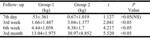
In group I, the bite force measurement at the left molar area had increased progressively at each of the postoperative follow-up visits like 7th day, 3rd week, 6th week, 3rd month compared with the previously recorded preoperative visits Table 7. In group II, the bite force measurement at the left molar area had also increased progressively at each of the postoperative follow-up visits like 7th day, 3rd week, 6th week, 3rd month compared with the previously recorded preoperative visits Table 8. On the 7th day, no significant change was seen in the difference in the left molar area bite force between group I and group II. At 3rd week, 6th week and 3rd month, the difference in the left molar area bite force in each follow-up visit was notably better in group II compared to group I Table 9. No postoperative complications were found in both the groups except for one patient in group I in whom an infection was observed in the 3rd month in the form of purulent discharge and wound dehiscence. Drainage was instituted and the patient was put under empirical antibiotics
and observation until the infection subsided. Some of the
patients had discomfort postoperatively while recording the bite force but with subsequent appointments the discomfort reduced.
DISCUSSION
The three main principles of fracture management include anatomical reduction, fixation and immobilization. In this study, the clinical parameter being assessed, the maximum voluntary bite force measurement postoperatively was found to be greater in patients with 3D titanium plates with locking screws than those with titanium miniplates. The good biocompatibility of titanium as an implant device within the body has been well understood in the recent years as there is no necessity of plate removal at a later stage. Miniplates, in general, have the advantage of stability with only minimal surgical exposure necessary, and 3D titanium plates with locking screws, in particular, have the necessary geometric configuration to achieve maximum stability.
The locking system has been designed in a way by which the screws on placement are further held by a second thread below the screw’s head end in a way that the desired maximum stability is achieved. According to Champy’s lines of osteosynthesis, two plates should be placed in the anterior region of the mandible, but in case of 3D titanium plates with locking screws, a single plate provides sufficient stability, which is an added advantage. The other advantages include: (1) less technical sensitivity associated with both external and internal fixation method and (2) the reduced friction lock between the plate–screw interface resulting in decreased forces transmitted to the underlying bone. The placement of a single plate in the fracture of the anterior mandible instead of the two plates traditionally placed according to Champy’s principles also reduces the operating time for the surgeon (Virendra
Singh et al., 2012). According to a study conducted by
Dhananjay H Barde et al. (2014) for comparing the efficacy of
3D plates over Champy’s miniplates in mandibular anterior fractures, it was concluded that the fixation of fractures in the anterior region of the mandible with 3D plates provided better three-dimensional stability probably due to the geometric configuration and had lesser rates of infection and morbidity postoperatively. They also made a note that probably the most common limitation was the use of excessive implant material, but concluded that they are a comparatively better alternative to Champy’s miniplates.
In this present study, another disadvantage that was observed was the increased cost that had to be borne by the patient, but the surgeon was well aware of this extra cost that was borne by the patient and it was also taken into consideration while choosing the sample for this study. The other disadvantage was the broad size of the 3D titanium plate which makes it difficult to use in atrophic mandible, as manipulation and fixation of these plates could have possibly resulted in pathological fractures, but it was not encountered in our study. Virendra
Singh et al. in 2012, conducted a prospective randomized
study to compare the efficacy of conventional versus 3D miniplate in the management of mandibular fracture. They concluded that there was no significant difference in general terms of treatment outcome in both the systems, as both of them appeared equally effective in the management of mandibular fractures. However, in particular reference to the symphysis/parasymphysis, region, 3D miniplate fixation was an easy-to-use alternative compared to conventional miniplates as the surgical site was markedly reduced by the use of only a single plate for stabilization at both the superior and inferior borders.
They concluded that the surgical trauma was also comparatively lesser. A similar conclusion was drawn in
another study conducted by R. Balakrishnan et al (15) who
analysed the efficacy of using 3D titanium miniplates for the management of mandibular fractures. They concluded that 3D titanium miniplates were a viable option for management of mandibular fractures with the advantages of placement through an intraoral approach minimizing surgical time and trauma with added advantage of simple techniques for adaptation to
the bone. In another study conducted by Bipin S. Sadhwani et
al. in 2014, comparing conventional 2.0 mm miniplates and 3D
Graph 1. Etiology of fracture
Graph 2. Comparison of incisor bite force change in group 1 and group 2
Graph 3. Comparison of right molar bite force change in group 1 and group 2
[image:5.595.39.289.47.409.2]Graph 4 comparison of left molar bite force change in group 1 and group 2
Table 1. Incisor bite force in group 1 (ns-not significant)
Follow- up Bite force (kg)
Change (kg)
t P
Value
Pre-op 0.15±0.266 _ _ _
7th day 0.35±0.267 0.2±0.001 1.757 ˃0.05(NS)
3rd week 1.01±0.649 0.86±0.383 3.061 ˃0.05(NS)
6th week 1.73±0.478 1.58±0.212 4.592 ˂0.05
[image:5.595.311.552.79.150.2]3rd month 3.86±0.865 3.71±0.599 6.766 ˂0.05
Table 2. Incisor bite force in group 2
Follow- up Bite force (kg)
Change (kg)
t P
Value
Pre-op 0.25±0.551 _ _ _
7th day 0.41±0.358 0.16±0.193 _ _
3rd week 1.63±1.160 1.38±0.609 4.346 ˂0.05
6th week 4.31±1.227 4.06±0.676 5.293 ˂0.05
[image:5.595.314.554.188.257.2]3rd month 10.03±2.629 9.78±2.078 7.386 ˂0.05
Table 3. Comparison of incisor bite force change in group 1 and group 2
(ns-not significant)
Follow- up Group 1 (kg)
Group 2 (kg)
t P
Value 7th day 0.2±0.001 0.16±0.193 .446 ˃0.05(NS)
3rd week 0.86±0.383 1.38±0.609 1.475 ˃0.05(NS)
6th week 1.58±.212 4.06±0.676 6.190 ˂0.05
3rd month 3.71±.599 9.78±2.078 7.050 ˂0.05
Table 4 right molar bite force in group 1. (ns-not significant)
Follow- up Bite force (kg)
Change (kg)
t P
Value
Pre-op 0.15±0.084 _ _ _
[image:5.595.307.559.408.479.2]7th day 0.71±0.512 0.56±0.428 3.193 ˃0.05(NS) 3rd week 1.71±1.984 1.56±1.9 2.085 ˃0.05(NS) 6th week 4.71±1.425 4.56±1.341 4.095 ˂0.05 3rd month 13.79±0.865 13.64±2.74 10.704 ˂0.05
Table 5. Right molar bite force in group 2. (ns-not significant)
Follow- up Bite force (kg)
Change (kg)
t P
Value
Pre-op 0.46±0.599 _ _ _
[image:5.595.304.562.505.577.2]7th day 1.52±2.675 1.06±2.076 3.193 ˃0.05(NS) 3rd week 4.56±3.026 4.1±2.427 2.085 ˃0.05(NS) 6th week 9.43±3.443 8.97±2.884 4.095 ˂0.05 3rd month 30.71±9.101 30.25±8.502 10.704 ˂0.05
Table 6. Comparison of right molar bite force change in group 1 and group 2. (ns-not significant)
Follow- up
Group 1 (kg)
Group 2 (kg)
t P
Value
7th day .56±.428 1.06±2.076 .941 ˃0.05(NS)
3rd week 1.56±1.9 4.1±2.427 2.491 ˂0.05
[image:5.595.48.282.618.767.2]6th week 4.56±1.341 8.97±2.884 4.013 ˂0.05 3rd month 13.64±2.74 30.25±8.502 5.617 ˂0.05
Table 7. Left molar bite force in group 1 (ns-not significant)
Follow- up
Bite force (kg)
Change (kg)
t P
Value
Pre-op 0.12±0.058 _ _ _
[image:5.595.311.558.728.799.2]Table 8. Left molar bite force in group 2 (ns-not significant)
Follow- up
Bite force (kg)
Change (kg)
t P
Value
Pre-op 0.96±1.704 _ _ _
7th day 1.63±2.723 0.67±1.019 .818 ˃0.05(NS) 3rd week 4.00±3.081 3.04±1.377 6.716 ˂0.05 6th week 9.34±3.404 8.38±1.7 4.621 ˂0.05 3rd month 31.93±10.556 30.97±8.852 7.540 ˂0.05
Table 9 Comparison of left molar bite force change in group 1 and group 2. (ns-not significant)
Follow- up Group 1 (kg)
Group 2 (kg)
t P
Value 7th day .53±.361 0.67±1.019 1.127 ˃0.05(NS) 3rd week 1.66±1.487 3.04±1.377 2.041 ˂0.05 6th week 4.44±1.058 8.38±1.7 4.217 ˂0.05 3rd month 13.04±1.975 30.97±8.852 5.520 ˂0.05
reduced and the postoperative complications were also reduced as evidenced by the observation that none of the patients in Group II had any postoperative infection or wound dehiscence. The parameter measured in the present study was the maximum voluntary bite force for estimating the masticatory
efficiency. In a study conducted by Rajesh Kshirsagar et al.
(2011). for bite force measurement in mandibular
parasymphyseal fractures in a preliminary clinical study, the maximum voluntary bite force measured in patients treated by open reduction and rigid internal fixation was estimated in 60 volunteers. The bite force was recorded in a standardized position in the incisor and molar region at regular intervals and the measured values were compared in the same standardized manner and with the same equipment in six patients with isolated parasymphyseal fractures in whom fixation had been done using two miniplates.
The results showed that in the volunteer group, the bite forces ranged from 22 to 50 kg in the molar region and 3–27 kg in the incisor region, whereas in mandibular parasymphysis fractures managed by internal fixation, the incisor bite forces were significantly reduced. In 2011, Mohit Agarwal et al. (2011) conducted a study comparing the efficacy of 2-mm locking miniplates and 2-mm standard miniplates in the management of mandibular fractures based on improvement in various clinical parameters and bite force measurement. They concluded that the use of locking miniplates for the purpose of fixation in the management of mandibular fractures was comparatively more efficacious in terms of better plate adaptation, stability, minimal compromise of the periosteal blood supply, along with the advantage of increased postoperative bite force measurements. The disadvantage – the increased cost of the locking system – comparatively is considerable. There is not much difference in the armamentarium for the locking system as it requires only few minor additions. In this study, the clinical parameter being assessed – the maximum voluntary bite force measurement – was done in relation to the incisor, right molar and left molar region with follow up done at 1, 3, 6 weeks and 3 months. The forces measured were the maximum voluntary bite forces capable by the patient and there was significant variation between these and the bite force produced during functional activities. However, the measurement of the bite force is relatively easy and is an important indicator of the efficacy of the masticatory system. There is an expected reduction in the bite force postoperatively which can be attributed to protective
neuromuscular mechanisms in which there is selective activation or deactivation of some of the components of the neuromuscular system chiefly to relieve the damaged skeleton
(Tharani Kumar et al., 2013). Gupta et al. (2012) conducted a
study evaluating bite force measurements in mandibular fractures treated by using microplates and miniplates. They concluded that replacing an upper miniplate with a microplate significantly improved masticatory efficiency in fractures of the anterior region of the mandible. Similarly, in this present study, a statistically significant result was seen in each of the postoperative follow-up visits and the bite force for the incisor and molar regions in the fractured segments. In group I, 7th day, 3rd week, 6th week and 3rd month incisor bite force was 0.35, 1.01, 1.73 and 3.86 kg, respectively, postoperatively. In group II, 7th day, 3rd week, 6th week and 3rd month incisor bite force at 0.41, 1.63, 4.31 and 10.03 kg, respectively, postoperatively. At 7th day and 3rd week, the incisor bite force was statistically not significant after surgery. From the 6th week to 3rd month, a significant difference was present in the incisor bite forces recorded postoperatively. With these results, it is observed that the bite force measured in the incisor area is significantly better in group II compared to group I Graph 2.
In this study, in group I, 7th day, 3rd week, 6th week and the 3rd month right molar area bite force was at 0.71, 1.71, 4.71 and 13.79 kg, respectively, postoperatively. In group II, 7th day, 3rd week, 6th week and the 3rd month right molar area bite force was at 1.52, 4.56, 9.43 and 30.71 kg, respectively, postoperatively. On the 7th day, the right molar area bite force was statistically not significant postoperatively. From 3rd week, 6th week and the 3rd month, the difference in the right molar area bite force in each follow-up visit was significantly better in group II compared to group I Graph 3. In group I, 7th day, 3rd week, 6th week and 3rd month left molar area bite force was at 0.65, 1.78, 4.56 and 13.16 kg, respectively, postoperatively. In group II, 7th day, 3rd week, 6th week and the 3rd month, left molar area bite force was at 1.63, 4.00, 9.34 and 31.93 kg, respectively, postoperatively. On the 7th day, the left molar area bite force was statistically not significant after surgery. Statistically no significant left molar bite force was found at 7th day and 3rd week after surgery. From the 3rd week, 6th week and 3rd months there was significant difference present between the left molar bite forces postoperatively. These results show that the difference in the left molar bite force in each of the follow-up visits was significantly better in group II compared to group I (Graph 4).
Conclusion
[image:6.595.37.288.185.249.2]REFERENCES
Agrawal M., Shadab M. and Singh RK. 2011. Prospective randomized clinical trial comparing bite force in 2-mm locking plates versus 2-mm standard plates in treatment of
mandibular fractures. J Oral Maxillofac Surg., 69:1995–
2000
Anand Gupta, Vibha Singh, Shadab Mohammad 2012. Bite
force evaluation of mandibular fractures treated with
microplates and miniplates. J Oral Maxillofac Surg.,
70:1903–1908
Bakke M. 1993. Mandibular elevator muscles: physiology, action, and effect of dental occlusion. Scand J Dent Res., 101:314–331
Balakrishnan R., Vijay Ebenezer Abu Dakir, 2014. Three dimensional titanium mini plates in management of mandibular fractures. Biomed Pharmacol J 7(1):241–246
Bipin S Sadhwani, Sonal Anchlia, 2013.Conventional 2.0 mm
miniplates versus 3-D plates in mandibular fractures. Ann
Maxillofac Surg., July–December:3(2)
Cawood JL. 1985. Small plate osteosynthesis of mandibular
fractures. Br J Oral Maxillofac Surg., 23:77–91
Champy M., Lodde JP., Schmitt R. 1978. Mandibular osteosynthesis by miniature screwed plates via buccal
approach. J Maxillofac Surg., 6:14
Dhananjay H Barde, Anupama Mudhol, Fareedi Mukram Ali, Madan RS., Sanjay Kar, Farheen Ustaad 2014. Efficacy of 3-dimensional plates over Champys miniplates in
mandibular anterior fractures. J Int Oral Health., 6(1):20–
26
Donald f. Huelke, 1961. Study of mandibular fractures
stresscoat. J Dental Res., 40(5):1042
Farmand, M. 1995. Three-dimensional plate fixation of
fractures and osteotomies. Facial Plast Surg Clin North
Am., 3(1):39–56
Gutwald R., Alpert B., Schmelzeisen R. 2003 Principle and
stability of locking plates. Keio J Med ., 52(1):21–24
Hagberg C. 1987. Assessments of bite force: a review. J
Craniomandib Disord., 1:162–169
Heath Stacey, D. John F. Doyle, Delora L. Mount, Mary C.
Snyder, Karol A. Gutowsk 2006. Management of
mandibular fractures, Plast Recon str Surg., 117: 48e.
Hodgson 1967. Tolerance of the facial bones to impact. Am j
of Anat., 120.
Michelet FX., Deymes J., Dessus B. 1973. Osteosynthesis with
miniaturized screwed plates in maxillofacial surgery. J
Maxillofac Surg., 1:79
Rajesh Kshirsagar, Nitin Jagg, Rajshekhar Halli 2011. Bite
force measurement in mandibular parasymphyseal
fractures: a preliminary clinical study. Craniomaxillofac Trauma Reconstr 4(4).
Rowe and Williams, 2009. Maxillofacial injuries. Volume 1: page no 3.
Tharani Kumar S, Saurabh Saraf, Prasanna Devi S. 2013. Evaluation of bite force after open reduction and internal
fixation using microplates. J Dentistry., 10(5):466–477
Virendra Singh, Puneet Puri, Sanjay Arya, Sunita Malik, Amrish Bhagol 2012. Conventional versus 3-dimensional miniplate in management of mandibular fracture: a
prospective randomized study. Otolaryngol Head Neck.,
Surg 147(3):450–455