CONVENTIONAL VERSUS MODIFIED
1
Dr. Anupam
1
BDS, MDS (Perio). Lecturer, Department
2
BDS, MDS (Perio), Ph D (UK). Professor
Regional Dental
ARTICLE INFO ABSTRACT
Background:
occurring due to frictional forces encountered during mastication and dissipate the pull on th
created by the muscles of the adjacent alveolar mucosa. It also facilitates subgingival plaque formation and results in loss of attachment and soft tissue recession and a shallow vestibular fornix that in turn favours food accumulation.
Apically Repositioned Flap is a predictable method of increasing the zone of attached gingiva. This Conventional Apically Repositioned Flap
retained as a marginal collar, referred to as Modified Apically Repositioned Flap. was conducted to compare the
Repositioned Flap in terms of changes in width of attached gingiva, apparent gingival recession, sulcus depth and clinical attachment level.
sites were referred to as Experimental Site ‘A’ and ‘B’, referring to as the Conventional Apically Repositioned Flap and Modified Apically Repositioned Flap, respectively. The clinical parameters were recorded preoperatively on day 0 and again postoperatively
Results:
effective and efficient methods for widening the attached gingiva.
effective in increasing the zone of attached gingiva, though Modified Apically Repositioned Flaps may be preferred due to its advantages.
Copyright © 2019, Anupam Deka and Swarga Jyoti Das
permits unrestricted use, distribution, and reproduction
INTRODUCTION
Attached gingival extends from the free gingival groove to
mucogingival junction (Newman et al., 2006).
buffer zone between two movable tissues, namely the marginal gingiva and alveolar mucosa. An adequate width of attached gingiva is considered as critical for maintenance of periodontal tissue health by many distinguished researchers (Friedman and Levine, 1964; Nabers, 1966; Lang and Löe, 1972; Bernimoulin and Mühlemann, 1973; Ochsenbein and Maynard, 1974; Hall, 1981; Matter, 1982), as inadequate zone
protect the periodontium from injury occurs due to frictional forces encountered during mastication and dissipate the pull on the gingival margin created by the muscles of the adjacent alveolar mucosa (Friedman, 1999; Ochsenbein, 1960). Insufficient attached gingiva also facilitates subgingival plaque formation and results in loss of attachment and soft tissue recession (Ruben 1979; Stern 1981) and a shallow vestibular fornix that in turnfavours food accumulation (Gottsegen, 1954;
Corn, 1962; Rosenberg et al., 1981; Carranza and Carraro,
1970). Lang and Loe (1972) demonstrated persistent
ISSN: 0975-833X
Article History:
Received 20th March, 2019
Received in revised form 28th April, 2019
Accepted 23rd May, 2019
Published online 30th June, 2019
Citation: Dr. Anupam Deka and Dr. Swarga Jyoti Das
study”, International Journal of Current Research, 11, (
Key Words:
Microorganism, Corrosion, MIC, Biofilm.
*Corresponding author: Dr. Swarga Jyoti Das
ORIGINAL ARTICLE
MODIFIED TECHNIQUE OF THE APICALLY REPOSITIONED
COMPARATIVE STUDY
Anupam Deka and
2, *Dr. Swarga Jyoti Das
Department of Periodontics and Oral Implantology, Regional
Guwahati-781032, Assam, India
Professor and Head, Department of Periodontics
Dental College, Guwahati-781032, Assam, India
ABSTRACT
Background: Inadequate zone of attached gingiva is not sufficient to protect the periodontium from injury occurring due to frictional forces encountered during mastication and dissipate the pull on th
created by the muscles of the adjacent alveolar mucosa. It also facilitates subgingival plaque formation and results in loss of attachment and soft tissue recession and a shallow vestibular fornix that in turn favours food accumulation. Thus, an adequate zone of attached gingiva is essential for maintenance of periodontal health. The Apically Repositioned Flap is a predictable method of increasing the zone of attached gingiva. This Conventional Apically Repositioned Flap method has been modified by Carnio in 1996, where the existing keratinized tissue is retained as a marginal collar, referred to as Modified Apically Repositioned Flap.
was conducted to compare the results of Conventional Apically Repositioned F
Repositioned Flap in terms of changes in width of attached gingiva, apparent gingival recession, sulcus depth and clinical attachment level. Methods: The study comprised of 28 sites in 14 subjects, in a split mouth design. The es were referred to as Experimental Site ‘A’ and ‘B’, referring to as the Conventional Apically Repositioned Flap and Modified Apically Repositioned Flap, respectively. The clinical parameters were recorded preoperatively on day 0 and again postoperatively on day 90 and 150.The data obtained was analyzed statistically.
Results: The results showed that both Conventional and Modified Apically Repositioned Flapsare effective and efficient methods for widening the attached gingiva. Conclusion
effective in increasing the zone of attached gingiva, though Modified Apically Repositioned Flaps may be preferred due to its advantages.
Swarga Jyoti Das. This is an open access article distributed under the Creative in any medium, provided the original work is properly cited.
extends from the free gingival groove to , 2006). It acts as a buffer zone between two movable tissues, namely the marginal gingiva and alveolar mucosa. An adequate width of attached maintenance of periodontal tissue health by many distinguished researchers (Friedman and Levine, 1964; Nabers, 1966; Lang and Löe, 1972; Bernimoulin and Mühlemann, 1973; Ochsenbein and Maynard, 1974; Hall, is insufficient to protect the periodontium from injury occurs due to frictional forces encountered during mastication and dissipate the pull on the gingival margin created by the muscles of the adjacent alveolar mucosa (Friedman, 1999; Ochsenbein, 1960). ent attached gingiva also facilitates subgingival plaque formation and results in loss of attachment and soft tissue recession (Ruben 1979; Stern 1981) and a shallow vestibular food accumulation (Gottsegen, 1954; ., 1981; Carranza and Carraro, 1970). Lang and Loe (1972) demonstrated persistent
inflammation in areas with less than 2 mm keratinized gingiva in spite of effective oral hygiene measures, thus,
keratinized gingiva of 2 mm width as
gingival health. Considering the importance of attached gingiva for maintenance of gingival health, various surgical techniques are indicated to increase the width of attached gingiva, namely Push-back technique (Gujar and Kathariya, 2014), vestibular extension technique (Bohannan, 1962) apically repositioned flap (Freidman and Levine, 1964), and free autogenous gingival grafts (Pennel
Conventional apically repositioned flap involves displacement of sof
suturing, leaving 3-5mm of alveolar bone denuded in the coronal part of surgical area.
width of gingiva can be predicted with this technique. Considering the post surgical gingival recession of technique, a modified form of the conventional partial thickness ARF (MARF) has been described by Carnio in
199620, where marginal gingiva is preserved unlike the original
technique. The other advantages of MARF are minimal surgical trauma with no prerequisite of sutures and thereby,
International Journal of Current Research Vol. 11, Issue, 06, pp.4441-4445, June, 2019
DOI: https://doi.org/10.24941/ijcr.35383.06.2019
Dr. Swarga Jyoti Das. 2019. “Conventional versus modified technique of the apically repositioned flap: a comparative , 11, (06), 4441-4445.
REPOSITIONED FLAP: A
Regional Dental College,
and Oral Implantology,
Inadequate zone of attached gingiva is not sufficient to protect the periodontium from injury occurring due to frictional forces encountered during mastication and dissipate the pull on the gingival margin created by the muscles of the adjacent alveolar mucosa. It also facilitates subgingival plaque formation and results in loss of attachment and soft tissue recession and a shallow vestibular fornix that in turn favours food Thus, an adequate zone of attached gingiva is essential for maintenance of periodontal health. The Apically Repositioned Flap is a predictable method of increasing the zone of attached gingiva. This Conventional odified by Carnio in 1996, where the existing keratinized tissue is retained as a marginal collar, referred to as Modified Apically Repositioned Flap. Objective: The present study results of Conventional Apically Repositioned Flap and Modified Apically Repositioned Flap in terms of changes in width of attached gingiva, apparent gingival recession, sulcus depth and The study comprised of 28 sites in 14 subjects, in a split mouth design. The es were referred to as Experimental Site ‘A’ and ‘B’, referring to as the Conventional Apically Repositioned Flap and Modified Apically Repositioned Flap, respectively. The clinical parameters were recorded on day 90 and 150.The data obtained was analyzed statistically. The results showed that both Conventional and Modified Apically Repositioned Flapsare equally
Conclusion: Both the procedures are equally effective in increasing the zone of attached gingiva, though Modified Apically Repositioned Flaps may be
Creative Commons Attribution License, which
inflammation in areas with less than 2 mm keratinized gingiva in spite of effective oral hygiene measures, thus, recommended keratinized gingiva of 2 mm width as adequate to maintain gingival health. Considering the importance of attached gingiva for maintenance of gingival health, various surgical techniques are indicated to increase the width of attached back technique (Gujar and Kathariya, 2014), vestibular extension technique (Bohannan, 1962) apically repositioned flap (Freidman and Levine, 1964), and
free autogenous gingival grafts (Pennel et al., 1965).
Conventional apically repositioned flap (CARF) procedure of soft tissue flaps apically during 5mm of alveolar bone denuded in the A postsurgical increase in the width of gingiva can be predicted with this technique. post surgical gingival recession of CARF a modified form of the conventional partial thickness ARF (MARF) has been described by Carnio in , where marginal gingiva is preserved unlike the original technique. The other advantages of MARF are minimal requisite of sutures and thereby,
INTERNATIONAL JOURNAL OF CURRENT RESEARCH
time requirement is less (Carnio and Miller, 1999). Both CARF and MARF techniques were described as effective measures in increasing the zone of attached gingiva. However, comparative assessment of CARF and MARF are not found in the literature. Considering this, a clinical study was carried to compare the CARF with MARF technique in terms of changes in the width of attached gingiva, gingival recession (apparent), sulcus depth and clinical attachment level.
MATERIALS AND METHODS
The present study was conducted in the Department of Periodontics and Oral Implantology, Regional Dental College, Guwahati. A total no of 14 patients (11 males and 3 females), mean age being 32.14±6.48 (ranges from 23-42 years) involving 28 sites comprised the study samples. The study was carried out in split mouth design and the sites were designated as
Site ‘A’: treated with conventional apically repositioned flap (CARF)
Site ‘B’: treated with modified apically repositioned flap (MARF)
The inclusion criteria were as follows:
At least 2 sites with Miller’s Class I recession with
sulcus depth of at least 0.5mm
Adequate vestibular depth
Positive tension test
No periapical pathology, dehiscence, trauma from
occlusion and within normal arch form
Healthy, nonsmoker with no systemic diseases
The clinical parameters were:
Width of attached gingiva
Gingival recession (apparent)
Sulcus depth
Clinical attachment level
The clinical parameters were assessed by UNC-15 periodontal probe. The working end of this probe is 15 mm long with
markings at each mm and colour coding at 5th, 10th and 15th
mm. The cementoenamel junction (CEJ) was used as a fixed reference point. A stent was fabricated with self cure acrylic
resin covering the occlusal/incisal 1/3rdboth buccally and
lingually of the teeth to be recorded extending to two adjacent teeth, one on mesial and other on distal side. Vertical groove corresponding to the midline on the facial aspect of the tooth to be treated was made on the stent to guide the probe during measurements at different time points to make sure that all measurements were made at the same orientation to avoid any discrepancy. The surgical sites were stained with an iodine solution (Betadine, 0.5% w/v iodine) to differentiate between alveolar mucosa and keratinized gingiva. This procedure done in each measurement highlighted the MGJ and facilitated the measurement.
The following measurements were taken in relation to each tooth (Figure 2)
CEJ to base of sulcus (A)
CEJ to gingival margin (B)
CEJ to mucogingival junction ( C)
From these measurements, the following clinical parameters were calculated:
Width of Attached Gingiva: Distance between base
of sulcus to MGJ (C minus A)
Apparent Gingival Recession: CEJ to gingival
margin(B)
Sulcus Depth: Measured from the crest of the gingival
margin to the base of sulcus (A minus B)
Clinical Attachment Level: Distance from CEJ to
base of sulcus (A)
All the parameters were recorded preoperatively on day 0 and post operatively on day 90 and 150. The entire procedure was explained in detail to each patient and written consent was obtained from them. Oral hygiene instructions and brushing technique demonstration were done. Intraoral periapical radiographs of selected teeth was obtained. Scaling and root planning were performed. Haematological investigation included total leucocyte count and differential leucocyte count, bleeding and clotting time, haemoglobin percentage and random blood sugar. The subjects with haematological investigations falling within the normal range were selected. The surgery was performed 6 weeks after the initial preparation. A broad spectrum antibiotic (Cap. Amoxycillin 500 mg, 8 hourly) was prescribed 2 days prior to the surgery and asked to continue for 3 more days post surgically along with an NSAID in SOS. Patients were asked to rinse the oral cavity with 0.2 % Chlorhexidine gluconate mouthwash half an hour before surgery.
Surgical procedure: The Conventional Apically Repositioned
Flap (Figure 2): Conventional apically repositioned flap surgery for increasing the attached gingiva was carried out on the facial gingiva and alveolar mucosa in relation to the selected tooth. Anaesthesia of the surgical site was achieved with 2% Lignocaine hydrochloride with adrenaline at a concentration of 1:80,000, through local infiltration. A Bard-Parker (BP) No. 15 blade fitted to a BP handle was used to make a horizontal internal bevel incision from the gingival margin and directed towards the crest of the alveolar bone (Figure3, B). The incision was made following the existing scalloping. The mesial and distal extensions of this initial incision was determined by the size of the tooth and the gingival contour. A crevicular incision was then made and the wedge of tissue containing the sulcular epithelium was removed with curettes. Two vertical releasing incisions were then made at the mesial and distal ends connecting the horizontal incision. These vertical incisions were extended beyond the mucogingival junction to release it adequately so that it could be moved apically and is positioned at the desired level and in a slightly divergent direction so that the base of the flap is wider than its coronal end. A partial thickness flap was now elevated using sharp dissection with BP blades and scissors without denuding the alveolar bone. A layer of connective tissue, including the periosteum was left intact on the bone (Figure 3, B, C). The flap was then held in position by applying gentle pressure with a moist gauze for 3 to 5 minutes or until bleeding stopped. The flap was stabilized with 4-0 black silk suturing thread at its proximal and coronal margins. Interrupted sutures were placed and additional periosteal suturing was done in cases where frenal and muscle pull was likely to cause increased tension.
Table 1. Changes in Clinical
CLINICAL PARAMETERS
Days
Width of Attached Gingiva (Ranges in bracket)
Gingival (Ranges Site A
Mean ± SD (in mm)
Site B Mean ± SD ( in mm)
Site Mean (in 0
(Baseline)
1.21 ± 0.25 (1.00 -1.50)
1.0 ± 0.23 (0.50-1.50)
2.18 (1.50
90
2.93 ± 0.42 (2.00 - 3.50)
3.07 ± 0.37 (2.00 - 3.50)
2.54 (2.00
150
3.36 ± 0.29 (2.50 - 3.50)
3.25 ± 0.37 (2.50 - 3.50)
2.68 (1.50 0 vs 90 1.72* 2.07* 0.36 90 vs 150 0.43* 0.18ns 0.14
0 vs 150 2.15* 2.25* 0.50
[image:3.595.57.484.95.503.2]*= significant (p <0.01) ns = not significant
Figure 1. Schematic representation of Conventional Apically Repositioned Flap (CARF) and Modified Apically (MARF). Note that marginal gingiva is kept intact in MARF
Figure 2. Landmarks considered CEJ to gingival
Parameters at Site A (CARF) and Site B (MARF) at various
Gingival Recession (Ranges in bracket)
Sulcus Depth (Ranges in bracket) Site A
Mean ± SD (in mm)
Site B Mean ±SD ( in mm)
Site A Mean ±SD (in mm)
Site B Mean ±SD ( in mm) 2.18 ± 0.62
(1.50 - 3.0)
1.53 ± 0.76 (1.50 - 3.0)
0.50 ± 0.00 (0.50 - 0.50)
0.50 ± 0.00 (0.50 - 0.50) 2.54 ± 0.61
(2.00 - 3.50)
1.96 ± 0.44 (1.50 - 3.00)
0.93 ± 0.26 (0.50 - 1.50)
0.61 ± 0.22 (0.50 - 1.00) 2.68 ± 0.64
(1.50 - 3.50)
1.89 ± 0.59 (1.50 - 3.0)
1.11 ± 0.18 (1.00 - 1.50)
0.71 ± 0.25 (0.50 - 1.00) 0.36* 0.43* 0.43* 0.11 0.14ns 0.07ns 0.18ns 0.10ns
0.50* 0.36* 0.61* 0.21*
Schematic representation of Conventional Apically Repositioned Flap (CARF) and Modified Apically (MARF). Note that marginal gingiva is kept intact in MARF, denoted by arrows
considered for measurements of Clinical Parameters: CEJ to base gingival margin (B), CEJ to mucogingival junction (C)
various time points
Clinical Attachment Level (Ranges in bracket) Site A
Mean ± SD (in mm)
Site B Mean ± SD ( in mm) 2.75 ± 0.59
(2.00 - 3.50)
2.46 ± 0.46 (2.00 - 3.50) 3.46 ± 0.67
(2.50 - 4.50)
2.82 ± 0.49 (2.00 - 4.00) 3.79 ± 0.67
(2.50 - 4.50)
2.79 ± 0.59 (2.00 - 4.00) 0.71* 0.36ns 0.33* (-) 0.03ns
1.04* 0.33ns
Schematic representation of Conventional Apically Repositioned Flap (CARF) and Modified Apically Repositioned Flap , denoted by arrows
[image:3.595.182.418.545.789.2]The surgical site was covered with Coe-Pak, a non-eugenol periodontal dressing, which was removed after 1 week following removal of the sutures.
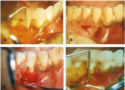
The Modified Apically Repositioned Flap (Fig 3): CARF
modified by Carnio was carried out in relation to those teeth, which were located contralateral to the site where the CARF surgery was performed. The basic procedure was same with certain modifications as described below. The initial horizontal internal bevel incision was made in the attached portion of the keratinized gingiva slightly apical to the alveolar crest retaining the tissue coronal to the horizontal incision as a marginal collar (Figure. 1 B and 4B,C). Suturing was done for adequate stability of the flap, though not considered by Carnio, Professional plaque control care was performed weekly during the first 8 weeks. Henceforth, the patients were recalled on days 90 and 150 for evaluation and postoperative measurements. The data were analyzed statistically using
paired ‘t’ test and was considered as significant when p value
found to be equal or < 0.05.
RESULTS AND OBSERVATIONS
Changes in Width of Attached Gingiva: In Site A, the mean
width of attached gingiva on day 0 and 90 was 1.21 ± 0.25 mm and 2.93 ± 0.42 mm, respectively. Thus, the width of attached gingivawas found to be increased by 1.72 mm on day 90, which was statistically highly significant. The width of attached gingiva was further increased by 0.43 mm (mean width 3.36 ± 0.29 mm) on day 150. Thus, the total gain in width of attached gingiva from day 0 to 150 was 2.15 mm, which was found to be statistically highly significant. In Site B, the mean width of attached gingiva on day 0 and 90 was 1.00 ± 0.23 mm and 3.07 ± 0.37 mm, respectively. Thus, it wasfound to be increased by 2.07 mm on day 90, which was statistically highly significant. The width of attached gingiva was further increased by 0.18 mm (mean 3.25 ± 0.37 mm) on day 150, though it was not found to be statistically significant. The total gain of attached gingiva from day 0 to 150 was 2.25 mm and was found to be statistically highly significant. The gain in attached gingiva in Site B was more than that of Site A at different time points.
Figure 3. Conventional Apically Repositioned Flap. (A) Preoperative clinical measurements with UNC -15 probe with stent in positionafter stained using iodine, (B) Crevicular incision in horizontal direction and vertical incisions at both ends, (C)Reflection of Partial-thickness flap and placed apically, (D) Postoperative view on day 150. Note the increased zone of attached gingiva postoperatively
Changesin Gingival Recession: In Site A, the mean gingival recession (apparent) on day 0 and 90 was 2.18 ± 0.62 mm and 2.54 ± 0.61 mm, respectively. Thus, gingival recession increased by 0.36 mm on day 90, which was found to be statistically highly significant. Gingival recession was further increased by 0.14 mm (mean 2.68 ± 0.64 mm) on day 150,though not statistically significant. Thus, the total increase in gingival recession between day 0 and 150 was 0.50 mm, which was statistically highly significant. In Site B, the mean gingival recession (apparent) at day 0 and 90 was 1.53 ± 0.76 mm and 1.96 ± 0.44 mm, respectively. Thus, the gingival recession increased by 0.43 mm at day 90 which was found to be statistically highly significant. This was further increased by 0.07 mm on day 150 (mean 1.89 ± 0.59 mm) which was not found to be statistically significant. Thus, the total increase in gingival recession between day 0 and 150 was 0.36 mm. It was found to be statistically highly significant. On comparison of Site A and Site B, the difference of mean gingival recessions between day 0 and 90 was found to be statistically highly significant, though not so between day 90 and 150. On day 150, the mean difference in the amount of gingival recession in Site B was less than that of Site A, the difference being 0.14 mm.
Figure 4. Modified Apically Repositioned Flap. (A) Preoperative clinical measurements with UNC-15 probe with stent in position after stained using iodine, (B) Internal bevel incision in horizontal direction to leave the marginal gingiva intact, (C) Reflection of Partial-thickness flap and placed apically, (D) Postoperative view on day 150. Note the increased zone of attached gingiva postoperatively
Changes in Sulcus Depth: In Site A, the mean sulcus depth at
day 0 and 90 was 0.50 ± 0.0 mm and 0.93 ± 0.26 mm, respectively. Thus, the sulcus depth increased by 0.43 mm at day 90, which was statistically highly significant. The sulcus depth was further increased by 0.18 mm (mean 1.11 ± 0.18 mm) at day 150. The overall increase of sulcus depth from day 0 to 150 by a mean difference of 0.61 mm was found to be highly significant. In Site B, the mean sulcus depth at day 0 and 90 was 0.50 ± 0.0mm and 0.61 ± 0.22 mm, respectively. Thus, the sulcus depth increased by 0.11 mm to at day 90. It further increased by 0.10 mm to 0.71 ± 0.25 mm at day 150. Both these changes were not found to be statistically significant. However, the overall increase of sulcus depth from day 0 to 150 was 0.21 mm, which was found to be statistically significant. The comparative changes in sulcus depth at Experimental Site A and B at different time intervals was not found to be statistically significant from day 0 to 90, but was
[image:4.595.312.558.329.512.2] [image:4.595.40.287.547.724.2]highly significant on day 150. Between day 0 and 150, the total increase of sulcus depth in Site A was more than that of Site B; the mean difference being 0.40 mm.
Comparative Changes in Clinical Attachment Level: In Site
A, the mean clinical attachment level on day 0 and 90 was 2.75 ± 0.59 mm and3.46 ± 0.67 mm, respectively. Thus, the mean clinical attachment level was increased by 0.71 mm on day 90. It was further increased by 0.33 mm (mean 3.79 ± 0.67 mm) on day 150. Both these increases were statistically significant. Thus, the overall increase of clinical attachment level from day 0 to 150 was 1.04 mm, which was statistically highly significant. In Site B, the mean clinical attachment level on day 0 and 90 was 2.46 ± 0.46 mm and 2.82 ± 0.49 mm, respectively. Thus, the mean clinical attachment level was increased by 0.36 mm on day 90, which was found to be not statistically significant. However, the mean clinical attachment level was reduced by 0.33 mm from day 90 (mean 2.79 ± 0.59 mm) on day 150. This was not found to be statistically significant. The overall increase in clinical attachment level from day 0 to 150 was 0.33 mm, which was statistically not significant. Thus, there was gain in clinical attachment from day 90 to 150 in Site B. On comparison of Site A and Site B, the difference of mean clinical attachment level between day 0 and 90 was not found to be statistically significant but was highly significant between day 90 and 150. On day 150, the mean clinical attachment level in Site A was more than that of Site B; the difference being 0.71 mm. No complications were observed in any patients regardless of the sites and postoperative healing was found to be uneventful.
DISCUSSION
The present study was carried out to compare CARF and MARF procedures in increasing the width of attached gingiva. The MARF technique is similar to that of ARF, except that the existing marginal gingiva is not detached from the tooth, retained it as a marginal collar. The study comprised of 28 sites in 14 subjects, in a split mouth design. The sites were referred to as Experimental Site ‘A’ and ‘B’. ‘A’ and ‘B’indicate the CARF and MARF, respectively. The clinical parameters, such as width of attached gingiva, gingival recession (apparent), sulcus depth and clinical attachment level were recorded preoperatively on day 0 and again postoperatively on day 90 and day 150. The CARF has wide general application to widen the zone of attached gingiva. The repositioned attached gingiva acts as a barrier to creeping return of the mucogingival junction towards the teeth and results in a permanent gain in width of attached gingiva and vestibular depth. The findings of this study reveal that both CARF and MARF are equally effective and efficient in increasing the width of keratinized tissue and attached gingiva, as shown by Carnio (1996) and Fagan and Freeman (1974).
However, MARF technique may be preferred over the former considering the fact that it is less time consuming with minimal trauma and excellent esthetic results with less/ no resultant gingival recession. Both these techniques allow the clinician to gain a new band of attached gingiva, eliminating the need for second surgical site. Considering the sample size and duration of the study period, further study involving larger sample size and long duration, is required to assess the effects of these techniques.
Conclusion
Both CARF and MARF are found to be equally effective in increasing the zone of attached gingiva. However, MARF has certain advantages, such as no further attachment loss, easy to execute, predictable results, minimal surgical trauma, less time consuming, better esthetics and less or no gingival recession.
REFERENCES
Bernimoulin, JP., Mühlemann, HR. 1973. Is there any new
development in gingival transplantation? Schweiz Monatsschr
Zahnheilkd., 83:505-514.
Bohannan, H. 1962. Studies in the alteration of vestibular depth. I
Complete denudation. JPeriodontol., 33:120-129.
Carnio, J. 1996. The modified technique of apically repositioned
flap. Periodonto., 46:1-6.
Carnio, J., Miller, PD Jr. 1999. Increasing the amount of attached
gingiva using a modified apically repositioned flap. J
Periodontol., 70:1110-1117.
Carranza, FA Jr.,Carraro JJ. 1970. Mucogingival techniques in
periodontal surgery. J Periodontol., 41:294-299.
Corn, H. 1962. Periosteal separation –Its clinical significance.
JPeriodontol., 33:142-153.
Fagan, F., Freeman, E. 1974. Clinical comparison of the free
gingival graft and partial thickness apically positioned flap. J
Periodontol., 45:3-8.
Freedman, AL., Green, K., Salkin,LM., Stein, MD., Mellado, JR. 1999. An 18-year longitudinal study of untreated
mucogingival defects. J Periodontol., 70:1174-1176.
Friedman, N., Levine HL. 1964. Mucogingival surgery, current
status. J Periodontol., 35:5-21.
Gottsegen, R. 1954.Frenum position and vestibular depth in
relation to gingival health. Oral Surg Oral Med Oral
Pathol., 7:1069-1078.
Gujar, D., Kathariya, R. 2014. Modified apically repositioned flap: A novel technique for increasing the width of attached
gingiva: A case series. J Dent Orofac Res., 10:25-28
Hall, WB. 1981. Can attached gingiva be increased nonsurgically?
QuintInt., 13: 455-462.
Lang, NP.,Loe, H.1972. The relationship between the width of the
attached gingiva and gingival health. JPeriodontol.,
43:623-627.
Matter, J. 1982. Free gingival grafts for the treatment of gingival
recession. A review of some techniques. J Clin Periodontol.,
9:103-114.
Nabers, JM. 1966. Extension of the vestibular fornix utilizing a
gingival graft – Case history. Periodontics., 4:77-79.
Newman, MG., Takei, HH., Klokkevold, PR. Carranza’s Clinical
Periodontology. 10th ed., Philadelphia: PA Saunders; 2006.
Ochsenbein, C., Maynard, JG. 1975. The problem of attached
gingiva in children. J Dent Child., 41:263-272.
Ochsenbein, C.1960. Newer concepts of mucogingival surgery. J
Periodontol., 31:175-185.
Pennel, BM.,Higgason, JD., Towner, JD., King, KO., Fritz, BD., Sadler, F. 1965. Retention of periosteum in mucogingival
surgery. JPeriodontol., 36:39-43.
Rosenberg, E., Garber, DA., Malinowski, A. 1981. Considerations
in the utilization of the free gingival graft. Quintessence Int
Dent Dig., 12:927-932.
Ruben, MP. 1979. A biologic rationale for gingival reconstruction
by grafting procedures. Quintessence Int Dent Dig., 10:47-55.
Stern, IB. 1981. Current concepts of the dentogingival junction: The epithelial and connective tissue attachments to the tooth.
J Periodontol., 52:465-476.