University of Pennsylvania
ScholarlyCommons
Publicly Accessible Penn Dissertations
1-1-2015
Adaptations of the Supraspinatus Tendon and
Muscle in a Rat Model of Exercise
Sarah Ilkhanipour Rooney
University of Pennsylvania, [email protected]
Follow this and additional works at:http://repository.upenn.edu/edissertations Part of theBiomechanics Commons, and theBiomedical Commons
This paper is posted at ScholarlyCommons.http://repository.upenn.edu/edissertations/1984 For more information, please [email protected].
Recommended Citation
Rooney, Sarah Ilkhanipour, "Adaptations of the Supraspinatus Tendon and Muscle in a Rat Model of Exercise" (2015).Publicly Accessible Penn Dissertations. 1984.
Adaptations of the Supraspinatus Tendon and Muscle in a Rat Model of
Exercise
Abstract
Rotator cuff tendon and muscle can beneficially adapt to exercise, but it is unknown how acute responses following a single bout of exercise lead to chronic, beneficial adaptations. To develop new prevention and treatment strategies for overuse injuries, we must first characterize how tissues adapt to non-injurious loading. One feature that may distinguish beneficial and detrimental responses are pathways associated with
inflammation and tissue healing, including activation of the arachidonic acid cascade and matrix turnover facilitated by matrix metalloproteinases (MMPs). The objective of this dissertation was to characterize temporal, acute and chronic responses of the rat supraspinatus to non-injurious exercise and examine the roles of cyclooxygenases-1 and -2 (arachidonic acid cascade components) and MMPs in governing these responses by using pharmacologic inhibition. Using a rat model of non-injurious exercise, we showed that tendon adapted chronically by enhancing its mechanical properties. Following a single bout of exercise, tendon showed mild trends toward reduced mechanical properties, which may initiate the beneficial adaptations detected chronically. Exercised tendon also had decreased MMP activity. We administered ibuprofen, an inhibitor of cyclooxygenases-1 and -2, to understand how this commonly used over-the-counter drug affects muscle and tendon responses to exercise. Ibuprofen did not detrimentally affect tendon mechanical properties but did result in decreased muscle fiber cross-sectional area. Taken together, these findings imply that the arachidonic acid cascade is not a primary modulator of tendon adaptations to non-injurious exercise but is important for muscle growth. Finally, we studied the effects of doxycycline, a broad-spectrum MMP inhibitor, on supraspinatus muscle and tendon adaptations to exercise. Doxycycline enhanced tendon mechanical properties and collagen organization from sedentary animals but not in exercised animals, which supports the increased MMP activity detected in sedentary tendons. Doxycycline reduced muscle fiber cross-sectional area specific to exercised animals. We conclude that the pathways responsible for muscle and tendon adaptations to exercise may be tissue-dependent, and drugs such as ibuprofen and doxycycline also have activity-dependent effects. These studies provide a foundation for future work to develop more efficient physical training regimens and new prevention, treatment, and rehabilitation modalities for overuse injuries.
Degree Type
Dissertation
Degree Name
Doctor of Philosophy (PhD)
Graduate Group
Bioengineering
First Advisor
Louis J. Soslowsky
Keywords
Doxycycline, Ibuprofen, Inflammation, Matrix Metalloproteinases, Rotator Cuff, Shoulder
Subject Categories
Biomechanics | Biomedical
ADAPTATIONS OF THE SUPRASPINATUS TENDON AND MUSCLE IN A RAT
MODEL OF EXERCISE
Sarah Ilkhanipour Rooney
A DISSERTATION
in
Bioengineering
Presented to the Faculties of the University of Pennsylvania
in
Partial Fulfillment of the Requirements for the
Degree of Doctor of Philosophy
2015
Supervisor of Dissertation
___________________________
Louis J. Soslowsky, PhD, Fairhill Professor, Orthopaedic Surgery
Graduate Group Chairperson
___________________________
Jason A. Burdick, PhD, Professor, Bioengineering
Dissertation Committee Dana T. Graves, DDS, DMSc
Professor, Periodontics, University of Pennsylvania
Tejvir S. Khurana, MD, PhD
Professor, Physiology, University of Pennsylvania
Andrew F. Kuntz, MD
Assistant Professor, Orthopaedic Surgery, University of Pennsylvania
Beth A. Winkelstein, PhD (Committee Chair)
ii
Acknowledgements
I could not have gotten to this point without the support of many people, a series
of fortunate events, a bit of good luck in circumstance and timing, hard work, and my
belief in something greater than myself. I want to thank the many people who prepared
me for and carried me along in this journey. First, I would like to thank my advisor, Dr.
Lou Soslowsky, for his mentorship, practicality, and patience. With purpose and
compassion he guided me through difficult research decisions and the inevitable highs
and lows. I would also like to thank my committee members for their individual
contributions to my graduate education and dissertation. Specifically, I would like to
thank Dr. Beth Winkelstein for her support, which began when I first interviewed for the
Bioengineering graduate program, long before I started my dissertation work. I thank Dr.
Andrew Kuntz for his clinical expertise as I developed my project and his encouragement
through the process. I thank Dr. Teji Khurana for his insights into exercise and muscle;
his interest in my project and advice from the very beginning motivated me to pursue this
research topic. Finally, I thank Dr. Dana Graves, who provided valuable feedback and
suggestions that significantly shaped my study designs for this project.
I would also like to thank the community of the McKay Orthopaedic Research
Laboratory. I am privileged to have had the opportunity to collaborate and surround
myself with such talented and inquisitive individuals. This is a community that was
incredibly willing to help when I asked to be taught a new technique, needed
troubleshooting advice, or wanted to borrow reagents. The office staff keeps McKay
running smoothly. I would like to especially thank Susan Dinella for her kind heart, help
iii
the Pennsylvania Muscle Institute, another wonderful research community, for their
support and advice. I thank Dr. Michael Ostap for taking a leap of faith and selecting a
tendon bioengineer with a mechanics background to be a part of a muscle biology NIH
T32 training grant; I learned so much that I was able to apply to this dissertation. I would
also like to thank two individuals specifically who spent many hours teaching me about
muscle. First, I thank Dr. Emanuele Loro for his unwavering belief in me and incredible
patience over the past three years. Second, I thank Dr. Lucas Smith for his willingness to
help me throughout this project. In addition to these two research communities, the
University of Pennsylvania has many excellent core facilities that I had the privilege of
being able to use during my time here. I want to especially thank Dr. John Tobias, who
was instrumental in helping develop and analyze my screening study.
I want to especially thank my project helpers: Rachel Baskin, Dr. Dan Torino, and
Rameen Vafa. Their hard work, positive attitudes, and commitment made this project
possible. I would like to thank Pooja Khandekar for the hours she spent helping me
analyze data, Pankti Bhatt for her tendon smashing skills and encouragement, and Jennica
Tucker for her assistance with time-sensitive dissections and friendship. I want to thank
the large group of Rat Runners, past and present, who coached my rats through their
treadmill exercise protocols. I would also like to thank my fellow graduate students for
their support and friendship: Brianne Connizzo, Ben Freedman, Cori Riggin, and Adam
Pardes. I would like to especially thank Brianne Connizzo for being my partner-in-crime,
office-mate, and friend since the beginning. I would also like to thank Dr. Katie Reuther
for her pep talks, friendship, and mentorship every step of the way. I would like to
iv
members of our lab for building such a great work environment. I want to thank Dr.
Spence Szczesny for mentoring me during my first semester and providing hours of great
conversation. Outside of our lab, I was honored to get to know many of my fellow
bioengineering graduate students through GABE, the Graduate Association of
Bioengineers. In addition, I want to thank our janitor, Scott, who welcomed me every
morning with a greeting and a smile to help me start my day. Similarly, I want to thank
the animal facility staff for their friendliness, especially when I needed it most during
early mornings and weekends. Finally, I want to thank my 528 pet rats for their cuddles
and noble contributions to research.
Apart from research, while at the University of Pennsylvania, I spent significant
time developing my teaching skills. I want to sincerely thank Dr. LeAnn Dourte Segan,
who began as a fellow graduate student and later became my teaching mentor. I
appreciate her advice and encouragement and for allowing me the opportunity to become
involved in her and Dr. Dave Meaney’s Introduction to Biomechanics course. Similarly, I
want to thank the University of Pennsylvania’s Center for Teaching and Learning for
their workshops, mini-courses, seminars, and overall commitment to developing
reflective educators. Specifically, I want to thank Dr. Julie McGurk and Dr. Emily Elliott
for teaching me about active learning and inviting me to help them train other teaching
assistants.
This milestone could only be achieved because of many prior experiences that
helped shape me. First, I want to acknowledge my PreK-12 teachers who prepared me for
college, particularly those who inspired curiosity in me and my fellow classmates. I want
v
engineer, and the Epeians, Society of Women Engineers, and Women in Science and
Engineering Residential Program for helping me develop many organizational and
interpersonal skills during my time there. I want to thank Dr. James Ashton-Miller, my
research advisor at the University of Michigan, for his mentorship and for providing me
my first research experience. I also want to thank two of my long-term friends from home
for their years of support since elementary school: Dr. Grace Huang Amadi and Virginia
Calkins. Finally, I want to thank my many gymnastics coaches (particularly Coach Brian
and Jen Lewis) for instilling in me resilience, persistence, ambition, work ethic, and
confidence (or at least how to fake it til you make it).
Finally, I am eternally grateful for my family, who raised me to be the woman I
am today and have always provided support and love throughout my life. I thank my
parents, Roz Ilkhani-Pour and Dr. Mehdi Ilkhani-Pour, for their sacrifices and for being
my number one role models. They taught me hard work and morals; they gave me
strength. I thank my sister, Arianna Ilkhani-Pour, for being the yin to my yang and for
being my favorite hair stylist and nail artist. I thank my grandparents for raising my mom
and dad’s families, and I especially thank my grandma Susie Bailey for her prayers. I feel
very fortunate to have also married into a loving family, and I thank my mother- and
father-in-law, Judy Rooney and Dr. Michael Rooney, and my sister- and brother-in-law,
Elise Rooney and (soon-to-be-Dr.) Ed Rooney for their support. Finally, I thank my
husband, Nick, for his selflessness, patience, understanding, and love. His unwavering
support and compassion (in addition to cuddles from my cat, Coco Chanel) helped me
through my most raw moments. I am so thankful for the partnership we share. This
vi
ABSTRACT
ADAPTATIONS OF THE SUPRASPINATUS TENDON AND MUSCLE IN A RAT
MODEL OF EXERCISE
Sarah Ilkhanipour Rooney
Dr. Louis J. Soslowsky
Rotator cuff tendon and muscle can beneficially adapt to exercise, but it is
unknown how acute responses following a single bout of exercise lead to chronic,
beneficial adaptations. To develop new prevention and treatment strategies for overuse
injuries, we must first characterize how tissues adapt to non-injurious loading. One
feature that may distinguish beneficial and detrimental responses are pathways associated
with inflammation and tissue healing, including activation of the arachidonic acid
cascade and matrix turnover facilitated by matrix metalloproteinases (MMPs). The
objective of this dissertation was to characterize temporal, acute and chronic responses of
the rat supraspinatus to non-injurious exercise and examine the roles of
cyclooxygenases-1 and -2 (arachidonic acid cascade components) and MMPs in governing these responses
by using pharmacologic inhibition. Using a rat model of non-injurious exercise, we
showed that tendon adapted chronically by enhancing its mechanical properties.
Following a single bout of exercise, tendon showed mild trends toward reduced
mechanical properties, which may initiate the beneficial adaptations detected chronically.
vii
inhibitor of cyclooxygenases-1 and -2, to understand how this commonly used
over-the-counter drug affects muscle and tendon responses to exercise. Ibuprofen did not
detrimentally affect tendon mechanical properties but did result in decreased muscle fiber
cross-sectional area. Taken together, these findings imply that the arachidonic acid
cascade is not a primary modulator of tendon adaptations to non-injurious exercise but is
important for muscle growth. Finally, we studied the effects of doxycycline, a
broad-spectrum MMP inhibitor, on supraspinatus muscle and tendon adaptations to exercise.
Doxycycline enhanced tendon mechanical properties and collagen organization from
sedentary animals but not in exercised animals, which supports the increased MMP
activity detected in sedentary tendons. Doxycycline reduced muscle fiber cross-sectional
area specific to exercised animals. We conclude that the pathways responsible for muscle
and tendon adaptations to exercise may be tissue-dependent, and drugs such as ibuprofen
and doxycycline also have activity-dependent effects. These studies provide a foundation
for future work to develop more efficient physical training regimens and new prevention,
viii
TABLE OF CONTENTS
Acknowledgements ... ii
ABSTRACT ... vi
TABLE OF CONTENTS ... viii
LIST OF TABLES ... xiii
LIST OF FIGURES ... xiv
Chapter 1: Introduction ... 1
A. Introduction ... 1
B. Background ... 3
1. Rotator Cuff Anatomy ... 3
2. Supraspinatus Tendinopathy ... 3
a. Prevalence ... 3
b. Presentation ... 4
c. Proposed Etiology ... 6
d. Conservative Treatments ... 6
3. Exercise ... 7
4. Tendon Properties ... 8
a. Biology ... 8
b. Mechanics ... 9
c. Adaptations to Beneficial Exercise ... 9
5. Skeletal Muscle Properties ... 10
ix
b. Adaptations to Beneficial Exercise ... 11
6. Rat Rotator Cuff Model ... 13
7. Rat Model of Supraspinatus Non-Injurious Exercise ... 14
8. Inflammation ... 19
a. Arachidonic Acid Cascade and MMPs ... 19
b. Roles of Inflammation in Tendon Adaptations ... 20
c. Roles of Inflammation in Muscle Adaptations ... 23
9. Nonsteroidal Anti-Inflammatory Drugs (NSAIDs) ... 24
a. Prevalence of Use ... 24
b. Effect of NSAIDs on Tendon ... 25
c. Effect of NSAIDs on Muscle ... 26
10. Doxycycline ... 27
a. Background ... 27
b. Effects on Tendon and Muscle Healing ... 27
C. Specific Aims ... 28
D. Study Design ... 30
E. Chapter Overview ... 32
F. References... 34
Chapter 2: Rat Supraspinatus Tendon Responds Acutely and Chronically to Exercise ... 51
A. Introduction ... 51
B. Methods ... 52
x
2. Tendon Mechanical Testing ... 55
3. Tendon Histology... 57
4. MMP Activity Assay ... 57
5. Statistics ... 58
C. Results ... 59
1. Tendon Mechanics ... 59
2. Tendon Histology... 61
3. MMP Activity Assay ... 64
D. Discussion ... 64
E. References ... 69
Chapter 3: Ibuprofen Differentially Affects Supraspinatus Muscle and Tendon Adaptations to Exercise in a Rat Model ... 73
A. Introduction ... 73
B. Methods ... 74
1. Study Design and Treadmill Protocols ... 74
2. Tendon Mechanical Testing ... 77
3. Tendon Histology... 78
4. Muscle Histology ... 78
5. Statistics ... 79
C. Results ... 80
1. Tendon Mechanics ... 80
2. Tendon Histology... 81
xi
D. Discussion ... 87
E. References ... 92
Chapter 4: Doxycycline Improves Sedentary, but not Exercised, Supraspinatus Tendon and Muscle in a Rat Model ... 97
A. Introduction ... 97
B. Methods ... 98
1. Study Design and Treadmill Protocols ... 98
2. Tendon Mechanical Testing ... 101
3. Tendon Histology... 102
4. Muscle Histology ... 103
5. Statistics ... 104
C. Results ... 104
1. Tendon Mechanics ... 104
2. Tendon Histology... 107
3. Muscle Histology ... 109
D. Discussion ... 112
E. References ... 117
Chapter 5: Conclusions and Future Directions... 121
A. Introduction ... 121
B. Rat Supraspinatus Tendon Responds Acutely and Chronically to Exercise .. 121
xii
D. Doxycycline Improves Sedentary, but not Exercised, Supraspinatus Tendon
and Muscle in a Rat Model ... 124
E. Final Conclusions ... 126
F. Future Directions ... 126
1. Additional Biologic Assays ... 126
2. Additional Muscle Assays ... 128
3. Other Loading Conditions... 130
4. Age ... 131
5. Gender ... 132
6. De-Training ... 133
7. Acute Responses to Exercise in Trained Animals ... 134
8. Recovery ... 134
9. Acute Responses Lead to Chronic Adaptations ... 135
10. Temporal Drug Administration... 136
11. Prevention of Overuse Injuries ... 137
12. Fatty Atrophy ... 138
13. Systemic Diseases ... 138
14. Other Tendons and Muscles... 138
15. Genome Analysis for Predisposition ... 139
16. Final Conclusions... 139
G. References ... 141
xiii
LIST OF TABLES
Table 1.1: Chronic Tendinopathy Characteristics ... 5
Table 1.2: Properties of Skeletal Muscles ... 11
Table 1.3: Shoulder Adaptations to Exercise Protocol ... 17
Table 1.4: Genes Altered Acutely and Chronically in Supraspinatus Tendon Following Non-Injurious Exercise ... 23
Table 1.5: Genes Altered Acutely and Chronically in Supraspinatus Muscle Following Non-Injurious Exercise ... 24
Table 3.1: Centrally Nucleated Fibers ... 85
Table 3.2: Fiber Type Distribution ... 87
Table 3.3: Fiber Type Cross-Sectional Area... 87
Table 4.1: Centrally Nucleated Fibers ... 109
Table 4.2: Fiber Type Distribution ... 111
xiv
LIST OF FIGURES
Figure 1.1: Shoulder Anatomy ... 4
Figure 1.2: Tendon Stress-Strain Curve... 9
Figure 1.3: Weight Changes with Exercise ... 16
Figure 1.4: Oxidative Phosphorylation Proteins ... 18
Figure 1.5: Arachidonic Acid Cascade ... 20
Figure 1.6: Aim 1- Acute Effects of Exercise Study Design ... 31
Figure 1.7: Aim 1- Chronic Effects of Exercise Study Design ... 32
Figure 1.8: Aims 2 and 3 Study Designs ... 33
Figure 2.1: Acute (A) and Chronic (B) Study Designs ... 55
Figure 2.2: Tendon Mechanical Properties ... 60
Figure 2.3: Tendon Dynamic Mechanics ... 61
Figure 2.4: Tendon Histology ... 62
Figure 2.5: Tendon Organization ... 62
Figure 2.6: Representative Tendon Histology Images ... 63
Figure 2.7: Injured Tendon Histology Images ... 63
Figure 2.8: MMP Activity... 64
Figure 3.1: Acute (A) and Chronic (B) Study Designs ... 76
Figure 3.2: Tendon Mechanical Properties ... 81
Figure 3.3: Dynamic Modulus ... 82
Figure 3.4: tan(δ)... 83
Figure 3.5: Tendon Histology ... 83
xv
Figure 3.7: Representative Tendon Histology Images ... 84
Figure 3.8: Average Muscle Fiber Cross-Sectional Area ... 85
Figure 3.9: Deep and Superficial Region Supraspinatus Muscle Fiber Type Distribution ... 86
Figure 4.1: Acute (A) and Chronic (B) Study Designs ... 100
Figure 4.2: Tendon Mechanical Properties ... 105
Figure 4.3: Dynamic Modulus ... 106
Figure 4.4: tan(δ)... 107
Figure 4.5: Tendon Histology ... 108
Figure 4.6: Tendon Organization ... 108
Figure 4.7: Representative Tendon Histology Images ... 109
1
Chapter 1: Introduction
A. Introduction
Rotator cuff tendinopathy, which primarily affects the supraspinatus tendon, is
estimated to affect 6.1% of women and 4.5% of men ages 25-64 years in the general
population.113 If left untreated, the tendon degeneration can progress to a rotator cuff tear,
which affects up to 50% of asymptomatic people over the age of 50 years.72,102,117 These
injuries are especially common in laborers and athletes that repeatedly use overhead
activity. The proposed etiology is accumulated microtrauma in which the tendon is
unable to properly adapt to the loading, resulting in a chronic overuse injury. These
injurious changes are in contrast to those found following non-injurious exercise, in
which the tissue is able to beneficially adapt to the loading. The fine line between
beneficial and detrimental loading conditions has not been elucidated.
One feature that may distinguish beneficial and detrimental adaptations to loading
is the role of pathways associated with inflammation. Acute inflammation is a series of
biologic events following injury. Two important processes related to inflammation are
activation of the arachidonic acid cascade and matrix turnover facilitated by a class of
enzymes called matrix metalloproteinases (MMPs). Previous studies suggest that
activation of these inflammatory pathways is important for enabling tendon and muscle
regeneration following acute injury; however, the mechanisms governing the role of these
inflammatory pathways in the adaptation of tendon and muscle to physiologically
2
The use of pharmacologic inhibitors of these two inflammation-related pathways
(arachidonic acid cascade and MMPs) may help identify their roles in tissue adaptations
to load. Ibuprofen, one of the top three consumed drugs in the United States, is an
inhibitor of cyclooxygenases-1 and -2, components of the arachidonic acid cascade.
Current literature exploring the effects of ibuprofen on tissue adaptations to loading is
unclear, despite its prevalent use. Doxycycline, in addition to being an antibiotic, is an
MMP inhibitor that has been suggested as a treatment to improve tissue healing following
acute injury. The effect of doxycycline on muscle and tendon adaptions to load has not
been explored. Administration of ibuprofen and doxycycline, inhibitors of two pathways
associated with inflammation, may highlight the mechanisms that manage tissue
responses to loading.
The original objective of this dissertation was to investigate and compare an
overuse and non-injurious exercise model to distinguish pathologic from physiologic
adaptations to load; however, only the non-injurious exercise model was used here.
Therefore, the goal of this dissertation is to use an animal model to characterize how
supraspinatus tendon and muscle adapt to non-injurious exercise over time and the roles
of two inflammation-related pathways (arachidonic acid cascade and MMPs). Aim 1
characterizes the natural, temporal response of tendon to non-injurious loading. Aim 2
helps to answer the question “Will ibuprofen adversely affect muscle and tendon
adaptations to exercise?” Finally, Aim 3 explores the role of MMPs in sedentary and
exercised supraspinatus tendon and muscle by using doxycycline as a pharmacologic
3
Before addressing the role of inflammation in overuse injuries, we must first
understand its role in tissue adaptations to non-injurious exercise. This research is
innovative in that it directly investigates the supraspinatus tendon and muscle response to
non-injurious loading acutely and chronically using clinically relevant drugs. These
studies will identify the key processes associated with beneficial tissue responses to
exercise to inform physical training regimens. Importantly, results will reinforce or
challenge the regular use of anti-inflammatory drugs. Finally, these studies will also
provide a foundation to support development of new prevention, treatment, and
rehabilitation modalities for tendinopathy.
B. Background
1. Rotator Cuff Anatomy
The rotator cuff refers to the four muscles and associated tendons that stabilize the
shoulder: supraspinatus, infraspinatus, subscapularis, and teres minor. These muscles
originate on the scapula and insert (through their tendons) on the humerus. Of these four
tendons, the supraspinatus is most commonly injured with tendinopathy (tendon
degeneration) or tear. The supraspinatus passes underneath the coracoacromial arch, a
bony-ligamentous arch formed by the acromion and coracoid processes of the scapula
and the coracoacromial ligament that spans the two processes (Figure 1.1).
2. Supraspinatus Tendinopathy
a. Prevalence
Rotator cuff tendinopathy, which primarily affects the supraspinatus tendon, is a
common clinical condition. In the general population, the prevalence of this injury is
4
complaints, 29% were due to rotator cuff tendinopathy.112 This injury is frequent in
overhead laborers and athletes. For example, 18% of industrial welders suffered from
rotator cuff tendinopathy.39 Degeneration precedes spontaneous tendon rupture in 97% of
patients,44 and if left untreated, chronic tendinopathy may progress to a full tendon tear.
Up to 50% of asymptomatic people over the age of 50 years suffer from a rotator cuff
tear.72,102,117 These tears can be difficult to treat, and tendinopathy can be painful and
debilitating; therefore, strategies to prevent the onset of supraspinatus tendinopathy are
needed.
b. Presentation
Tendinopathy is a clinical diagnosis characterized by focal tenderness,
activity-related pain, weakness, and increased size. Ultrasound shows hypoechoic regions, and
MRI shows increased signal due to fluid retention. “Tendinosis” is confirmed upon Figure 1.1. Shoulder Anatomy
The supraspinatus passes underneath a bony-ligamentous arch formed by the acromion, coracoid, and coracoacromial ligament.
5
histological evaluation of a tendon biopsy showing increased cellularity, rounding of
nuclei, increased mucoid ground substance (proteoglycans), loss of collagen organization,
and increased vascularity.43,46,61 These changes are generally heterogeneous and may be
present without clinical symptoms. Inflammation is not seen in the late stages of
degeneration when biopsies are taken, and early stages have not been studied since
patients typically present to the clinic only once tendon degeneration has progressed to a
chronic stage. Animal models would allow investigation of these early stages, but few
studies have yet been performed. Chronic tendinopathy is often described as a “failed
healing” response,35
with increases in both matrix synthesis and degradation that result in
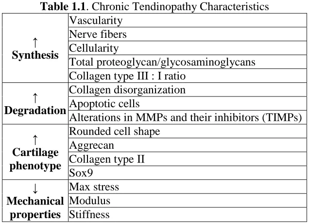
net matrix catabolism. A summary of changes associated with chronic tendinopathy is
presented in Table 1.1.
Table 1.1. Chronic Tendinopathy Characteristics
↑ Synthesis Vascularity Nerve fibers Cellularity Total proteoglycan/glycosaminoglycans Collagen type III : I ratio
↑ Degradation
Collagen disorganization Apoptotic cells
Alterations in MMPs and their inhibitors (TIMPs)
↑ Cartilage phenotype
Rounded cell shape Aggrecan
6
c. Proposed Etiology
The proposed etiology of supraspinatus tendinopathy is gradual accumulation of
microtrauma, resulting in an overuse injury. Overuse injuries account for approximately
29% of all injuries in college athletes.119 Over one-third of these overuse injuries could be
attributed to inflammation or tendinopathy.119 Highlighting the role of overuse in the
initiation and progression of tendon degeneration, supraspinatus tendinopathy in elite
swimmers correlated with the hours and kilometers swum per week.92 Overuse injuries
occur due to maladaptations to loading: while beneficial exercise results in matrix
turnover and net anabolism, overuse injuries result in unbalanced degeneration-repair
processes creating net catabolism. In order to understand the initiation and progression of
these injuries, we must first identify the physiologic adaptations to beneficial,
non-injurious exercise.
d. Conservative Treatments
When a patient presents with tendinopathy, the first treatments prescribed are
conservative: e.g., administration of nonsteroidal anti-inflammatory drugs (NSAIDs)
such as ibuprofen, physical therapy, corticosteroid injections, extracorporeal shock wave
therapy, and/or sclerotherapy.6 NSAIDs and corticosteroid injections appear to provide
short-term pain relief but no long-term benefits.4,6 Other potential treatment mechanisms
targeting biologic pathways include modulating substance P,5,16,31 administration of
glyceryl trinitrate or L-NAME to alter nitric oxide,22,77,105 anti-TNF-α,85,1 NMDA
antagonist to target glutamate signaling pathways,85 and anakinra (IL-1 antagonist).1 The
7
treatment strategy is use of MMP inhibitors. Several studies have shown alterations in
MMP expression with tendinopathy. It is believed that an imbalance in these enzymes
and their inhibitors leads to the advanced degeneration of the tendon. Aprotinin (MMP
inhibitor) injections to the Achilles tendon are promising in treatment of chronic
tendinopathy in humans,76 though another study has found no benefit over placebo.15
While changes in MMP expression are characteristic of chronic tendinopathy,
non-damaging exercise may also produce changes in MMP expression indicative of matrix
turnover and beneficial adaptation. Exploring effects of potential “treatments” such as
NSAIDs and MMP inhibitors in sedentary and non-injurious exercise models would help
elucidate the differences between beneficial adaptations and maladaptations to loading.
3. Exercise
Exercise has both local and systemic effects on the body. Locally, exercise affects
the muscle, tendon, and bone of the region (among other tissues not discussed here).
Systemically, exercise affects the cardiac and metabolic systems (among other systems
not discussed here). The effects of exercise are both acute, wherein the
tissue/organ/system shows an immediate response to the altered loading or stress, and
chronic, wherein the tissue/organ/system shows adaptations with repeated exercise
sessions. These acute responses and chronic adaptations to exercise are tissue- and
time-dependent in addition to exercise protocol-time-dependent. Defining whether a tissue has been
“exercised” requires the analysis of a combination of features. Importantly, no single
metric can define exercise; therefore, a number of measurements must be made to
understand the entirety of the response. In the following sections, tendon- and
8
4. Tendon Properties
a. Biology
Tendons are tissues that connect muscle to bone, allowing transfer of loads. The
primary constituents are tenocytes (tendon fibroblasts), extracellular matrix, and water.
The extracellular matrix is composed of collagens (types I, II, III, V, IX, X), which are
the main structural proteins, with smaller amounts of proteoglycans, elastin, and
fibronectin.33 Type I collagen is the primary component, comprising approximately 60%
of the tendon dry weight and 95% of the total collagen.114 The collagen in healthy
tendons is arranged longitudinally along the axis of loading to facilitate efficient load
transfer. With injury, this collagen becomes more disorganized. The extracellular matrix
varies regionally along the tendon and is believed to be defined by the loading profile
experienced in that location. Healthy tendon is specifically adapted to its required
function. The biology and composition of tendon is influenced by age, loading
(unloading, exercise, overloading), gender, disease, and injury. These changes in
composition can lead to changes in tissue function, and alternately, changes in function
can lead to changes in composition. For example, eliminating compressive loading in a
rabbit flexor tendon led to changes in tendon thickness, nuclear density,
glycosaminoglycan content, collagen organization, and type II collagen.63 These biologic
9
b. Mechanics
Tendons are characterized by having non-linear, strain-dependent viscoelastic
behavior. The unique crimping pattern of the tendon results in a toe region during which
the fibers straighten before entering the linear region of the stress-strain curve (Figure
1.2).114 Furthermore, tendons exhibit stress-relaxation, creep, and unique recovery
properties.28,27 The viscoelasticity of tendon has been attributed to accumulation of highly
polarized glycosaminoglycans that attract water.
c. Adaptations to Beneficial Exercise
As previously described, tendons alter their composition and structure to meet the
demands of their function, such as adaptations to non-injurious exercise. These changes
occur over time with chronic, non-injurious exercise. Compared to more metabolically
active tissues (such as muscle or bone), tendon adaptations to exercise may be mild and
difficult to detect, which results in unclear descriptions of exercise effects in the
literature. For example, tendon cross-sectional area is often unchanged32,51,57 with chronic Figure 1.2. Tendon Stress-Strain Curve
10
exercise but some studies find increases.21,93 Similarly, tendon stiffness and modulus may
increase21,32,51,93 or remain unchanged49,57,62 with chronic exercise. Importantly, the
mechanical properties do not decrease, as this would be indicative of injury. Increased
collagen content is likely the compositional change that supports these functional
adaptations.10,99 The temporal, chronic adaptations to exercise have not been
well-defined, making it difficult to draw conclusions from studies that investigate a single time
point. In addition to chronic adaptations, tendons respond acutely to a change in loading,
as in response to a single bout of exercise.101 These changes include increased collagen
turnover,37,50,53,55,98 blood flow,14,89 and evidence of inflammation-related
components.52,54 Again, these changes are time-dependent, and it is unknown how acute
responses to exercise translate to chronic adaptations.
5. Skeletal Muscle Properties
a. Biology
Skeletal muscle is a voluntarily controlled, striated tissue. It has a hierarchical
structure composed of numerous muscle fibers. Muscle fibers (also called muscle cells or
myocytes) are cylindrical structures that have multiple nuclei. In mature, healthy tissue,
these nuclei are located on the periphery of the muscle fibers. During myogenesis or fiber
regeneration, myoblasts differentiate into myotubes, which contain centrally located
nuclei before maturing. Muscle fibers are composed of myofibrils which primarily
contain actin and myosin filaments in a repeating pattern, forming the contractile unit
(sarcomere). Muscle fibers come in a variety of “types” that result in different functional
capacities, summarized in Table 1.2. Myosin heavy chain (MHC) fiber type can be
11
Depending on the function of the muscle, different muscles have different compositions
of fiber type.
Table 1.2. Properties of Skeletal Muscles
Type I Type IIa Type IIb
Also known as Slow oxidative
(slow twitch)
Fast oxidative (fast twitch A)
Fast glycolytic (fast twitch B)
Color Red fibers Red fibers White fibers
Fatigue Fatigue
resistant Fatigue resistant Fatigable
Myoglobin ↑ ↑ ↓
Mitochondria ↑ ↑↑ ↓
Oxidative capacity ↑ ↑ ↓
Glycolytic capacity
and Glycogen levels ↓ ↑ ↑↑
Storage fuel Triglycerides
Creatine phosphate Glycogen ATP Creatine phosphate Functional Examples Aerobic activities (long run) Middle-distance
aerobic activities Sprinting
b. Adaptations to Beneficial Exercise
Skeletal muscle adapts to both resistance and endurance exercise. In response to
resistance training, the most notable adaptation is skeletal muscle hypertrophy which
leads to increased maximal force production. In response to endurance training, muscle
enhances its capacity to efficiently create energy by increasing the number of
mitochondria and their oxidative phosphorylation proteins. This response leads to
decreased fatigability of the trained muscle. These hypertrophic and aerobic metabolism
changes can be independent of each other. Furthermore, these adaptations may be fiber
type-specific.29Whether fibers change type as a result of exercise training (e.g., type I
12
result in transition towards increased proportion of oxidative fiber types with endurance
training. Glycogen is the main energy storage form in muscle. With endurance training,
glycogen content within the resting muscle is increased compared to untrained
muscle.120,56 Additionally, the trained muscle is depleted of glycogen more slowly than
untrained.29 Gene expression and protein transcription is altered with exercise training.
PPARβ/δ transcription factor and PGC-1α and -1β transcriptional co-activators play a
major role in controlling the oxidative metabolism of skeletal muscle fibers.90 PGC-1α
overexpression in skeletal muscle is associated with resistance to fatigue and a shift to
oxidative phenotype.64 AMP-activated protein kinase (AMPK) activation can lead to
increased levels of mitochondrial oxidative enzymes.90 Exercise training induces a higher
basal AMPK activity level.34 It has been shown that AICAR, an AMPK agonist,
improves running endurance.73 AMPK and PPARδ regulate type I muscle fiber
specification and endurance adaptations during exercise.73 Finally, Myo1c is an
actin-based motor protein that acts in GLUT4 translocation. GLUT4, the major glucose
transporter protein expressed in skeletal muscle, is increased in highly oxidative
muscle.106 Endurance training has been shown to significantly increase the GLUT4
expression and Myo1c abundance.106
Muscle also exhibits acute responses to a single bout of exercise, suggesting
matrix turnover and influences of growth factors.36-38,57 After an acute bout of exercise,
glycogen content is diminished. Although muscle responses to exercise are much better
defined than tendon, it is still debated what role inflammatory processes play in initiating
13
6. Rat Rotator Cuff Model
The rat has previously been identified as a model of the human rotator cuff.96
Apart from non-human primates, the rat was the only species to satisfy a checklist of
criteria important to model the human shoulder. Importantly, the rat supraspinatus passes
through a bony tunnel similar to that in humans. Furthermore, during normal walking, the
rat supraspinatus passes repeatedly underneath the coracoacromial arch, mimicking the
movements of the human supraspinatus during lateral arm raise and overhead activity.
The rat has also been used to develop a model of supraspinatus tendinopathy.97 In
this model, rats run on a 10º decline treadmill at a speed of 17 m/min, 5 days per week.
After as early as 4 weeks, the tendon shows degenerative changes consistent with injury,
including inferior mechanical properties, disorganized collagen matrix, hypercellularity,
and rounding of cell nuclei. The rat overuse model has since been used in several studies.
Similar to human tendinopathy, changes to the tendon matrix are evident in specific
alterations of MMPs,8 their inhibitors TIMPs,8,7 ADAM-10,7 proteoglycans (decorin,
biglycan, versican, aggrecan),7,9 and collagens (I, II, III, VI).9,7 Rounded cell shape and
increased aggrecan, type II collagen, and Sox9 are indicative of a shift toward a
fibrocartilaginous phenotype. Apoptosis may be present,69,91 and increased expression of
nitric oxide synthases100 and heat shock proteins69 are indicative of cellular stress.
Inflammatory regulators have also been identified in this model. Gene expression
quantified using PCR shows temporal changes in five-lipoxygenase activating protein
(FLAP), cyclooxygenase-2 (COX-2), vascular endothelial growth factor (VEGF), and
von Willebrand factor (VWF) over the span of 3 days to 16 weeks, indicating that
14
Molloy and co-authors70 showed that inflammatory and apoptotic genes are altered after 4
weeks of overuse. In a model of human rotator cuff tendinopathy, genes IL-15, -18, -6,
MIF, TNF-α, caspase 3, and caspase 8 were upregulated compared to control, similar to
the findings in the rat model.68 More recently, a slight progressive increase in TNF-α
protein expression from 2 to 4 weeks of overuse with no increase in ED1
(pro-inflammatory) or ED2 (anti-(pro-inflammatory) macrophages in that time frame was shown in
the rat overuse model.8 Inflammatory cytokines including MCP-1, IL-1, IL-6, and IL-13
were decreased at 2 but not 4 weeks of overuse.8 TGF-β3 has also been shown to
decrease at 2 and 4 weeks of overuse.7 These studies suggest a role of inflammation in
the early stages of supraspinatus tendinopathy in the rat overuse model, but whether this
response is pathologic or physiologic is unknown. Until recently, a model of
non-injurious exercise to the rat supraspinatus did not exist.
7. Rat Model of Supraspinatus Non-Injurious Exercise
To address the need for a model to allow distinctions between detrimental and
beneficial adaptations to exercise, we developed a non-injurious exercise model to which
the rat supraspinatus overuse model could be compared, as described in the published
study by Rooney et al., 2015.88 In this initial study, adult, male Sprague-Dawley rats were
divided into exercise (EX; n=8) and control cage activity (CA; n=8) groups (IACUC
approved). EX rats ran on a flat treadmill at 10 m/min, 1 h/day, 5 days/wk, for 12
weeks104 while CA rats maintained normal cage activity. Upon completion, rats were
euthanized, weighed, and stored at -20°C.
Tissue Harvest: Rats were thawed, and the right supraspinatus (supra) muscle and
15
measured with a custom laser device.79 Tendon at the insertion site was isolated from the
muscle for o-Hydroxy-proline (OHP) assay. The superficial and deep regions of the supra
muscle11 were collected separately for protein analysis. The heart and retroperitoneal and
epididymal fat pads were dissected and weighed. Tendon Mechanics: Rats were thawed,
and the left supra tendon was dissected and prepared for tensile mechanical testing with
preconditioning and ramp to failure.80Tendon Collagen: OHP, a measure of collagen
content, was determined26,74 and normalized to tendon wet weight. Muscle Protein: A
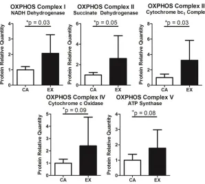
Western blot was performed to quantify mitochondrial proteins. Total protein from the
supra muscle superficial region was probed for oxidative phosphorylation complexes I-V
using Total OXPHOS antibody cocktail (MitoSciences). Bands were visualized by
chemiluminescence, imaged, and analyzed with commercial software. Mitochondrial
proteins were normalized to α-tubulin, and the relative quantity of target protein in the
EX group compared to CA was calculated. Humerus Bone μCT: μCT (Scanco
VivaCT40) was used to determine trabecular and cortical bone structure. For trabecular
bone, a 2mm region just distal to the growth plate was scanned (15μm isotropic voxels);
for cortical bone, a 1.5 mm region at 60% of the humerus length was scanned (35μm
isotropic voxels). Bone was segmented from marrow using a global thresholding
technique and then subjected to standard microstructural analysis. Humerus Bone 3-Point
Bending: A custom fixture attached to an Instron created a 3-point bend in the humeral
shaft until fracture.84Statistics: Comparisons between EX and CA were made with t-tests
16
EX rats had reduced body (7%) and fat pad (retroperitoneal: 45%, epididymal:
-39%) mass. An 8% increase in supra muscle mass was measured with no change in heart
mass (Figure 1.3). Supra muscle CSA significantly increased 10% with exercise (Table
1.3). Tendon Mechanics: Supra tendon CSA and elastic modulus were not different
between groups (Table 1.3). Tendon Collagen: Collagen content of the supra tendon
normalized to wet weight trended toward increased in the EX group (Table 1.3). Muscle
Protein: Supra muscle had increased oxidative phosphorylation proteins (Figure 1.4).
Bone μCT: Trabecular bone in the EX group demonstrated trends toward increased
trabecular number (Tb.N) and connectivity (Conn.D) and decreased spacing (Tb.Sp) as
well as significantly greater degree of anisotropy (DA, Table 1.3). No differences were
found in trabecular bone volume, structure model index, thickness, bone mineral density Figure 1.3. Weight Changes with Exercise. Consistent with exercise,
treadmill-trained rats trended toward decreased body weight over time and had significantly decreased fat pad weight, increased supraspinatus muscle weight, and no change in heart weight. Mean + StDev
17
(BMD), or tissue mineral density (TMD, not shown). Cortical bone in the EX group had
significantly increased BMD and TMD (Table 1.3) with no changes in volume or cortical
thickness (not shown). Bone 3-Point Bending: No differences were found for second
moment of area, max load, max displacement, modulus, flexural rigidity, or max stress
(not shown).
Table 1.3.Shoulder Adaptations to Exercise Protocol. Treadmill-trained rats showed adaptations consistent with exercise in supraspinatus tendon and muscle, and trabecular (Tb) and cortical (Ct) humerus bone. Mean ± StDev
Tissue Measurement CA EX p-value
Muscle CSA (mm2) 34.3 ± 3.9 37.7 ± 2.6 *0.04
Tendon
CSA (mm2) 1.97 ± 0.42 1.97 ± 0.28 0.5
Failure Load (N) 22.3 ± 5.8 26.8 ± 3.0 *0.04 Failure Stress (MPa) 11.6 ± 3.4 14.0 ± 3.2 +0.09
Modulus (MPa) 100 ± 42 101 ± 24 0.5
Collagen/Wet Weight (%) 25 ± 5 28 ± 1 +0.07
Bone
Tb Connective Density (1/mm3) 60 ± 12 70 ± 12 +0.07
Tb Number (1/mm) 3.1 ± 0.7 3.5 ± 0.4 +0.10 Tb Spacing (mm) 0.34 ± 0.09 0.28 ± 0.04 +0.08
Tb Degree of Anisotropy 1.79 ± 0.07 1.86 ± 0.07 *0.03 Ct Thickness (mm) 0.77 ± 0.02 0.80 ± 0.03 *0.05
18
After 12 weeks of treadmill training, rats showed systemic (decreased body and
fat pad mass) and local shoulder (tendon, muscle, bone) changes consistent with exercise.
Heart mass did not change, indicating that this protocol does not tax the cardiovascular
system. No changes were seen in tendon elastic properties, but stress-relaxation
significantly increased. Other studies on tendon mechanics after training show mixed
results but often find no differences in modulus or CSA.e.g.,57 Unlike the established supra
overuse model, tendons did not have decreased mechanics, indicating that this protocol is
non-injurious to the tendon.97 Supra muscle showed hypertrophy (increased weight and
CSA), indicative of a response to loading, and increased oxidative phosphorylation
proteins, indicative of endurance training. Humerus trabecular bone had increased Figure 1.4. Oxidative Phosphorylation Proteins. Consistent with
19
anisotropic orientation, consistent with load-induced bone remodeling. Cortical bone
showed increased BMD and TMD with no volume change, suggesting that bone mass
changes are due to tissue mineralization. Other studies have also shown no changes in
humerus mechanics following moderate treadmill training.e.g.,116 Although these
adaptations to exercise are mild, they are consistent and present across multiple tissues
using multiple assays. This study88 is limited by the quality of tissue available, sample
size, and single time point investigated. In conclusion, this is the first non-injurious rat
shoulder exercise model, which can be compared to the previously established overuse
model97 to differentiate beneficial adaptations and maladaptations to loading.
8. Inflammation
Wound healing is a structured, temporal response that consists of four phases:
hemostasis, inflammation, proliferation, and remodeling. Inflammation is a complex
biologic event that aims to protect and repair tissue. Acute inflammation is an immediate
(minutes to days) response to tissue insult. Two pathways associated with inflammation
are activation of the arachidonic acid (AA) cascade and matrix turnover facilitated by
matrix metalloproteinases (MMPs).
a. Arachidonic Acid Cascade and MMPs
An important pathway in the acute inflammatory response is the acrachidonic acid
cascade (Figure 1.5).40 In this system, arachidonic acid is converted by cyclooxygenase
(COX) to prostaglandins, prostacyclins, or thromboxane or by 5-lipoxygenase (LOX) to
leukotrienes. COX-1 is constitutively expressed, and COX-2 is induced.60 Prostaglandins,
synthesized through a COX-2 specific mechanism, can mediate blood flow to the tissue48
20
Figure 1.5. Arachidonic Acid Cascade (Figure from Hertel 1997)
carefully balanced with their inhibitors can result in excess fibrosis or degeneration.78
b. Roles of Inflammation in Tendon Adaptations
Tendon is a dynamic tissue that responds biologically to mechanical loads. In
vitro experiments show that cultured human tendon fibroblasts, known as “tenocytes,”
respond to strain by modulating production of prostaglandin E2 (PGE2), 1,
COX-2,115 and leukotriene B(4),58 components of the arachidonic acid pathway. Direct
administration of PGE2 to these cells reduced proliferation and increased MMP-1 and
MMP-3 protein levels with no effect on collagen type I gene expression.103 Similarly,
PGE2, collagenase, and cell death increased in in vitro loaded tendon explants with no
change in collagen or sulfated GAG.25 Increases in PGE2, collagenase, and cellular
turnover were dependent on both duration and magnitude of loading.24In vivo, patellar
and Achilles tendons of mice run to exhaustion also showed increased PGE2, which
decreased tendon stem cell proliferation and increased differentiation into
21
histological changes mimicking tendon degeneration in rabbits.47
Despite the implications that PGE2 may be a contributor to early tendinopathy,
repeated, direct injections improved mechanical properties in another study.30 The
presence of PGE2 may indicate tendon remodeling, which is supported by increased
levels in humans following a bout of exercise.54,55 Also immediately following exercise, a
marker of collagen synthesis and an indicator of collagen breakdown were decreased,
with a rise in collagen synthesis seen 72 hours later.55 PGE2 and inflammatory cytokines
may be involved in converting mechanical load to type I collagen synthesis, the primary
component of tendon.48 Similar to in vitro experiments,118 tendons from mice undergoing
moderate and intense loading protocols demonstrate distinct biologic responses,
suggesting that appropriate loads could be beneficial while intense loads could lead to
tendon degeneration.121 Whether inflammation is a physiologic response to load or
pathologic in early tendon degeneration is unknown.
In an initial study, we examined the response at a molecular level of the rat
supraspinatus tendon following acute and chronic exercise.87 Twenty male,
Sprague-Dawley rats were divided into cage activity (CA) or acute or chronic exercise (EX)
groups. Rats in the acute group ran a single bout of non-injurious exercise on a flat
treadmill and were euthanized 12 or 24 hours after. Rats in the chronic group ran 5
days/week for 1 or 8 weeks and were euthanized 72 hours after their final bout.88 CA
animals maintained normal cage activity for 5 weeks to be within 10% body weight of
the other groups. Supraspinatus muscle and tendon were harvested for RNA extractions
(n=4 per group), and a custom Panomics QuantiGene 2.0 plex array was used to
22
components, matrix turnover enzymes, and factors associated with tendinopathy. Results
were log2 transformed and normalized by the geometric mean of the 3 housekeeping
genes for each sample. Principal components analysis (PCA) confirmed distinctions
between tissues and time points, supporting the study design. Results of this assay
support the hypothesis that a physiologic, mild, acute inflammatory response occurs in
tendon following a single bout of exercise. Specifically, changes in arachidonic acid
cascade components were detected (Table 1.4), supporting investigation of this pathway
and effects of nonsteroidal anti-inflammatory drugs that inhibit this pathway.
Additionally, results support the hypothesized adaptive matrix changes following chronic
loading. Increased collagen expression was detected, consistent with beneficial
adaptation to exercise. Mmp14 and TIMP expression were altered acutely and
chronically, indicative of matrix turnover (Table 1.4). This finding supports investigation
of the role of MMPs in acute and chronic responses of tendon to exercise, as well as
investigation of drugs that inhibit these enzymes. Finally, results suggest that tendon
response to chronic, beneficial exercise is distinct from overuse. Unlike overuse, we did
not find increased expression of cartilage markers (Sox9, Acan, Col2a1), heat shock
proteins (Hspa2, Hspb1), or nitric oxide synthases (Nos2, Nos3) in tendon. This study
provides direct evidence for inflammation (arachidonic acid cascade) and matrix turnover
(MMPs/TIMPs) responses of supraspinatus tendon and muscle to acute and chronic
exercise.
23
Table 1.4. Genes altered acutely and chronically in supraspinatus tendon following non-injurious exercise
Acute Tendon Chronic Tendon Inflammation Ptges Ptger4 Tnf Alox5ap Matrix Mmp14 Timp3 Col1a1 Col3a1 Dcn Mmp14 Timp1 Timp3 Growth Factors Igf1 Ctgf Igf1 Tgfb3 Vegfa
Tissue-Specific Bmp7
Sox9 Tendon Degeneration: Heat Shock Proteins
Tendon Degeneration: Apoptosis Casp3
Remodeling and Adhesion Tnc Fn1
c. Roles of Inflammation in Muscle Adaptations
This initial, mild inflammatory response is also believed to be important for
skeletal muscle adaptation to load. At rest, healthy people almost exclusively express
COX-1, but COX-2 appears to play a role in response to muscle injury. PGE2 is the
dominant prostaglandin in human skeletal muscle, and PGF2α is thought to be a regulator
of muscle protein synthesis, contributing to hypertrophy.107 This response to exercise is
intensity-dependent.
In the same gene screening study described previously, we also investigated
changes in rat supraspinatus muscle following non-injurious exercise.87 Similar to the
changes found in tendon, our results supported a mild, acute inflammatory response as
24
response, as indicated by matrix protein changes (Table 1.5).
Table 1.5. Genes altered acute and chronically in supraspinatus muscle following non-injurious exercise
Acute Muscle Chronic Muscle Inflammation Ptges Ptger4 Ptgfr Ptgfr Matrix Col1a1 Timp3 Timp4 Col1a1 Col3a1 Fmod Mmp14 Growth Factors Ctgf Tgfb1 Tgfb3 Ctgf Tgfb3
Tissue-Specific Bmp7
Ppargc1a
Tendon Degeneration: Heat Shock Proteins Hspb1 Hspa2 Tendon Degeneration: Apoptosis
Remodeling and Adhesion Tnc
9. Nonsteroidal Anti-Inflammatory Drugs (NSAIDs)
Nonsteroidal anti-inflammatory drugs (NSAIDs) are a class of drugs that are
either nonspecific COX-1 and COX-2 inhibitors or COX-2 specific inhibitors.
Administration of these drugs inhibits production of prostaglandins and thromboxanes.
Examples include naproxen, aspirin, ibuprofen, indomethacin, meloxicam, and celecoxib.
Ibuprofen, an over-the-counter, nonspecific COX inhibitor, is one of the mostly
commonly used NSAIDs.
a. Prevalence of Use
In the United States, acetaminophen, ibuprofen, and aspirin are the top 3
25
NSAIDs.41 75% of high school football players had used NSAIDs in the previous 3
months and 15% used them daily, while NSAID use among Olympic athletes was second
only to vitamins.19 Clearly, many people frequently use these over-the-counter drugs
following injury or prophylactically, so the drug’s effects on musculoskeletal tissues
should be examined.
b. Effect of NSAIDs on Tendon
In vitro studies on tenocytes have shown that administration of NSAIDs reduces
PGE2115,58,2 and DNA2 but increases protein synthesis2 and leukotriene B(4),58 suggesting
that drug administration may have negative effects during the proliferative phase of
healing but beneficial effects during remodeling.2 In accordance with these findings, our
lab has shown that administration of ibuprofen following an acute supraspinatus
detachment and repair produced inferior mechanical properties and fiber organization at
an early time point compared to control.20 Other studies have shown that tenocytes
cultured with ibuprofen had decreased proliferation111 and migration110,111 and increased
MMP expression with no effect on collagen I or III.109 These studies begin to identify
ibuprofen’s effects on tendon; however, they were not designed to replicate an adaptive
response of the tendon to load.
The literature on the effects of NSAIDs on in vivo tendon is equivocal. In
response to an acute bout of exercise in humans, a non-specific COX inhibitor decreased
PGE218,52 abolished the adaptive increase in collagen synthesis,18 and decreased blood
flow52 in tendons. In contrast, NSAID administration in Achilles tendinopathic patients
that underwent an acute bout of running decreased IL-10, an anti-inflammatory cytokine,
26
ibuprofen administration did not affect tendon cross-sectional area, strain, or ultrasound
readings.17 Few other studies have investigated the effects of chronic ibuprofen
administration on tendon. Overall, little is known about the interaction between ibuprofen
and the response of tendons to mechanical loading.
c. Effect of NSAIDs on Muscle
NSAID effects on skeletal muscle have been more widely studied than tendon but
again reveal contradicting results. Following acute loading in humans, NSAID
administration has been shown to reduce the number of muscle satellite cells,66 increase
COX-2 mRNA,67 have no effect on myofibrillar and collagen protein synthesis,67,82 have
no effect on COX-1, HGF, bFGF, VEGF, ColI, Col3, tenascin c, TGF-β, PPARγ,
myostatin, or PGC-1α mRNA,67 and decrease circulating PGF2α.82 In studies on older
adult adaptation to chronic training, ibuprofen surprisingly increased muscle volume and
strength, had no effect on muscle protein content, water content, myosin heavy chain
distribution, COX-1, or COX-2,17 increased PGF2α receptor, and prevented the
characteristic increase of IL-6 and IL-10 that was seen with the placebo group.108 A
review is provided by Trappe and colleagues.107 Overall, muscle protein synthesis
appears to be acutely inhibited by ibuprofen, but this effect is not detected chronically. To
gain insight into the mechanisms behind these findings, animal studies have been
undertaken.
Acute muscle strain injuries in rats administered a non-selective COX inhibitor
demonstrated a delayed inflammatory reaction and muscle regeneration.3,75 Also
worrisome, ibuprofen administration in mice undergoing 4 weeks of wheel running
27
Frequently, rat muscle overload is used to model hypertrophy. In these studies, the
gastrocnemius and/or soleus muscles are transected, significantly increasing the load seen
by the remaining plantaris or soleus, causing these muscles to hypertrophy.
Administration of NSAIDs inhibited plantaris hypertrophy by 50% in one study,95 but
had no effect in another study.65 A non-specific COX inhibitor administered to rat soleus
muscles decreased the number of MyoD-positive and satellite cells.71 Although the
plantaris overload model is frequently used, it may not mimic the human hypertrophy
scenario: hypertrophy takes place at rates of 25-40% per week in the overloaded rat
plantaris, in contrast to the human hypertrophy rate of 0.75-1%/week.17 The use of an
appropriate animal exercise model that better mimics the human scenario could promote
advancements in the field.107 The inconsistent findings in human and animal studies
could be due to age, timing of NSAID administration, dose, specific NSAID used, and
animal model-human differences. Overall it is unknown how changes in inflammatory
activity are important for mediating the exercise-induced changes in circulation,
metabolism, and matrix of the tendon and muscle.48
10. Doxycycline
a. Background
Doxycycline is a broad-spectrum antibiotic in the tetracycline class. Apart from
its antimicrobial properties, it is also a broad inhibitor of matrix metalloproteinases.94
MMP inhibition through drugs like doxycycline has been suggested as a treatment for a
variety of diseases ranging from periodontal disease to cancer.42
b. Effects on Tendon and Muscle Healing
28
tendon healing.23 Aprotinin, an MMP inhibitor, injected into the Achilles tendon has been
shown to improve chronic tendinopathy in human patients in one study,76 but had no
benefit over placebo in another study.15 For healing rat supraspinatus tendons following
an acute transection and repair, both doxycycline and α-2-macroglobulin (another MMP
inhibitor) showed some beneficial effects, though effects depended on the timing of the
drug administration post-repair.13,12 Doxycycline also reduced the severity of skeletal
muscle reperfusion injury86. Exploring effects of doxycycline in sedentary and exercised
muscle and tendon, which has not been done previously, would help elucidate the role of
MMPs in tissue adaptations to load.
C. Specific Aims
During exercise, muscle and tendon must adapt in order to benefit from the
training. This adaptation may present in the form of protein or organizational changes
that improve the mechanics of the tissue in the desired loading condition. Acute
inflammation is a complex biologic event that aims to protect and repair tissue. Two
important processes related to inflammation are activation of the arachidonic acid
cascade and degradation of matrix proteins by matrix metalloproteinases (MMPs).
Previous studies suggest that inflammation plays an important role in the regeneration of
muscle and tendon following injury; however, the mechanisms governing the role of
inflammation in the adaptation of muscle and tendon to beneficial loading conditions
have not been identified. If the appropriate balance in acute inflammation is not achieved,
the tissue may not be able to adapt, resulting in injury.
The overall objective of this study is to elucidate the role of inflammation in the
29
condition. My global hypothesis is that a mild inflammatory response is a physiologic
requirement for muscle and tendon to adapt to load and perturbation of this inflammatory
response will prevent the tissue from beneficially adapting.
SPECIFIC AIM 1: Define the temporal, acute responses and chronic adaptations
of supraspinatus tendon following non-injurious loading to understand how the tissue
responds to a single physical training session and adapts to exercise over time.
Hypothesis 1a- Acutely, beneficial exercise induces an increase in MMP activity
that quickly resolves by 24 hours in supraspinatus tendon. Tendon mechanical properties
are unaffected 24 hours after a single bout of exercise. Tendon cells will respond to this
exercise by increasing in number.
Hypothesis 1b- Chronically, MMP activity will be increased with greater changes
at earlier time points. Non-injurious exercise produces adaptive matrix changes in the
tendon over time, leading to increased mechanical properties.
SPECIFIC AIM 2: Determine effects of ibuprofen, a nonspecific COX inhibitor,
on tendon and muscle response to non-injurious loading to understand how it affects
tissue adaptations.
Hypothesis 2- Chronic ibuprofen administration will detrimentally affect the
tendon and muscle, impairing its ability to adapt to exercise. Ibuprofen will have no
effect on tissue from sedentary animals.
SPECIFIC AIM 3: Determine effects of doxycycline, an MMP inhibitor, on
tendon and muscle response to non-injurious loading to understand how it affects tissue
30
Hypothesis 3- Chronic doxycycline administration will detrimentally affect the
tendon and muscle, impairing its ability to adapt to exercise. Doxycycline will have no
effect on tissue from sedentary animals.
These studies help to define how muscle and tendon beneficially adapt to exercise
and the potential roles for MMPs and the arachidonic acid cascade in these beneficial
adaptations. These studies investigate both acute responses and chronic adaptations,
providing a comprehensive investigation of exercise effects on tendon and muscle.
Furthermore, administration of ibuprofen and doxycycline create clinically relevant
scenarios. These studies will help define the beneficial adaptations to exercise so that
future studies may be better informed when investigating maladaptations to exercise. The
results from these studies provide a foundation for designing more efficient exercise
training protocols, distinguishing beneficial and detrimental effects of exercise, and
developing strategies for prevention of overuse injuries.
D. Study Design
308 male, Sprague-Dawley rats (400-450g at arrival) were included in these
studies, approved by the University of Pennsylvania Institutional Animal Care and Use
Committee (IACUC). In these studies, animals were divided into exercise and cage
activity groups for acute and chronic time points and received ibuprofen (Aim 2),
doxycycline (Aim 3), or no drug (Aim 1 and comparison groups for Aims 2 and 3).
Animals in the acute exercise groups underwent 2 weeks of progressive, downhill
treadmill training followed by 72 hours of rest and then a single bout of the exercise
protocol.87,88 These animals were then euthanized 3, 12, 24, 48, or 72 hours after