MATRIX METALLOPROTEINASES IN EXPERIMENTAL COLONIC HEALING
Thesis submited for the degree of Doctor of Philosophy
to the
University of London
by
Domingos Lourenço Penna Lacombe MD
Universidade Federal do Rio de Janeirio
Department of Surgery, Faculty of Clinical Sciences,
ProQuest Number: 10046078
All rights reserved
INFORMATION TO ALL USERS
The quality of this reproduction is dependent upon the quality of the copy submitted.
In the unlikely event that the author did not send a complete manuscript and there are missing pages, these will be noted. Also, if material had to be removed,
a note will indicate the deletion.
uest.
ProQuest 10046078
Published by ProQuest LLC(2016). Copyright of the Dissertation is held by the Author.
All rights reserved.
This work is protected against unauthorized copying under Title 17, United States Code. Microform Edition © ProQuest LLC.
ProQuest LLC
789 East Eisenhower Parkway P.O. Box 1346
ABSTRACT
Matrix metalloproteinases in experimental colonic healing
Indirect evidence has suggested that collagenase, an enzyme that specifically degrades collagen is responsible for colonic dehiscence. This protease is only one member of a family of enzymes, the matrix metalloproteinases (MMPs), which between them degrade all the components of the extracellular matrix. The aim of this thesis is to investigate the distribution of MMPs and their inhibitor, tissue inhibitor of m etalloproteinases, TIM P-1, in colonic anastom osis in rabbit m odels using immunohistochemistry.
Anastomotic healing in three different conditions has been investigated following; (i) transection of normal colon; (ii) colonic obstruction for 24h then resection; and (iii) ischaemia for 12h then resection; all carried out in the distal colon. To study different phases of the healing process, tissue from rabbits killed 12h, 1, 3 and 7 days following anastomosis was examined by indirect immunofluorescence. Sections were stained with haematoxylin and eosin and with antisera to collagenase, stromelysin, gelatinase and TIMP-1.
In normal healing the distribution of the MMPs was limited to the suture line with TIM P-1 always present at the same site. In an anastomosis following ischaemia MMPs and TIM P-1 were not restricted to the suture line but spread out proximally and distally. In an anastomosis following obstruction a similar pattern was observed but there was also extensive expression of MMPs beyond the anastomotic segment involving the entire colon. The distribution of MMPs was related to the extent of histological damage with extracellular MMPs, mainly collagenase earlier and stromelysin later after injury usually
î TABLE OF CONTENTS
Abstract ? 2
Table of contents 4
List of Tables ; 7
List of Figures 8
Abbreviations 12
Acknowledgements 14
Dedication 15
Statement of originality 16
CHAPTER 1
INTRODUCTION 17
1 . 1 Historical development of colonic anastom osis 18
1 . 1 . 1 Sutures 20
1 . 1 . 2 Surgical techniques 21
1 . 2 W ound healing 25
1 . 2 . 1 Description of colonic healing 25
1 . 2 . 2 Biochemical aspects of wound healing 28
1 . 3 Colonic anastom osis dehiscence 29
1 . 3 . 1 Clinical studies 29
1 . 3 . 2 Experimental studies 35
1 . 4 Extracellular matrix and basement membrane 43
1 . 4 . 1 Collagen 44
1 . 4 . 1 .i Molecular structure 45
1 . 4 . 1 . ii Genetic types of collagen 46
1 . 4 . 1 . iii The fibrillar collagens 48
1 .4 . 1 . iv Collagen biosynthesis 50
1 . 4 . 1 . V Collagen degradation 52
1 . 4 . 2 Proteoglycans 53
1 . 4 . 2 . i Extracellular proteoglycans 54
1 . 4 . 2 . ii Cell surface proteoglycans 57
1 . 4 . 2 . iii Intracellular proteoglycans 58
1 . 4 . 2 . iv Biosynthesis and degradation of proteoglycans 58
1 . 4 . 3 Glycoproteins 59
1 . 4 . 3 . i Fibronectin 59
1 . 4 . 3 . ii Laminin 61
1 . 5 M a trix m etallo p ro tein ases 62
1 . 5 . 1 The structure of metalloproteinases 63
1 . 5 . 2 Synthesis and secretion of metalloproteinases 66 1 . 5 . 3 Inhibition of matrix metalloproteinase activity 68 1 . 5 . 4 Connective tissue degradation by matrix
metalloproteinases 72
1 . 6 A nim al m odels of colonic anastom osis 73
1 . 6 . 1 The rabbit model 74
1 . 6 . 2 Suturing material and technique 76
1 . 6 . 3 Intestinal obstruction and colonic anastomosis 78 1 . 6 . 4 Intestinal ischaemia
CHAPTER 2
PURPOSE AND PLAN OF THE STUDY 84
CHAPTER 3
MATERIAL AND METHODS 89
3 . 1 M aterials 89
3 . 2 M eth o d s 89
3 . 2 . 1 Operative technique for colonic anastomosis 90
3 . 2 . 2 Sampling and preparation of tissue 90
3 . 2 . 3 Histological examination 92
3 . 2 . 4 Immunolocalisation 93
3 . 2 . 5 Histochemistry 95
CHAPTER 4
DEVELOPMENT OF MODELS 97
4 . 1 N orm al healing model 97
4 . 1 . 1 Sham operation 100
4 . 2 . In te stin a l o b stru ctio n model 100
4 . 2 . 1 Development of the obstruction model 100
4 . 2 . 2 The obstruction model 102
4 . 2 . 3 Ring resection and anastomosis 103
4 . 2 . 4 Release of obstruction without anastomosis 103
4 . 2 . 5 Placement of a large ring 104
4 . 3 Ischaem ic model 104
4 . 3 . 1 Development of the ischaemic model 104
CHAPTERS
RESULTS 125
5 . 1 Normal healing model 125
5 . 1 . 2 Tissue from sham operations 125
5 . 1 . 3 Tissue from anastomotic operations 125
5 . 1 . 4 Summary of results in the normal healing model 130
5 . 2 Obstruction model 130
5 . 2 . 1 Tissue from the obstruction site (ring site) 131 5 . 2 . 2 Tissues from animals with anastomosis 131
5 . 2 . 3 Placement of a large ring 141
5 . 2 . 4 Summary of results in the group of obstruction and anastomosis 5 . 2 . 5 Tissues from animals without anastomosis 142 5 . 2 . 6 Summary of results in the group of obstruction without
anastomosis 148
5 . 3 Ischaemic model 148
5 . 3 . 1 Ischaemic segment 149
5 . 3 . 2 Tissues from animals after resection and anastomosis 151 5 . 3 . 3 Summary of the results in the ischaemic model 155 CHAPTER 6
DISCUSSION
6 . 1 Introduction 190
6 . 1 . 1 Methodology 191
6 . 2 Normal healing model 192
6 . 3 Obstruction model 196
6 . 3 . 1 Obstruction only 196
6 . 3 . 2 Obstruction and anastomosis 199
6 . 4 Ischaemia model 202
6 . 4 . 1 The ischaemic segment 202
6 . 4 . 2 Ischaemia and anastomosis 204
6 . 5 Comparison of the models 205
6 . 6 The interplay of MMPs and TIMP-1 207
6 . 6 . 1 Introduction 207
6 . 6 . 2 Collagenase 208
6 . 6 . 3 Stromelysin 209
6 . 6 . 4 Gelatinase 210
6 . 6 . 5 TIMP-1 211
6 . 7 Possible regulatory m echanism s 211
6 . 8 C on clu sion s 213
List o f Tables
1 . 1 Collagen types
5 . 1 Distribution of MMPs and TIMP-1 after anastomosis in the normal healing model
5 . II Distribution of MMPs and TIMP-1 in the animals killed after 24 h of obstruction only
5 . Ill Distribution of MMPs and TIMP-1 in the animals killed 24 h after releasing the obstruction
5 . IV Distribution of MMPs and TIMP-1 in the animals killed 3 days after releasing the obstruction
5 . V Distribution of MMPs and TIMP-1 immediately after formation of an anastomosis following an obstruction
5 . VI Distribution of MMPs and TIMP-1 12 h after resection and anastomosis following obstruction
5 . VII Distribution of MMPs and TIMP-1 24 h after resection and anastomosis following an obstmction
5 .
Vni
Distribution of MMPs and TIMP-1 3 days after resection and anastomosis following an obstruction5 . IX Distribution of MMPs and TIMP-1 7 days after formation of an anastomosis following an obstruction
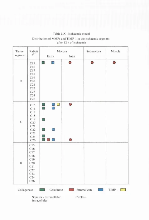
5 . X Distribution of MMPs and TIMP-1 in the ischaemic segment after 12 h of ischaemia
5 . XI Distribution of MMPs and TIMP-1 12 h after resection and anastomosis following ischaemia
5 . XII Distribution of MMPs and TIMP-1 24 h after resection and anastomosis following ischaemia
List o f F ig u res
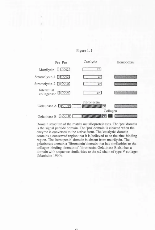
1 . 1 Domain structure of the matrix metalloproteinases
4 . 1 Diagram of blood supply to distal colon in rabbit showing location of transection and anastomosis
4 . 2 . a Tissue segnients taken for immunohistochemistry and H&E in the normal healing model
4 . 2. b Tissue segments taken for immunohistochemistry and H&E in the obstruction + anastomosis group
4 . 3 . a Diagram of distal colon in rabbit showing site of ring and resection used in obstruction model
4 . 3 . b Figure showing the ring in place
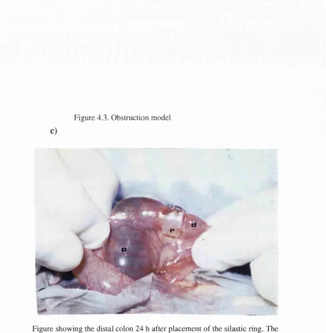
4 . 3 . c Figure showing the obstructed bowel 24 h after placement of the ring 4 . 3 . d Photograph showing the resected segment containing the ring
4 . 3 . e The same segment after remove the ring and opening the bowel 4 . 3 . f Photograph showing an anastomosis 24 h after resection of the ring
in the obstruction model
4 . 4 . a Tissue segments taken for immunohistochemistry and H&E in the obstruction only group
4 . 4 . b Tissue segments taken for immunohistochemistry and H&E in the ischaemic model
4 . 5 . a Photograph showing the normal blood supply to the distal colon 4 . 5 . b Aspect of the distal bowel after 2 h of ischaemia
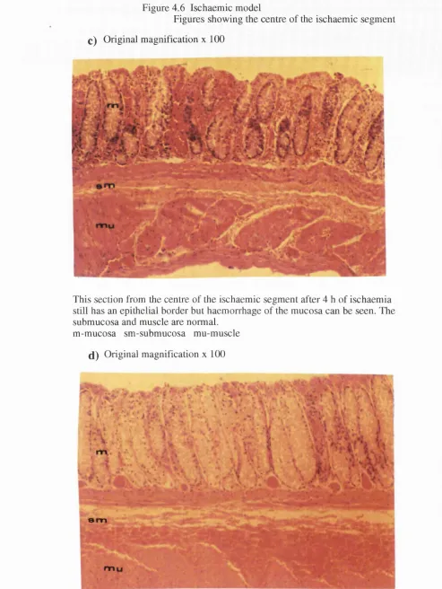
4 . 5 . c View of the distal colon after 4 h of ischaemia 4 . 5 . d Photograph of the distal colon after 6 h of ischaemia 4 . 6 . a Histology of the bowel after 2 h of ischaemia
4 . 6. b H&E staining of the ischaemic segment 4 h after tying the vessels 4 . 6. c Section of the centre of the ischaemic segment 4 h after ischaemia 4 . 6 . d Section showing the tissue damage after 6 h of ischaemia
4 . 7 View of the necrotic segment after 12 h of ischaemia
4 . 8 . a,b Two H&E staining of the necrotic bowel 12 h after ischaemia 4 . 8 .c,d Details of fig 4 .8 . b showing the muscle layer
4 . 9 . c,d Sections of the distal ischaemic segment showing in c the region near the distal border and in d the region near the centre
4 . 1 0 Diagram showing the blood vessels tied to produce an ischaemic segment in the distal colon
5 . 1 Section showing the rabbit normal distal colon at the site of anastomosis 5 . 2 . a H&E staining of the anastomotic segment 12 h after anastomosis in the
normal healing model
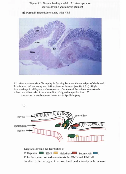
5 . 2 . b Diagram showing the distribution of MMPs and TIMP-1 12 h after transection and anastomosis
5 . 2 . c,d Details of figure 5 . 2 . a showing inflammatory cell infiltration 5 . 2 . e,f Immunolocalisation of collagenase in the mucosa and muscle of the
anastomotic segment 12 h after transection and anastomosis
5 . 2 . g,h Sections at the same region of figure 5 . 2 . e incubated with antiserum to gelatinase and stromelysin
5 . 3 . a H&E stained section showing the anastomotic segment 24 h after transection and anastomosis
5 . 3 . b Diagram showing the distribution of MMPs and TIMP-1 24 h after anastomosis in the normal healing model
5 . 4 . a H&E stained section showing the anastomotic segment 3 days after transection and anastomosis
5 . 4 . b Diagram showing the distribution of MMPs and TIMP-1 3 days after anastomosis in the normal healing model
5 . 5 . a H&E stained section showing the anastomotic segment 7 days after transection and anastomosis
5 . 5 . b Diagram showing the distribution of MMPs and TIMP-1 7 days after anastomosis in the normal healing model
5 . 5 . c,d Details of figure 5 . 5 . a showing inflammatory cell infiltration in the fibrous tissue
5 . 5 . e,f Sections of the fibrous tissue at the anastomotic segment incubated with antiserum to gelatinase and stromelysin demonstrating intracellular enzymes
5 . 5 . g Section of the mucosa at the same segment of figure 5 . 5 . e incubated with antisemm to stromelysin showing extracellular secretion
5 . 6 . a H&E stained section showing the anastomotic segment 12 h after resection of the segment containing the ring and anastomosis formation 5 . 6 . b Diagram showing the distribution of MMPs and TIMP-1 in the
5 . 6 . c,d Details of figure 5 . 6 . a showing inflammatory cell infiltration 5 . 6 . e,f Section of the mucosa of the segment of the figure 5 . 6 . a incubated
with antiserum to collagenase and TIMP-1
5 . 6 . g,h Section of the mucosa of the segment of the figure 5 . 6 . a incubated with antisemm to gelatinase and stromelysin
5 . 6 . i j Section of the segment of figures 5 . 6 . a in the submucosa stained with antisemm which recognise Mr 95000 form of gelatinase 5 . 6 . k Section of the segment of figures 5 . 6 . a in the submucosa stained
with antisemm which recognise Mr 72000 and Mr 95000 forms of gelatinase
5 . 7 . a,b Section show typical histological abnormalities 12 h after resection of the ring and anastomosis formation
5 . 8 . a H&E stained section showing the anastomotic segment 24 h after resection of the segment containing the ring and anastomosis formation 5 . 8 . b Diagram showing the distribution of MMPs and TIMP-1 in the
anastomotic segment of figure 5 . 8 . a
5 . 8 . c Detail of figure 5 . 8 . a showing stitches surrounded by large number of inflammatory cells
5 . 9 . a H&E stained section showing the anastomotic segment 3 days after resection of the segment containing the ring and anastomosis formation 5 . 9 . b Diagram showing the distribution of MMPs and TIMP-1 in the
anastomotic segment of figure 5 . 9 . a
5 . 9 . c,d Section of the mucosa of the segment of the figure 5 . 9 . a incubated with antiserum to collagenase and TIMP-1
5 . 9 . e,f Section of the mucosa of the segment of the figure 5 . 9 . a incubated with antisemm to gelatinase and stromelysin
5 . 10 . a H&E stained section showing the anastomotic segment 7 days after resection of the segment containing the ring and anastomosis formation 5 , 10 . b Diagram showing the distribution of MMPs and TIMP-1 in the
anastomotic segment of figure 5 . 10 . a
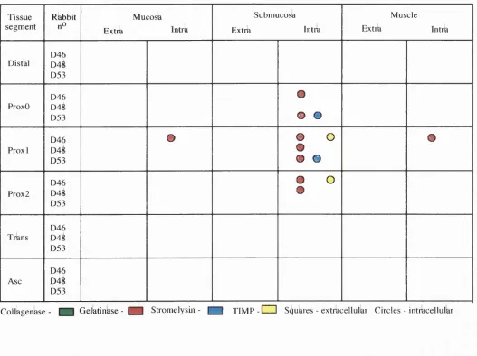
5 . 11 . a Section of ProxO segment after 24 h of obstmction stained with H&E 5 . 11 . b Diagram showing distribution of MMPs and TIMP-1 in the segment of
figure 5. 11 . a
5 . 11 . c,d Details of figure 5 . 11. a showing extensive mucosal damage and inflammatory cell infiltration
5 . 11 . e Section of the segment of figures 5 . 1 1 . a,c,d in the submucosa stained with antisemm which recognise Mr 72000 and Mr 95000 forms of gelatinase
5 . 12 . b Diagram showing distribution of MMPs and TIMP-1 in the segment of figure 5. 12 . a
5 . 12 . c,d Details of figure 5 .1 2 . a showing extensive mucosal damage and inflammatory cell infiltration
5 . 13 . a Section of the mucosa of ProxO segment 12 h after anastomosis formed after 12 h of ischaemia incubated with antiserum to stromelysin
5 . 13 . b Section of the distal segment of the same animal of figure 5 .1 3 . a incubated with antiserum to TIMP-1
5 . 14 . a H&E stained section showing the anastomotic segment 24 h after resection of the ischaemic segment and anastomosis formation 5 . 14 . b Diagram showing the distribution of MMPs and TIMP-1 in the
anastomotic segment of figure 5 . 14. a
5 . 15 . a H&E stained section showing the anastomotic segment 3 days after resection of the ischaemic segment and anastomosis formation 5 . 15 . b Diagram showing the distribution of MMPs and TIMP-1 in the
A bbreviations  - ângstrôm
BAR - biofragmentable anastomosis ring BM - basement membrane
C - centigrade cm - centimetre
CNP30- 1,1,1 -trichloroethane and 1,2-epoxypropane CO2 - carbon dioxide
DPX - mounting medium ECM - extracellular matrix
ES AF - endothelial cell stimulating angiogenic factor F(ab) - monovalent antibody fragment
Fig - figure
FTTC - fluorescein isothiocyanate g -gram
Gly - glycine h - hours
H&E - haematoxylin and eosin
HEPES - 4-(2-hydroxyethyl)-piperazine-ethane sulphonic acid IgG - immunoglobulin G
mmHg - millimetres of mercury MMP - matrix metalloproteinase Mr - molecular weight
mRNA - messenger ribonucleic acid |iM - micromolar
nm - nanometre
NSS - normal sheep serum PBS - phosphate buffered saline PFU - perfusion units
PMN - polymorphonuclear leukocyte
SDS-PAGE - sodium dodecyl sulphate polyacrylamide gel electrophoresis p0 2 - oxygen tension
RNA - ribonucleic acid
TIMP - tissue inhibitor of metalloproteinases U.K. - United Kingdom
A C K N O W L E D G E M E N T S
This thesis would not have been possible without the help of several members of the Department of Surgery, University College London Medical School.
I am sincerely grateful to Mr Paul Boulos, Reader in Surgery, who enrolled me into his research programme, for his guidance and support and for supervising the project.
A very special acknowledgement to Dr Felicity Savage who devoted considerable time to teaching me the scientific principles and techniques, for her constant help and advice and for her tolerance in supervising my laboratory work.
I thank Professor Michael Hobsley and Professor Irving Taylor for their constant interest in my project.
I am indebted to Dr J.J. Reynolds and Dr R.M. Hembry of Strangeways Research Laboratory, Cambridge for supplying the antisera, crucial for this work, and for availing us of their expertise whenever needed.
My sincere thanks to Dr Virginia Sams, Senior Lecturer in the Department of Histopathology who developed my skills in histopathology and advised on the histopathology of the models investigated in this thesis.
I am thankful to Marilena Loizidou for her constructive criticism, to Daniel Anthony who instructed me in the use of computers and to Abdul Gafur for his friendship and help with the photographs.
STA TEM EN T O F O R IG IN A L IT Y
The studies presented in this thesis were designed and personally carried out by the author. There has been no similar study on metalloproteinases in experimental colonic healing.
CHAPTER 1
INTRODUCTION
The first account of a bowel wound, with subsequent death, appeared in the Bible (Judges 3: 16-25).
... and the fat closed over the blade, so that he could not draw the dagger out of his belly; and the dirt came o u t.... And behold their lord was fallen down dead on the earth.
1 . 1 Historical development of colonic anastomosis
Since ancient times spontaneous recovery from bowel wounds has infrequently been described probably because in rare cases peritoneal adhesion to the injured intestine prevents the leakage of contents into the peritoneal cavity. The first surgeon to advocate intestinal suturing was Celsus in his book "De medicina" edited in ancient Rome (Spencer WG, English translation, 1938). Celsus believed that if the wound was in the small bowel it could be left but if it was in the large bowel it should be sutured because spontaneous healing was unlikely to occur. Reviews by Kerr (1923) and Wangensteen (1978) have covered early accounts of the evolution of intestinal suturing. In 1396 Lanfranck suggested suturing the viscera into the parietal peritoneum to encourage fistula formation into the skin surface. The earliest report of bowel excision and suturing was from Ramdohr in 1727. He excised two feet of gangrenous small bowel with its mesentery, then invaginated the proximal end of the bowel into the lumen of the distal segment and fixed it with a few sutures. The patient survived. Duverger in 1747 carried out the same procedure but rejoined the ends with a few sutures over a piece of animal trachea, before placing the bowel in the peritoneal cavity. The trachea was expelled in the stool on the twenty-first postoperative day. In 1812 Travers reported his experiments in dogs and horses showing that an intestinal wound could be repaired without the support of the parietal peritoneum provided that in suturing the bowel the visceral peritoneum was tightly approximated sealing the lumen completely before returning the gut into the abdomen. The Russian surgeon, Pirogoff, also employed intestinal suturing successfully during the Crimean war in 1856.
catgut or interrupted silk, to allow good apposition of the cut edges and to secure haemostasis. The second layer was an outer seromuscular or a Lembert stitch, often interrupted and of non-absorbable material to produce inversion and bring the peritoneal coats together. Halsted (1887) was the first to carry out a systematic study of intestinal repair with experiments in dogs demonstrating that the submucosa was the most important layer of the bowel in determining the anastomotic strength. The work of Halsted was based on the monograph "Experimental and clinical enquiry into the nature and treatment of wounds of the intestine" by Samuel D. Gross in 1843, who pointed out that the submucosa is the strongest coat of the intestine. Halsted showed that it is impossible to suture the serosa alone without piercing the submucosa and that stitches which penetrate the lumen are undesirable and are apt to be associated with adhesions. In spite of advocating the use of a submucosal inverting suture, Halsted drew attention to the dangers of folding-in an unnecessarily excessive intestinal wall because this increases the tissue bulk undergoing sloughing thus augmenting the risk of infection. Also tissue inverted into the intestinal lumen could obstruct the passage of faeces increasing the tension on the sutures (Halsted 1887). Gambee (1951) also counselled against excessive inversion and described a single layer suture that allowed a secured apposition of the cut edges of the bowel with minimal inversion.
Despite the work carried out in the nineteenth century showing the advantages of inverted anastomoses, Getzen in 1966 presented experimental evidence favouring everted anastomoses and these findings were confirmed by Hamilton 1967; Ravitch et al 1967; Healey et al 1967; Loeb 1967 and Buyers and Meier 1968. However other experimental work from Canalis and Ravitch 1968; Hargreaves and Keddie 1968; Mellish et al 1968; Singleton et al 1968; Gill et al 1969; McAdam et al 1969; Orr 1969 and Rusca et al 1969 supported an inverted suture technique. Trueblood et al (1969) using an experimental model of anastomosis in rats provided definitive evidence that the inversion suture, when properly performed, provides greater tensile strength, a diminished tendency to leak and far less peritoneal adhesions than the everted one. His histological studies showed that with the inverted technique the healing is more rapid with earlier restoration of mucosal continuity than with an everted suture. Goligher et al (1970b) instituted a controlled clinical trial of everting versus inverting suture in large bowel surgery that had to be terminated prematurely because inverted, were significantly better than everted suturing. In this trial a higher proportion of patients with the everted technique developed wound infection or peritonitis and faecal fistulation was much more frequent. The principles of Travers and Lembert (apposition of cut edges and serosa) and Halsted (inverting sutures piercing the submucosa) were then re-established and are still valid.
1 . 1 . 1 Sutures
colonie anastomoses has expanded since the 1950s when many new suture materials became available and these will be discussed further in chapter 1 . 6.
1 . 1 . 2 Surgical techniques
Although the basic principles of intestinal anastomosis are undisputed there remains the problem of how to achieve the best result. Should the anastomosis be of one or two layers of sutures, a continuous or an interrupted stitch and which type of suture material should be used? The conventional two-layer anastomosis uses an inner full thickness continuous layer of absorbable sutures and an outer seromuscular layer of interrupted non absorbable sutures. The one-layer anastomosis is usually performed with interrupted sutures inserted through the serosal surface and brought out in the submucosal layer at the cut edge rather than through the full thickness of the bowel wall. A good approximation of the serosa, the muscle and submucosal layers with a minimum inversion of the mucosa, not included in the suture, is obtained by insertion of sutures very near the cut edge, resulting in a smooth anastomosis without occlusion of the lumen. A two-layer inverting anastomosis allows a good apposition of the cut edges of the bowel but reduces mucosal blood flow (Chung, 1987) and results in a larger degree of stenosis than the single layer technique (Sacko and Wangensteen, 1951, McAdams et al, 1970, Irvin and Edwards, 1973). Koruda and Rolandeli (1990) reviewing experimental studies on the healing of colonic anastomoses concluded that single layer closure in colonic anastomoses will allow improved healing provided that the amount of tissue inverted by the suture line is minimised to avert compromising the mucosal blood flow and the luminal diameter irrespective of the suture material and the suturing method used.
considerable risk of leakage and infection. Therefore in addition to attention to the suturing technique that allowed adequate healing without leakage, means of reducing the risk of faecal contamination were sought.
Wangensteen (1978) records Ivlurphy in 1892 as the surgeon who introduced a small circular metal button with two parts, the first ingenious device to avoid contamination of the peritoneum with an expedient anastomoses. Each part was inserted into a segment of the two hollow viscera to be approximated and fixed with a purse-string suture of catgut. The two buttons, male and female, were then pushed home: a spring held them firmly in position. A continuous over-and-over catgut suture or interrupted silk suture sufficed to terminate the operation. The button passed spontaneously distally into the intestine and was readily evacuated per rectum. The Murphy Button was widely used at the beginning of this century (Halsted 1922) and has recently been revived as the biofragmental anastomosis ring (Hardy 1985).
Lahey in 1932, Lloyd-Davies in 1945 broadened the scope of the operation for extended resections and for resecting tumours in the fixed segments of the colon after adequate mobilisation.
Another method that was developed to avoid the risk of infection after colonic anastomosis was the "aseptic" anastomosis. This attempted to resect and suture, delaying opening the bowel lumen until the anastomosis was almost complete. Usually two crushing clamps were used and the bowel divided between them. The two clamped bowel ends were opposed and turning the clamps away and towards the suture line, the anterior and posterior rows of Lembert sutures were inserted and tied. The clamps were then opened slightly and withdrawn and the defect in the row of seromuscular suture where the forceps had been placed was closed by placing few additional stitches. Palpating the anastomosis between finger and thumb separated the crushed edges and re-established the lumen at the site of the anastomosis. The technique reduced considerably the amount of soiling during the operation and was used by a large number of surgeons from the end of the 19th century to the first half of the 20th century (Halsted 1898, O'Hara 1900, Parker and Kerr 1908, Schoemaker 1921, Fraser and Dott 1924, Pringle 1924, Rankin 1928 and Wangensteen 1940).
After World War II advances in general anaesthesia, a better understanding of fluid and electrolyte balance and the use of blood transfusion offered the surgeon much better operating conditions. This allowed a more careful and meticulous operative technique when performing an anastomosis with minimal contamination. The introduction of sulphonamides and antibiotics in the 1940s had a considerable impact on colorectal surgery as they could be used both for systemic therapy and for sterilising the colon. The possibility of intestinal antisepsis and treatment of peritoneal infection encouraged surgeons to proceed with immediate resection and intraperitoneal anastomosis avoiding a colostomy and a delayed anastomoses. This approach has become the practice when it has been shown to be as safe as the staged procedures (Lloyd-Davies 1953).
1.2 Wound healing
-j
The healing process has been much more extensively studied in skin and fascia than in other less homogenous tissues, such as the colon, however it is generally assumed that the process is the same in all soft tissues (Herrmann et al 1964). Essentially wounds of soft tissues heal by a process of connective tissue repair and the formation of a fibrous scar. Two types of repair are seen in soft tissue wounds. Primary wound healing occurs in incised wounds when the edges of the wound are apposed, and this is the type of repair encountered in the majority of surgical wounds. Secondary wound healing occurs in wounds associated with tissue loss or when the wound edges are widely separated. This method of repair is mainly found in contaminated and burn wounds. Although the basic features are the same in bowel anastomoses, healing in the colon is disturbed by the large numbers of bacteria and bulky faeces in the bowel lumen and by a rapid turnover of collagen following injury when as much as 40% of normal colonic collagen in the anastomotic region is lost by the third day (Cronin et al 1968). These factors are at least partially responsible for an appreciable morbidity and mortality due to failure of the healing process following large bowel anastomosis.
1 . 2 . 1 Description of colonic healing
Histological observations show that three phases can be identified in intestinal healing which correlate with biochemical changes associated with wound healing (Hermann et al 1964):
Phase I - 0 to 4 days. Lag phase - inflammation and oedema.
The second phase of healing, proliferative fibrous repair, begins between the third and fifth day. Oedema and inflammation decrease, fibroblasts stimulated to migrate by macrophage products gradually predominate in the fibrin plug and synthesise proteoglycans and new collagen to form the fibrous tissue, initially an oedematous granulation tissue composed of new capillaries, fibroblasts, macrophages, mast cells, inflammatory cells and cellular debris. New vessels become canalised developing into arterioles or venules. Collagen production increases, in the mid period of the second phase, initially only reticulin fibres are demonstrable but these are quickly followed by collagen fibrils that are responsible for the intrinsic mechanical strength of the suture line. Low cuboidal mucosal epithelial proliferation over the surface of the invaginated edge and extensive mitoses of proliferating smooth muscle cells are seen at the end of the second phase. The appearance of the anastomosis at this time varies with the extent of sloughing of the invaginated mucosal margin which is dependent on the suture technique. With interrupted sutures close to the cut edges of the bowel there is little inversion of the margins, less necrosis followed by an orderly restorative process with minimal formation of granulation tissue. Excessive sloughing of the inverted edges results in the production of exuberant granulation tissue and healing by secondary intention. Waninger et al (1992) studied the influence of distance and tension of sutures on the healing of experimental colonic anastomoses in rats and found that a wide distance between the interrupted sutures was followed by inadequate apposition due to prolapse of the mucosa between the sutures. Comparing wide and narrow distance with three different tensions he demonstrated that best results were achieved with a narrow distance between sutures combined with a moderate suture tension.
cells or foreign body giant cells usually around buried suture material. The end result is
a dense and relatively avascular collagenous scar (Irvin 1984). From 17 to 21 days a
hyperplastic mucosa overlies the previous defect and there is a proliferation of smooth
muscle cells and collagen fibres at the suture line and the muscularis mucosa begins to
separate from the proliferating muscle mass. F rom three weeks to one year,
remodelling results in a thinning of the bowel wall as gradually the layers re-orientate.
However the muscularis mucosa remains incompletely formed at one year frequently
still indistinguishable from the muscularis propria so that the submucosa is not clearly
demarcated.
1 . 2 . 2 Biochemical aspects of wound healing
Several chemical mediators are involved in the vascular inflammatory response in
the early phase of wound healing. The vasodilatation that follows injury is caused by
the release of histamine by platelets and mast cells. The action of histamine is quite
brief and the maintenance of capillary engorgement and increased vascular permeability
which follows is mediated by serotonin, bradykinin, kallidin and prostaglandins.
Serotonin is released by platelets and mast cells. Bradykinin and kallidin are peptides
released from plasma globulins by the enzyme kallikrein. Several prostaglandins are
involved in the initial phase. These substances are derived from the tissue and from
neutrophilic leukocytes and cause vasodilatation and increase the permeability of
capillaries. They may also regulate the duration of wound inflammation (Irvin 1984).
A ctiv a tio n o f p la te le ts re le a s e s p la te le t- d e r iv e d g ro w th facto rs and
ch em o attracta n ts for fib ro b last and v a s c u la r en d o th e lia l cells. F ibrinogen
polymerisation and fibrin lysis releases chem oattractant fibrinopeptides. Blood and
tissue com plement factors are released which, in conjunction with fibrinopeptides,
attract inflam m atory cells, which in turn release their own growth factors and
chemoattractants. Local cells proliferate and move towards the source of the signals.
Leukocytes and macrophages accumulate, the former rapidly and the latter more slowly (Hunt 1984).
Cytokines, notably interleukin-1, are released by macrophages and have far- reaching effects on connective tissue formation by encouraging proliferation of fibroblasts and on increasing collagen and fibronectin synthesis. Interleukin- 1 acts also to promote connective tissue destruction by increasing collagenase, neutral protease and prostaglandin 2% production (Dayer 1987, Pasternak et al 1986).
1.3 Colonic anastomotic dehiscence
1 . 3 . 1 Clinical studies
having a staged resection than in those having a single operation for diverticular disease which he believed was related to a more severe disease and a technically more demanding procedure in those patients requiring a defunctioning colostomy. Phillips et al (1985) reviewing 713 patients with malignant large bowel obstruction observed a high clinical leak rate (18%) with primary anastomosis in the left colon compared to elective surgery (6%).
Since Dixon (1939) advocated anterior resection many controversial reports about the incidence of dehiscence of the suture-line in anterior resection have appeared in the medical literature, some reporting a low incidence but the majority of surgeons agree that there is a substantial risk (Mayo et al 1951; Goligher 1951, 1958, 1962; Goligher et al 1965, Muir 1958; Cullen and Mayo 1963; Whitaker 1968 ). Goligher et al (1970a) in what is now a classic paper evaluated the occurrence of dehiscence in 73 patients who underwent anterior resection for carcinoma or for benign tumour of the rectum or rectosigmoid and for diverticular disease. They employed not only the usual diagnostic criteria based on the clinical course of the patient, septic complications, digital and sigmoidoscopy examination, but introduced the use of contrast radiology to assess anastomotic integrity during the immediate post-operative period. Radiology revealed suture-line leaks that were not clinically apparent and the overall incidence of leakages recorded were 40% and 69% in high and low resection respectively, with 51% for the entire series. They stressed the high frequency of dehiscence and its adverse influence on the post operative course. A 2.7% mortality was attributed to this particular complication and the mean period in hospital after high anterior resection was increased from 14 days in patients whose anastomoses remained intact to 35 days in those whose anastomoses broke down.
there was one death (1.02%) and one pelvic abscess. They advocated a proximal decompression for low anastomosis since routine use of post-operative contrast enema prior to colostomy closure showed 8 asymptomatic anastomotic defects. They believed that some of the defects at suture lines were related to accumulation of blood and serum around an anastomosis which drained through the suture line into the lumen of the colon. To avoid this they advised draining the pelvis long enough to allow liquefaction of the clot, leaving the drain in place until the danger of leak has passed. Debas and Thomson (1972) in a critical review of colectomy in 838 patients reported an 8% anastomotic dehiscence which was responsible for a third of all the deaths in the series, mainly following emergency operation. Surprisingly they found a higher mortality in right sided emergency resections than in resections of the left colon. This was attributed to the application of a one stage resection in those patients with established peritonitis at the time of right hemicolectomy whereas left side emergency resections were managed by staged procedures. Morgenstern et al (1972) reviewing 301 cases, comprising 178 low anterior resections performed for carcinoma and 123 segmental sigmoid resections for diverticulitis, documented 70 cases of leakage (23%) with 8
muscular hypertrophy and even micro-abscess formation at the resection margin. These findings were present not only in diverticulitis but also in the vicinity of a necrosing invasive carcinoma.
Schrock (1973) in a comprehensive review of 1703 patients with colonic anastomosis over a twenty year period found 4.5% had clinical evidence of suture line disruption. There was a 33% mortality associated with dehiscence, compared to 2.6% when the anastomosis was intact. The incidence of leakage in left colorectal anastomoses was 9.2% and in low rectal anastomoses 10.4%; this was attributed to poor blood supply and exclusion from the peritoneal cavity. Even in anastomoses carried out in ideal circumstances the incidence of low anastomotic leakage was still 1 0%
compared to 1.7% in other colonic anastomosis. Factors which contributed to an increased risk of dehiscence were old age, anaemia, radiation therapy, carcinoma at the margins, infection, intra-operative hypotension, operations lasting more than 5 hours and massive haemorrhage, the latter was associated with a particularly high rate (31.8%) of anastomotic dehiscence. Elective resection of the left colon for diverticular disease resulted in only 0.6% leakage in the absence of infection but if the operation was carried out as an emergency there was disruption in more than 1 0% of cases irrespective of the associated sepsis.
factors, probably mitigated by the use of prophylactic broad-spectrum antibiotics and peritoneal lavage with antibiotic solutions.
Fielding et al (1980) reported a multicentre study of 1466 anastomoses in patients with colorectal cancer. The overall clinical dehiscence rate was 13%; 10.8% in intraperitoneal anastomosis and 18.7% in anterior resection of the rectum and the highest incidence was found in advanced tumours with métastasés. The clinical seriousness of anastomotic dehiscence was that the duration of hospital stay was doubled and there was a threefold increase in post-operative mortality when this complication occurred. The most important single factor influencing anastomotic integrity was the surgeon who performed the operation, the range of dehiscence for individual surgeons varying from 5 to 30%.
Recent evaluation has been directed towards the use of stapling devices, usually in low colorectal or coloanal anastomosis. Smith (1981) in a survey of the American Society of Colon and Rectal Surgeons reported that 10% of patients with circular stapled anastomosis had a clinical leak. Fazio (1988) used the circular stapler to perform low and high colorectal anastomosis and compared the leak rates using clinical and radiological criteria. He found of 67 patients having a high colorectal anastomosis rates of dehiscence were 1.5% and 0%, for radiological and clinical leaks respectively; however in 95 patients with a low colorectal anastomosis the rates increased to 8.4% and 5.3%, respectively. The use of staplers has decreased the incidence of low colorectal anastomotic dehiscence but has not abolished it.
randomised multicentre trial of 782 patients reported 3% anastomotic leak rate in sutured, 4% in stapled and 3% in BAR anastomosis however low colorectal or emergency anastomosis were not investigated.
As emphasised by Irvin and Goligher (1973) faecal loading is an important cause of leakage in colonic anastomosis and is a contributing factor in emergency colonic surgery. To obviate this problem surgeons since the end of the last century have used a three staged procedure, a defunctioning colostomy first, followed by bowel resection in the second stage and colostomy closure in the final stage (Devine 1931). In most instances the diseased segment of the colon was removed at the initial operation followed by delayed restoration of the bowel continuity. This two stage procedure is preferred because primary resection eliminates continued contamination of the peritoneal cavity when treating an inflammatory disorder such as acute diverticulitis and achieves more timely removal of the tumour when dealing with a neoplastic process. The efficacy of these diverting operations is questionable because they still carry a certain morbidity and mortality. A variety of technical modifications have been proposed to address some of the potential hazards associated with resection and primary anastomosis of the distal colon in emergency operations when preparation of the colon is not possible. Muir (1968) described a technique of intraoperative lavage of the unprepared colon to facilitate primary anastomosis and a recent report from Murray (1991) showed no anastomotic leakage in 31 patients after emergency left colectomies in the Lahey Clinic with intraoperative lavage of the unprepared bowel followed by primary anastomosis. Ravo and Ger (1984) introduced a technique for protecting colonic anastomoses using an intracolonic bypass tube and Rosati et al (1992) reported only one anastomotic leak using this technique in 29 emergency colonic resections.
radiological leak rate in 100 patients was 22% for the aprotinin group and 35% for the placebo group, and the overall clinical leak rate was 8% and 15% respectively. They concluded that this reduction in anastomotic failure rate could be of clinical relevance and deserved further studies.
Sheridan et al (1989) in a double-blind, multicentre, prospective trial have also explored the potential value of aprotinin in 216 patients. There was a marked reduction in the overall anastomotic leak rate with the use of aprotinin, in patients undergoing anterior resection who received aprotinin, when compared to those receiving placebo, but this did not reach statistical significance. However in the patients undergoing left hemicolectomy or sigmoid colectomy higher leakage rates, both clinical and radiological, were higher in patients treated with aprotinin. They believed that although aprotinin could not be recommended for general application in colonic surgery, its use might be of value in improving anastomotic healing in patients undergoing anterior resection.
These were disappointing attempts to manipulate the enzymatic system to improve colonic healing and reflects on a simplistic understanding of a far reaching complex mechanism.
1 . 3 . 2 Experimental studies
external factors, like anti-inflammatory agents, infection, bowel preparation, nutrition and blood transfusions.
As early as 1812 experimental work carried out by Travers in horses demonstrated the importance of sealing the anastomosis with the serosa to avoid leakage. The first systematic research of colonic anastomosis was by Halsted in dogs who demonstrated that the strength of the bowel wall is reliant on the submucosal connective tissue (Halsted 1887). As the strength of this submucosal tissue depends upon the quantity and quality of the collagen it contains (Hawley et al 1970b), further studies were driven in this direction. Mechanical strength (the resistance of the anastomosis to rupture) and collagen concentration were taken as the main parameters fox studying collagen
Mechanical strength was measured by the longitudinal breaking strength or the tension at which the anastomosis disrupts when the two ends of the anastomotic segment are pulled apart or by determining the resistance of the anastomosis to intraluminal pressure. The pressure at which leakage or rupture occurs is called the bursting strength or bursting pressure.
Other methods used to assess the quality of an anastomosis in experimental situations were local blood flow at the anastomosis (Leaper 1983), anastomotic integrity with contrast radiography (Bubrick 1981) and histological evaluation
(Herrmann et al 1964, Daly et al 1972, Trimpi et al 1977).
These changes in collagen content correlating with a decrease and subsequent rise in the bursting pressure of the anastomosis suggests that the mechanical strength of the wall is dependant on its collagen content.
Similar conclusions were reached by Hawley ct al (1970b) working in rabbits in the same laboratory. They found a 40 per cent decrease in the bursting pressure on the third post-operative day and by the seventh day the bursting wall pressure had risen to a mean value similar to that of the unoperated colon. The fall in the bursting strength on the third operative day was associated with a fall in the hydroxyproline content of the colon of 27 per cent. By the seventh post-operative day the total collagen content was restored to a mean value similar to the normal colon.
Hawley (1970b) studied the collagenolytic activity of viable explants of colon and other segments of the gastrointestinal tract by measuring the lysis of collagen. The explants were placed in contact with a collagen gel, incubated for 72 hours and the area of lysis determined. He demonstrated that the caecum and the small intestine produce less lysis than the colon and most explants from the stomach failed to produce any. Following colonic anastomosis the collagenolytic activity was enhanced not only in the colon but also in all the other segments of the gastrointestinal tract, including the stomach. These variations in activity in the gastrointestinal tract appeared to correlate with the risk of anastomotic dehiscense at different sites in normal clinical practice, the incidence of anastomotic dehiscense being higher in the distal colon than in the proximal one or the small bowel.
normal colon bursts before the anastomotic site partly because of the decreased distensibility of the suture line.
Irvin and Hunt (1974a) studied colonic anastomosis in rats measuring the bursting pressure, total collagen concentration and collagen metabolism with tritiated proline. They confirmed the findings of Cronin and Hawley demonstrating lysis of mature collagen in the early phase of colonic healing and active collagen synthesis in the first few days following anastomosis, however the change in colonic collagen was of smaller magnitude probably because they used a different technique, measuring collagen content in anastomosis dried to constant weight in an oven. The measurements of bursting pressure also showed a different picture as they found that the early phase of healing was not characterised by a loss of tensile strength and the anastomoses on the third postoperative day had gained a significant degree of tensile strength compared to measurements made on the day of the anastomoses. These differences in the results may be due to the measurements being made of total collagen, and tensile strength and bursting pressure being compared not to the normal colon, as were Cronin's, but with the measurements made on the day of the anastomosis.
Van der Stappen et al (1992) measured a higher collagenolytic activity in extracts from first post-operative day anastomotic segments compared to extracts from uninjured intestine of the rat. Anastomosis in the large bowel had higher collagenolytic activity than anastomosis in the small bowel. Analysis, by SDS-polyacrylamide gel electrophoresis, of the reaction products of the degradation of fibrillar type I collagen revealed the presence of many fragments, amongst them ones characteristic of mammalian collagenase activity. This was evidence of enhanced degradation of collagen in the anastomotic area during the first post-operative day and an indirect implication of true mammalian collagenase.
anastomosis of ischaemic colon in the rat. Anastomotic healing in unprepared ischaemic rat colon was severely impaired with an 83 per cent dehiscence rate, but this was not observed in the colon prepared with enteral antibiotics. Dunphy (1970), based on experiments by Hawley (1969), stated that bacterial growth on the surface of the anastomosis promoted collagenolytic activity and accelerated breakdown of the anastomosis.
Other factors influencing collagen metabolism were studied by Jiborn et al (1978, 1980a,b) and Jonsson et al (1985) who in a series of experiments in rats demonstrated that collagen concentration decreased in the vicinity of anastomoses by 30 per cent on the first 4 days and this decrease was higher when continuous suture was used or when colonic dilatation occurred proximal to the anastomoses. In these circumstances the collagen concentration was even 45 to 55 per cent lower on the fourth post-operative day. They found a higher collagenolytic activity proximal to the anastomosis than distal to it and also activity was higher in the colon than in the small bowel. After the first 2 to 4 days a gradual restoration of collagen concentration occurred when synthesis compensated for lysis. Tornqvist et al (1988, 1990a, 1990b) in a series of experiments in the rat investigated the effect on collagen turnover of obstructing the colon. An obstruction in the distal colon was found to stimulate collagen synthesis not only at the obstructed site but also proximal to it. This resulted in an increase in collagen content proximal to the obstruction but not at the obstruction site indicating that there must also be an increase in collagen degradation here. Collagen metabolism following an anastomosis either alone or preceded by obstructing the colon and resection of the obstructed segment was also studied. No difference in collagen content was measured between the groups, however in the obstructed group collagen synthesis proximal to the anastomosis, but not at or distal to it, was twice as high implying that collagen lysis must also be occurring at a much greater rate in this region. The major difference between the groups was in the complication rate which was 2 per cent in the
animals. This study shows that an obstruction does have a profound effect on collagen dynamics with both synthesis and degradation being affected.
However the first direct demonstration of collagenase in the anastomotic site was made by Chowcat et al (1988) using an immunohistochemical method. They localised collagenase and TIMP-1 in the suture line of colonic anastomoses formed in rabbits after resection of a small segment of distal colon. Collagenase and TIMP-1 were confined to a narrow margin at the cut ends of the bowel wall. The enzyme appeared in the mucosa within 12 hours of operation and gradually progressed more deeply through the damaged edges, initiating collagen breakdown in the tissue prior to its infiltration by granulocytes. By 24 hours collagenase secretion was already beginning to wane and TIMP-1 appeared, suggesting tight control of collagenase activity. Three days after operation much less collagenase was localised and TIMP was seen in the serosal sealing layer probably to prevent protease degradation and maintain the anastomotic integrity. After 7 days both enzymes and inhibitor were active only in small areas where dying tissue remained to be destroyed and where remodelling was taking place. The enzyme performed a controlled process of breakdown of unwanted tissue and there was no evidence for excessive collagenase activity in the normal healing of a colonic anastomosis. In another study Chowcat et al (1990) measured colonic secretion of collagenase in tissue culture and further demonstrated that three days after anastomosis the colon secreted more collagenase than explants of one day postoperative tissue. This coincided with a decrease in collagen concentration in the colon wall measured as hydroxyproline.
seem to support these changes. Thus collagen is the most important component of the extracellular matrix and the mechanical strength depends on its content in the bowel wall. Increased collagenolysis has been implicated by indirect studies and recent directed measurements have demonstrated the role of collagenase.
Increased knowledge of the biochemistry of collagen degradation and of the components of the extracellular matrix in particular of the enzymes that specifically degrade these components, deserve a note and point to the gaps in our understanding of colonic healing.
1 .4 Extracellular matrix and basement membrane
The extracellular matrix (ECM) is the structurally stable material that lies under epithelia and surrounds connective tissue cells. The ECM, however, is not an inert supporting material, it interacts with the m atrix cells and influences cellular proliferation, shape, polarity and differentiation (Hay 1991). Four major classes of macromolecules form the basic extracellular matrix, collagen, proteoglycans, elastin and glycoproteins (e.g. fibronectin, laminin and chondronectin).
glycoproteins, laminin and fibronectin. The BM is thought to serve as a scaffold for organising and anchoring epithelia. In spite of having a composition similar to the ECM and "connecting" epithelia and muscle to the ECM, the BM is not a component of the connective tissue (Hay 1981).
Study of the ECM must consider its synthesis, composition, organisation and degradation. The most important component of the matrix is collagen.
1 . 4 . 1 Collagen
Collagen is the most abundant protein in animals constituting more than half of the total protein in adult organisms and is the principal component of the ECM (Linsenmayer 1991). The collagen molecule gives support and tensile strength to skin, bone, tendon and cornea of vertebrates and is the main structural component of many tissues, forming 72% of the dry weight of skin and 8 6% of Achilles tendon (Uitto et al
1986); it is mineralised to produce a rigid skeleton in bone. In the intestine, where it constitute 15% of the dry weight (Gottrup 1981), it forms a flexible supporting layer which allows expansion of the lumen and movement of the contents without loss of the integrity or the elasticity of the wall (Jackson 1980).
Collagen molecules are formed of a-chains of which 24 different ones are known resulting in at least 15 types of collagen. The types found in a tissue are related to its structure and function. Collagen is not only an important structural component but can also promote cell attachment and differentiation, is a chemoattractant for both macrophages and fibroblasts, an antigen in immunological processes (Linsenmayer
1 . 4 . 1 . i Molecular structure
Triple helical region - Collagen molecules are rod-like structures usually 3000
 long and 15  in diameter {Gross 1981). In the fibril forming collagens (types I, II, III, V and XI) more than 95% of the molecule is composed of a single triple helical domain that gives collagen its characteristic shape and properties. This region consists of three separate a-chains, each of which contains approximately 1 0 0 0 amino acids, twisted in the form of left-handed helix. These three chains are then wrapped around one another to produce the triple helical structure of the molecule (Linsenmayer 1991). The triple helical conformation is such that the peptide bonds linking adjacent amino acids of the chains are buried within the interior of the molecule, making it highly resistant to attack by general proteases such as pepsin. The only enzymes able to efficiently degrade the helical portion of native collagen molecules are the collagenases (Gross et al 1980).
allow transparency and, in skin, a more random pattern gives flexibility (Pinnell 1983). The collagen fibres of the submucosa in the small intestine form a complex mesh, are straight, lie parallel to the serosal surface and are oriented in different directions with a criss-cross pattern that changes with the movements of the intestine, the angle formed by the two arrays of collagen fibrils with the length of the gut increases to 60°-65° when the radial distension predominates, while it is reduced to about 30° when the longitudinal distension predominates. This arrangement gives strength to the wall, allowing it to be stretched by a considerable amount both along and across the longitudinal axes of the intestine (Gabella 1987).
Non-triple helical region - In addition to the triple helical region, collagen
molecules in the ECM also have short non-helical extension of approximately 20 peptides at the NH2- and COOH-terminal ends of each component alpha chain. These terminal extension peptides play important functional roles, for they represent the sites where hydroxylysine-derived intramolecular and intermolecular cross-links are formed when molecules are arranged in fibrils. As the cross-linking containing extension peptides are not in the triple-helical conformation they are susceptible to proteolytic degradation by general proteases under conditions in which the triple-helical body of the molecule is left intact (Linsenmayer 1991).
1 . 4 . 1 . ii Genetic types of collagen
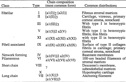
Table 1 . I. Collagens
Class
Chain composition
Type (most common form) Common distributions Fibrillar Fibril associated Network forming Filamentous Short chain Long chain I II ni V XI IX IV VI v n i
[al(I)]2 [a2(D] [al(II)]3
[al(in)]3
[al(5)]2 a2(V)
al(X I) a2(XI) a3(XI) al(IX) a2(IX) a3(IX)
[al(IV)]2 a2(rV) al(VI) a2(VI) a3(VI)
X
Vn [al(X)]3 [al(VII)]3
Fibrous stromal matrices Cartilage, vitreous, primary comeal stroma, notochord W ith type I in heteotypic fibrils
W ith type 1 in heterotypic fibrils; thin fibrils
with type II in heterotypic fibrils
Surface of type II collagen fibrils in cartilage; primary comeal stroma, notochord Basement membranes
1 0 0-nm beaded filaments of stromal matrices
molecule also includes the ratio in which the chains occur. Thus, the type II collagen molecule with its single type of chain is designated [ a 1(H) ] 3 and the type I collagen molecule with its two different chains is [ a l ( I)]2 a2(I). Collagens types are classified in six classes, determined either by the supramolecular forms in which they occur within extracellular matrices or by their molecular size as compared to the prototypical type I collagen, as seen in Table I.
The fibrillar collagens, types I, III and V, will be described in more detail because they are the most important ones in the study of wound healing. They are the major connective tissue collagens and are the proteins that provide the strength for the submucosal layer of the bowel. Type IV, the main component of basement membranes, will be also described.
1 . 4 . 1 . iii The fibrillar collagens
The fibrils of many tissues are heterotypic structures, composed of different combinations of fibril-forming collagens co-assembled with one another (Birk et al 1988, Linsenmayer 1991). The structural similarities of the fibrillar collagen molecules allow them to combine and this co-assembly of various collagens can determine certain properties of fibrils in different tissues, for example, fibril diameter (Birk et al 1990).
Junqueira 1982) and is also a major component of intestinal wall, chiefly in the submucosa (Orberg et al 1982, Fogdestam and Gottrup 1980). Type I molecules are 300 nm long and are composed of two a 1(1) chains and one a2(I) chain. Type II synthesised by chondroblasts is the major collagen in cartilage, having an intense interaction with proteoglycans, which is responsible for the resistance of cartilage to the intermittent action of pressure (Montes and Junqueira 1982). Type III has a tissue distribution similar to type I, except for bone, tendon and cornea where there are a very small amount. The use of specific antibodies to collagen suggests that type III is the principal component of the small argyrophilic collagen fibres that characterise reticular connective tissue, classically known "reticular fibres" of histologists (Linsenmayer 1991, Montes and Junqueira 1982). This type of collagen is usually found co assembled with collagen type I as in uterus, skin, arteries, lung, spleen, liver, kidney. It exists in many organs and is mainly, but not exclusively, related to smooth muscle cells. In intestine type III collagen is present in muscular layers and lamina propria. Collagen type III is produced by smooth muscle cells, fibroblasts, reticular cells of haemopoietic organs, liver cells and Schwann cells of the peripheral nervous system and it is always present in tissues and organs that need a mobile structural scaffolding, such as arteries, muscular layers of the intestine and uterus (Montes and Junqueira
1982).
cartilage (Van der Rest and Mayne 1988). Collagen type V may be involved in regulating fibril diameter, the greater the percentage of type V collagen in a heterotypic fibril, the smaller the diameter of fibril formed (Birk et al 1990). Type XI is the cartilage equivalent of type V.
Collagen type IV is synthesised in epithelial and endothelial cells and found in basement membranes. It has two main functions: the first as a structural support for epithelia and secondly for its filtering capacity in the renal glomerulus (Montes and Junqueira 1982). The helical region is much interrupted, so fibril formation does not take place and the molecule contains domains including collagenase sensitive triple helical ones, pepsin sensitive non-helical ones and ones that are resistant to both pepsin and collagenase (Linsenmayer 1991).
The fibril associated collagens type IX and XII can effect interactions between fibrils and between fibrils and other components of extracellular matrices (Anderson 1992). Type VI collagen has an anchoring function forming microfibrils linking major fibres (Keene et al 1988). Type VII forms anchoring fibrils to basement membranes (Lunstrum et al 1987); Type X is associated with calcifying cartilage (Thomas et al 1990) and collagens type VIII, XIII, XIV and XV are of unknown function (Anderson 1992).
1 . 4 . 1 . iv Collagen biosynthesis
transformed extracellularly to tropocollagen and incorporated into a stable cross-linked fibrillar structure (Montes and Junqueira 1982, Laurent 1987).
Collagen genes contain coding regions (exons) interrupted by non-coding intervening sequences (introns). The initial RNA transcripts of the genes are copies of both exons and introns. During processing of the RNA, splicing enzymes remove the intron sequences, producing mature mRNA molecules (Olsen 1991). The a-c h a in precursor (pre-pro-a) is synthesised on the rough endoplasmic reticulum and after several modifications by membrane bound enzymes (hydroxylation, glycosylation) and other enzymatic steps in the Golgi apparatus the pro-a-chains are completed and pass down the cisternae of the endoplasmic reticulum where three chains assemble to form a triple helix soluble procollagen (Olsen 1991).
Procollagen passes through the Golgi complex, is packaged for secretion and leaves the cell by exocytosis. In procollagen each chain has large additional extension- peptides, called propeptides, at both NH2 and COOH terminals (Tanzer et al 1974, 1976, Byers et al 1975). Prior to fibrillogenesis, at least two type-specific proteinases act: N- and C-terminal proteinases cleave their respective propeptides (Anderson 1992), leaving the long triple helix region with short non-helical ends characteristic of the collagen molecule, known at this stage as tropocollagen. Spontaneous self assembling into fibrils occurs due to the formation of a series of covalent bonds, creating the cross links which are responsible for the stability and strength of collagen (Montes and Junqueira 1982, Laurent 1987, Anderson 1992).