David L. Goldman,
Edward Nieves,
Antonio Nakouzi,
Johanna Rivera,
Ei Ei Phyu,
Than Htut Win,
Jacqueline M. Achkar,
a,dArturo Casadevall
a*
aDepartment of Microbiology and Immunology, Einstein College of Medicine, Bronx, New York, USA
bDepartment of Pediatrics, Einstein College of Medicine, Bronx, New York, USA
cDepartment of Biochemistry, Einstein College of Medicine, Bronx, New York, USA
dDepartment of Medicine at the Albert Einstein College of Medicine, Bronx, New York, USA
eThe Children’s Hospital at Montefiore, Bronx, New York, USA
ABSTRACT
Much of our understanding of the activity of anthrax toxin is based on
in vitro
systems, which delineate the interaction between
Bacillus anthracis
toxins
and the cell surface. However, these systems fail to account for the intimate
associa-tion of
B. anthracis
with the circulatory system, including the contribution of serum
proteins to the host response and processing of anthrax toxins. Using a variety of
immunological techniques to inhibit serum processing of
B. anthracis
protective
anti-gen (PA) along with mass spectrometry analysis, we demonstrate that serum digests
PA via 2 distinct reactions. In the first reaction, serum cleaves PA
83into 2 fragments
to produce PA
63and PA
20fragments, similarly to that observed following furin
di-gestion. This is followed by carboxypeptidase-mediated removal of the
carboxy-terminal arginine and lysines from PA
20.
IMPORTANCE
Our findings identify a serum-mediated modification of PA
20that has
not been previously described. These observations further imply that the processing
of PA is more complex than currently thought. Additional study is needed to define
the contribution of serum processing of PA to the host response and individual
sus-ceptibility to anthrax.
KEYWORDS
anthrax, proteases, toxin
B
acillus anthracis
is the causative agent of anthrax and is widely recognized for its
potential use as an agent of bioterrorism.
B. anthracis
secretes 2 bipartite toxins, the
lethal toxin and the edema toxin, that are essential for virulence. Both toxins require the
protective antigen (PA) component to mediate cell entry. PA is, therefore, essential to
the damaging effects of anthrax toxins, and PA-deficient mutants are avirulent (1).
The current paradigm of toxin pathogenesis posits that
B. anthracis
secretes the
proform of PA (PA
83), which binds to cell surface receptors (tumor endothelium
marker-8 or capillary morphogenesis protein-2), where it undergoes cleavage by
cell-associated furin into 2 fragments, PA
20and PA
63. PA
63subsequently undergoes
hep-tamerization to form a prepore structure that binds edema factor (EF) or lethal factor
(LF) and is internalized. Understanding the mechanism by which anthrax toxin is
processed is important because interference with the processing steps is the basis for
the development of therapeutics, including furin inhibitors (2). In addition, antibodies
(Abs) reactive to PA are protective in animal models of anthrax and one monoclonal
antibody, raxibacumab, has been licensed for clinical use (3–5).
Much of our understanding about toxin processing in anthrax pathogenesis is based
Received20 February 2018Accepted9 June 2018 Published27 June 2018
CitationGoldman DL, Nieves E, Nakouzi A, Rivera J, Phyu EE, Win TH, Achkar JM, Casadevall A. 2018. Serum-mediated cleavage ofBacillus anthracisprotective antigen is a two-step process that involves a serum carboxypeptidase. mSphere 3:e00091-18.
https://doi.org/10.1128/mSphere.00091-18.
EditorMichael Lorenz, University of Texas Health Science Center
Copyright© 2018 Goldman et al. This is an open-access article distributed under the terms of theCreative Commons Attribution 4.0 International license.
Address correspondence to David L. Goldman, [email protected].
*Present address: Arturo Casadevall, Department of Microbiology and Immunology, Johns Hopkins School of Medicine, Baltimore, Maryland, USA.
Serum carboxypeptidases cleave anthrax toxin component and could help in host defense
on September 8, 2020 by guest
on experiments using
in vitro
systems (reviewed in reference 6). Nonetheless, these
models fail to take into account the role of host serum proteins as part of the host
response to anthrax. During the course of anthrax,
B. anthracis
encounters serum
proteins at multiple stages, including invasion of the lymphatic system and high-level
bacteremia, which occurs in the context of sepsis. In late stages of experimental anthrax
in macaques, for example, lethal toxin concentrations on the order of 10
g/ml have
been reported (7). The intimate association between
B. anthracis
and serum is further
highlighted by the presence of pathogen-associated proteins that directly act on
elements within the circulation. This includes enzymes that digest host hemoglobin
and circulating lethal toxin, which interferes with neutrophil function (8, 9).
Several lines of evidence suggest that PA processing is more complex than is
apparent from the current model. Anthrax toxin is released from
B. anthracis
in vesicles
that contain all toxin components (10). Although these vesicles may be rapidly
dis-rupted by serum albumin-releasing toxin components (11), they are also released
intracellularly. In addition, PA circulating in the serum is found in animal models as a
complex of PA
63bound to LF or EF rather than as intact PA
83(12). In fact, serum from
humans and other species has been shown to contain proteolytic activity that digests
PA in a manner similar to that seen with furin (13–15). Our previous studies suggest a
correlation between serum-mediated digestion of PA and protection from the killing
effects of lethal toxin
in vitro
(15). In the current work, we found that serum-mediated
processing of PA is a 2-step reaction that involves carboxypeptidase (CP)-mediated
truncation of the PA
20fragment.
RESULTS
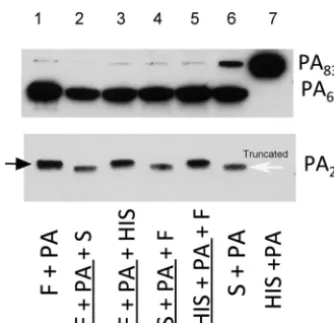
Serum-mediated digestion of rPA.
Serum treatment of recombinant PA
83(rPA
83)
produced 2 protein fragments, PA
63and a band that is slightly lower in molecular mass
than PA
20(Fig. 1; lane 6). The larger protein is similar in size to the PA
63protein
produced by furin digestion of rPA
83. However, the smaller protein is smaller than the
PA
20protein produced by furin digestion of rPA
83and is referred to as truncated PA
20.
Furthermore, serum treatment of rPA83 before or after furin digestion still produced
this truncated fragment (Fig. 1, lanes 2 and 4). Heat inactivation of serum prevented this
FIG 1 Serum-mediated digestion of rPA83 produces a truncated PA20 fragment compared withfurin-mediated digestion. Shown are the digestion fragments of PA83under conditions of incubation
with furin (F; lane 1), serum (S; lane 6), or heat-inactivated serum (HIS; lane 7). Treatment of rPA83 with serum after and prior to furin digestion (lanes 2 and 4, respectively) produced a truncated PA20fragment,
indicating that truncation is distinct from furin digestion. In contrast, incubation of furin-treated PA83
with HIS (lanes 3 and 5) failed to result in PA20truncation, suggesting that this process is heat labile. For
the purpose of the assay, serum was incubated with PA63for 1 h. MAb 10F4 (which recognizes domains
2 and 4 of PA83) was used to detect the PA63fragment, while MAb 19D9 (which recognizes domain 1)
was used to detect both the normal and truncated PA20fragments. The black arrow points to the normal
PA20fragment, while the white arrow points to the truncated PA20fragment. This experiment was done
2 times with similar results. Underlining indicates preincubation.
on September 8, 2020 by guest
http://msphere.asm.org/
truncation (Fig. 1; lanes 3 and 5), consistent with the idea that the enzyme responsible
for truncation is heat labile.
Inhibition of serum-mediated digestion of rPA.
To determine the precise site at
which serum cleaves rPA, we attempted to inhibit serum-mediated cleavage using a
library of overlapping peptides, which represent the PA sequence and antibodies that
recognize various PA sites. Preincubation of rPA with the 19D2 monoclonal antibody
(MAb), which recognizes an epitope immediately C terminal of the furin site (16),
prevented rPA digestion by serum and furin. This inhibition of digestion was not seen
with other PA-specific antibodies, including 7.5G, which recognizes domain 1 of PA
83.
Serum-mediated PA cleavage was also prevented by coincubation of serum with 3
overlapping peptides (D5, D6, and D7), which contain the furin digestion site, but not
with other peptides (including D12, E1, and E2, which represent PA sequences
approx-imately 30 amino acid [AA] residues C terminal to the furin site) (not shown).
Using chemical inhibitors while measuring PA
63formation, we found that the
serine/cysteine protease antipain partially inhibited the formation of PA
63. In contrast,
none of the other tested protease inhibitors, including bestatin, chymostatin, E-64,
leupeptin, pepstatin, phosphoramidon, Pefabloc SC, and aprotinin, prevented PA
63formation. As in previous studies, we found that EDTA was a potent inhibitor of
serum-mediated digestion of PA
83. In contrast, both competitive inhibitors of furin (I
and II) prevented serum-mediated digestion of PA. For furin inhibitor I, concentrations
as low as 0.001 mg/ml resulted in complete inhibition of serum digestion, whereas for
furin inhibitor II, concentrations as low as 0.010 mg/ml produced complete inhibition
of digestion (Fig. 2).
Truncated PA
20fragment.
To better identify the precise site of serum-mediated
digestion of rPA, the truncated PA
20fragment produced by serum digestion was
examined by mass spectrometry (MS). First, the intact-protein mass of this fragment
was measured and the experimental mass determined by liquid
chromatography-electrospray ionization mass spectrometry (LC-ESI MS) to be 23,600 Da (Fig. S1). Furin
cleaves at RXK/RR, which would correspond to a predicted molecular mass of 25,157 Da
for rPA (Fig. 3; N terminus to RKKR), which represents a difference of 1,557 Da (a value
far beyond the error of measurement). To determine the sequence of the truncated
PA
20fragment, in-gel trypsin digestion was performed. The liquid
chromatography-tandem mass spectrometry (LC-MS/MS) data identified the underlined tryptic peptides
shown in Fig. 3 (identified peptides from this tryptic digest are listed in Table S1). The
peptide sequence LLNES . . . GFIK is too large for fragmentation on an LTQ (linear ion
trap quadrupole) mass spectrometer and was not detected by MS/MS, but the
⫹
4,
⫹
5,
⫹
6,
⫹
7, and
⫹
8 charge states were detected (Fig. S2). The predicted protein mass from
the N terminus to the last tryptic peptide identified is 23,213 Da and if the next 4 amino
acids (SSNS) are included the predicted protein mass increases to 23,588 Da, a
differ-ence of 12 Da or 0.05% compared with the experimental intact-protein mass
(23,600 Da). These findings are consistent with serum-mediated cleavage of the basic,
Both furin inhibitor I and furin inhibitor II (F1 and F2) prevented serum digestion of PA83. PA83wasincubated for the serum for 30 min. This experiment was repeated 3 times with similar results.
on September 8, 2020 by guest
C-terminal arginine and lysine residues from the PA
20fragment produced by furin
digestion, possibly followed by carboxypeptidase.
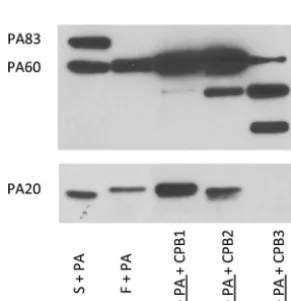
Carboxypeptidase treatment of rPA.
Given these results, we sought to determine
whether this truncated PA
20fragment could result from serum carboxypeptidase
digestion of PA
20. Carboxypeptidases are a family of enzymes that cleave residues from
the C-terminal end of a protein. This includes a group of enzymes that cleave basic
amino acid residues from the carboxy terminus. To determine if carboxypeptidase
could produce a truncated PA
20fragment, we conducted studies with a pancreatic
carboxypeptidase. The effects of carboxypeptidase B (CPB) treatment on furin-digested
rPA were dose dependent. At higher concentrations (250
g/ml) (Fig. 4, lane 5),
multiple digestion fragments of PA were observed and PA
20reactivity was completely
lost. A similar pattern was seen in the absence of furin and presumably relates to the
presence of contaminating trypsin in this pancreatic preparation. In contrast, at lower
concentrations of CPB (25
g/ml) (Fig. 4, lane 4), treatment produced a truncated PA
20fragment that was similar in size to that observed with serum digestion of PA (Fig. 4,
lane 1). Lower concentrations of CPB (2.5
g/ml) had no effect on the size of
furin-treated PA
20compared with the results seen with furin treatment alone.
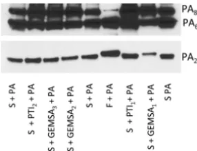
Inhibition of serum carboxypeptidase activity.
Next, we sought to determine
whether the ability of serum to produce a truncated PA
20fragment could be inhibited
FIG 3 Mass spectrometry of the serum-truncated PA20fragment. The predicted size of the fragment to
the SNSS amino acid sequence is 23,588 Da (thick arrow), a difference of 12 Da or 0.06% compared with the measured mass of 23,600 Da. In contrast, digestion at the furin consensus site should produce a PA20
fragment with a mass of 25,157 Da. Underlined peptide sequences were detected by MS analysis. The dotted box represents the consensus recognition site for furin.
FIG 4 Carboxypeptidase B (CPB) treatment of furin-digested PA produces a truncated PA20fragment.
Treatment of furin-digested rPA83with CPB from pig pancreas resulted in a dose-related truncation of the
PA20fragment. This was most apparent for CPB2 (25g/ml) compared to lower concentrations of CPB1
(2.5g/ml). Incubation with higher CPB3 concentrations (250g/ml) resulted in complete loss of PA20
reactivity and the appearance of multiple digestion fragments. Underlining indicates preincubation.
on September 8, 2020 by guest
http://msphere.asm.org/
by carboxypeptidase inhibitors. Both guanidinoethylmercaptosuccinic acid (GEMSA)
and potato tuber extract (PTI) are potent competitive inhibitors of carboxypeptidase,
though their inhibitory activity is not specific to any one class of carboxypeptidases.
Addition of GEMSA (500
g/ml) to serum prevented the formation of a truncated PA
20and resulted in a PA
20fragment that was more similar in size to that produced by furin
digestion (Fig. 5). In contrast, no inhibition was seen with lower concentrations of
GEMSA and for all concentrations of carboxypeptidase inhibitor (PTI) from potato tuber
extract.
DISCUSSION
B. anthracis
and the toxins that it secretes have an intimate association with the
circulation and with serum over the course of infection. Our studies confirm earlier
reports that both human and animal sera contain a furin-like enzyme, which digests PA
to produce PA
63and PA
20fragments (13–15). In our own studies, this activity was
associated with protection against lethal toxin
in vitro
(15). We now extend these
findings to demonstrate that human serum contains a carboxypeptidase which further
processes the PA
20fragment by removing the C-terminal basic amino acid residues,
resulting in a truncated PA
20fragment. These findings contrast with the current model
of anthrax toxin, which suggests that processing of PA occurs only at the cell surface,
and provide additional evidence for the complexity of anthrax toxin mechanisms of
action. However, we note that serum and cell surface PA processing are not mutually
exclusive events.
PA
20has been detected in the blood of
B. anthracis-infected animals, though its
contribution to anthrax pathogenesis is unknown (17). Nonetheless, several lines of
evidence suggest that it may play an active role in infection. For example, PA
20contains
a PA
14domain that is conserved among bacterial toxins and appears to play a role in
cell binding (18). Hammamieh et al. reported that exposure of human peripheral blood
mononuclear cells to PA
20induced a variety of genes related to the inflammatory
system and to cell migration and triggered apoptosis in these cells (17). Furthermore, PA
20has been reported to bind lethal factor (19). Although circumstantial, these findings are
consistent with a role for PA
20in the pathogenesis of anthrax.
Serum is known to contain 2 carboxypeptidases, CP-N and CPB
2(which is also
known as CPU, plasma carboxypeptidase B, and thrombactivatable fibrinolysis
in-hibitor). Both carboxypeptidases cleave carboxy-terminal arginine and lysine residues
from peptides/proteins and have been implicated in regulating inflammation through
their effects on serum protein cascades, such as the complement anaphylatoxins
and kinins (20). As members of the carboxypeptidase family, both CP-N and CPB
2contain a zinc-binding site that makes them susceptible to inhibition by metal
chelators. CP-N is constitutively produced by the liver, with serum concentrations
on the order of 30
g/ml (21). In contrast, CPB
2must be activated by fibrin and,
once activated, downregulates fibrinolysis by removing terminal lysines from fibrin and
20
Lower concentrations of GEMSA (50 and 5g/ml; GEMSA2 and GEMSA3) and PTI (PTI2; 125g/ml) did not prevent serum-mediated truncation of PA20. This experiment was done 3 times with similar results.
on September 8, 2020 by guest
is more susceptible to inhibition by potato carboxypeptidase inhibitor (31). Thus, our
findings are consistent with the hypothesis that
in vitro, CP-N is primarily responsible
for the observed truncation of PA
20. Nonetheless, the precise carboxypeptidase
respon-sible for the truncation of PA
20in vivo
(including during the sepsis of anthrax) is not
known and it is likely that there is redundancy to the process. Of note, macrophages
also express a membrane-associated carboxypeptidase (CP-M) that cleaves C-terminal
lysines and arginine residues from proteins (32). It is, therefore, likely that a similar form
of processing occurs at the surface of target cells.
In summary, we demonstrate that serum processing of PA is a 2-step process
that involves a furin-like digestion of the PA
83component followed by truncation of
the PA
20fragment by serum carboxypeptidases. The significance of these 2
serum-associated activities remains to be defined. On the basis of earlier studies that
associ-ated furin-like digestion with protection, we believe that this activity may in fact
contribute to the host response to anthrax. This would be consistent with the close
association of
B. anthracis
with the circulatory system. We also suggest that it is
possible that the variations in these serum proteolytic activities contribute to
differ-ences in individual susceptibilities to anthrax. Additional studies examining gain and
loss of function in the context of experimental infection may help further delineate the
importance of these processes.
MATERIALS AND METHODS
PA.Recombinant PA83(rPA) and its amino acid sequence were obtained from Wadsworth
Labora-tories, New York State Department of Health (Albany, NY).
Sera.Serum was obtained from laboratory volunteers and stored at⫺80°C with approval from the Committee of Clinical Investigations at Albert Einstein College of Medicine. In some experiments, pooled sera, processed to retain complement activity (Sigma, St. Louis, MO), was used. These commercial sera produced results comparable to those obtained with sera from human volunteers.
Antibodies and peptides.A library of 6 murine monoclonal antibodies (7.5G, 16A12, 10F4, 19D9, 20G7, and 2H9) that were previously generated and characterized was used both to define the digestion site and as detection reagents for immunoblot studies (33). Binding sites for these antibodies are provided in Table S2. A previously synthesized library of overlapping peptides which represents the PA sequence was used for inhibition studies (16).
Proteolytic digestion and fragment detection.Proteolytic digestion studies were performed as previously described (15). Briefly, rPA (2.5g) was incubated with 25l of serum, phosphate-buffered saline, or furin (Invitrogen) (0.5 units) at 37°C for 30 to 60 min. In some experiments, serum was heat treated at 56°C for 30 min prior to incubation with toxin. In other experiments, protease inhibitors (see below) or peptides at a concentration of 5g/ml were added to serum prior to incubation with rPA. Digested rPA was separated by SDS-electrophoresis and transferred to a nitrocellulose membrane. Membranes were blocked with 5% milk and then incubated with primary antibody. The following MAbs were used to characterize rPA cleavage: 10F4 (IgG1) and 7.5G (IgG2b). All MAbs were used at a concentration of 0.25g/ml. Primary antibody was detected with horseradish peroxidase-labeled goat isotype-specific antibody at a dilution of 1:25,000. Proteins were visualized by development with an ECL chemiluminescence kit (Pierce, Rockford, IL).
Inhibition studies. (i) Peptides.Serum (24l) was incubated with individual biotinylated peptides, peptide mixtures, or phosphate-buffered saline (PBS) for 2 h at room temperature. These peptides were chosen from a library of peptides representing the entire length of rPA and were synthesized as 15-mer, overlapping by 10 residues (16). This serum peptide mixture was then incubated with 1.5g of rPA for 30 min at 37°C, and the resulting mixture was subjected to separation by SDS-PAGE and detection by Western blotting.
(ii) MAbs.PA (1.5g) was incubated with one of several PA-specific MAbs (2g) (33) for 10 min at room temperature. This mixture was then added to 24l of serum, incubated at 37°C for 20 min, and then subjected to SDS-electrophoresis and immunoblotting.
(iii) Protease inhibitors.A volume of 10l of sera was preincubated with 1 of 9 protease inhibitors included in a commercially available protease inhibitor set (Roche) for 30 min at 30°C. Individual
on September 8, 2020 by guest
http://msphere.asm.org/
resin washed 5 times with binding buffer (Pierce). Following elution, the protein was separated in a nondenaturing gel and electroeluted for further analysis.
Mass spectrometry (MS) measurements and liquid chromatography (LC) separations were obtained on an LTQ (linear ion trap quadrupole) mass spectrometer (Thermo Scientific, San Jose, CA), an LC 3000 rapid-separation system (Dionex Corporation, Sunnyvale, CA) was used for processing of tryptic peptides, and an HP Agilent 1100 series system was used for intact-protein separation. For intact-protein molecular weight measurements of the electro-eluted protein, a C4 Vydac TP column (1 by 50 mm; 300 Å; 50l/min) was used. After desalting performed with 1% acetonitrile– 0.1% aqueous formic acid (FA) for 2 min, the protein was eluted after increasing the level of acetonitrile to 55% acetonitrile– 0.1% aqueous FA. The mass range from 600 to 1,800m/zwas acquired on the LTQ mass spectrometer, and the raw data were deconvoluted using MagTran (34) or ProMass (Thermo Fisher Scientific). Another aliquot of the electroeluted protein was separated on a one-dimensional (1D) SDS gel, and selected molecular weight bands were excised for in-gel tryptic digestion as described previously (35). After sample injection and LC peptide separation (using an acetonitrile gradient), the 10 most abundant ions obtained from the survey scan (300 to 1,600 m/z) were selected for fragmentation (MS/MS). Normalized collision energy of 35% and a 2m/zisolation width were used for MS/MS. The MS/MS data were converted to a text file for peptide/protein identification using Mascot (Matrix Science, Inc.).
Carboxypeptidase-mediated digestion of PA.To determine whether carboxypeptidase digestion of furin-treated rPA could produce a fragment similar in size to that seen with serum digestion of rPA, experiments were done with carboxypeptidase B (CPB) (Sigma). For these experiments, rPA was treated with furin for 10 min at 30°C and the mixture was incubated with CPB at different concentrations at 37°C. Proteins were separated by SDS-PAGE and detected by immunoblotting as described above.
SUPPLEMENTAL MATERIAL
Supplemental material for this article may be found at
https://doi.org/10.1128/
mSphere.00091-18
.
FIG S1,
TIF file, 1.2 MB.
FIG S2,
TIF file, 1.5 MB.
TABLE S1,
TIF file, 1.8 MB.
TABLE S2,
DOCX file, 0.04 MB.
ACKNOWLEDGMENTS
This work was supported, in whole or in part, by National Institutes of Health grants
AI33774-11, HL59842-07, AI33142-11, and AI52733-02 (to A.C.) and AI067665 and
AI127173 (to J.M.A.). This work was also supported by the Northeastern Biodefense
Center under grant U54-AI057158-Lipkin and by a NIH-funded Shared Instrumentation
grant (1S10RR019352) for the LTQ LC-MS/MS system.
REFERENCES
1. Pezard C, Berche P, Mock M. 1991. Contribution of individual toxin compo-nents to virulence of Bacillus anthracis. Infect Immun 59:3472–3477. 2. Shiryaev SA, Remacle AG, Ratnikov BI, Nelson NA, Savinov AY, Wei G,
Bottini M, Rega MF, Parent A, Desjardins R, Fugere M, Day R, Sabet M, Pellecchia M, Liddington RC, Smith JW, Mustelin T, Guiney DG, Lebl M, Strongin AY. 2007. Targeting host cell furin proprotein convertases as a therapeutic strategy against bacterial toxins and viral pathogens. J Biol Chem 282:20847–20853.https://doi.org/10.1074/jbc.M703847200. 3. Albrecht MT, Li H, Williamson ED, LeButt CS, Flick-Smith HC, Quinn CP,
Westra H, Galloway D, Mateczun A, Goldman S, Groen H, Baillie LW. 2007. Human monoclonal antibodies against anthrax lethal factor and protec-tive antigen act independently to protect against Bacillus anthracis
infection and enhance endogenous immunity to anthrax. Infect Immun 75:5425–5433.https://doi.org/10.1128/IAI.00261-07.
4. Brossier F, Lévy M, Landier A, Lafaye P, Mock M. 2004. Functional analysis of Bacillus anthracis protective antigen by using neutralizing monoclo-nal antibodies. Infect Immun 72:6313– 6317.https://doi.org/10.1128/IAI .72.11.6313-6317.2004.
5. Anonymous. 2013. Raxibacumab for anthrax. Med Lett Drugs Ther 55: 27–28.
6. Moayeri M, Leppla SH, Vrentas C, Pomerantsev AP, Liu S. 2015. Anthrax pathogenesis. Annu Rev Microbiol 69:185–208.https://doi.org/10.1146/ annurev-micro-091014-104523.
7. Boyer AE, Gallegos-Candela M, Quinn CP, Woolfitt AR, Brumlow JO, Isbell
on September 8, 2020 by guest
10. Rivera J, Cordero RJ, Nakouzi AS, Frases S, Nicola A, Casadevall A. 2010. Bacillus anthracis produces membrane-derived vesicles containing biologically active toxins. Proc Natl Acad Sci U S A 107:19002–19007. https://doi.org/10.1073/pnas.1008843107.
11. Wolf JM, Rivera J, Casadevall A. 2012. Serum albumin disrupts Crypto-coccus neoformans and Bacillus anthracis extracellular vesicles. Cell Microbiol 14:762–773.https://doi.org/10.1111/j.1462-5822.2012.01757.x. 12. Panchal RG, Halverson KM, Ribot W, Lane D, Kenny T, Abshire TG, Ezzell JW, Hoover TA, Powell B, Little S, Kasianowicz JJ, Bavari S. 2005. Purified Bacillus anthracis lethal toxin complex formed in vitro and during infec-tion exhibits funcinfec-tional and biological activity. J Biol Chem 280: 10834 –10839.https://doi.org/10.1074/jbc.M412210200.
13. Ezzell JW, Jr, Abshire TG. 1992. Serum protease cleavage of Bacillus anthracis protective antigen. J Gen Microbiol 138:543–549.https://doi .org/10.1099/00221287-138-3-543.
14. Moayeri M, Wiggins JF, Leppla SH. 2007. Anthrax protective antigen cleavage and clearance from the blood of mice and rats. Infect Immun 75:5175–5184.https://doi.org/10.1128/IAI.00719-07.
15. Goldman DL, Zeng W, Rivera J, Nakouzzi A, Casadevall A. 2008. Human serum contains a protease that protects against cytotoxic activity of Bacillus anthracis lethal toxin in vitro. Clin Vaccine Immunol 15:970 –973. https://doi.org/10.1128/CVI.00064-08.
16. Abboud N, De Jesus M, Nakouzi A, Cordero RJ, Pujato M, Fiser A, Rivera J, Casadevall A. 2009. Identification of linear epitopes in Bacillus anthra-cis protective antigen bound by neutralizing antibodies. J Biol Chem 284:25077–25086.https://doi.org/10.1074/jbc.M109.022061.
17. Hammamieh R, Ribot WJ, Abshire TG, Jett M, Ezzell J. 2008. Activity of the Bacillus anthracis 20 kDa protective antigen component. BMC Infect Dis 8:124.https://doi.org/10.1186/1471-2334-8-124.
18. Rigden DJ, Galperin MY. 2004. The DxDxDG motif for calcium binding: multiple structural contexts and implications for evolution. J Mol Biol 343:971–984.https://doi.org/10.1016/j.jmb.2004.08.077.
19. Chvyrkova I, Zhang XC, Terzyan S. 2007. Lethal factor of anthrax toxin binds monomeric form of protective antigen. Biochem Biophys Res Commun 360:690 – 695.https://doi.org/10.1016/j.bbrc.2007.06.124. 20. Djukanovic´ R, Wilson SJ, Kraft M, Jarjour NN, Steel M, Chung KF, Bao
W, Fowler-Taylor A, Matthews J, Busse WW, Holgate ST, Fahy JV. 2004. Effects of treatment with anti-immunoglobulin E antibody omalizumab on airway inflammation in allergic asthma. Am J Respir Crit Care Med 170:583–593.https://doi.org/10.1164/rccm.200312-1651OC.
21. Skidgel RA, Erdös EG. 2007. Structure and function of human plasma carboxypeptidase N, the anaphylatoxin inactivator. Int Immunopharma-col 7:1888 –1899.https://doi.org/10.1016/j.intimp.2007.07.014.
25. Kim PY, Kim PY, Hoogendorn H, Giles AR, Nesheim ME. 2008. Activated thrombin-activatable fibrinolysis inhibitor is generated in vivo at levels that can substantially affect fibrinolysis in chimpanzees in response to thrombin generation. J Thromb Haemost 6:1600 –1602.https://doi.org/ 10.1111/j.1538-7836.2008.03067.x.
26. Park R, Song J, An SS. 2010. Elevated levels of activated and inactivated thrombin-activatable fibrinolysis inhibitor in patients with sepsis. Korean J Hematol 45:264 –268.https://doi.org/10.5045/kjh.2010.45.4.264. 27. Bokisch VA, Müller-Eberhard HJ. 1970. Anaphylatoxin inactivator of
hu-man plasma: its isolation and characterization as a carboxypeptidase. J Clin Invest 49:2427–2436.https://doi.org/10.1172/JCI106462.
28. Campbell WD, Lazoura E, Okada N, Okada H. 2002. Inactivation of C3a and C5a octapeptides by carboxypeptidase R and carboxypeptidase N. Microbiol Immunol 46:131–134. https://doi.org/10.1111/j.1348-0421.2002 .tb02669.x.
29. Premanandan C, Storozuk CA, Clay CD, Lairmore MD, Schlesinger LS, Phipps AJ. 2009. Complement protein C3 binding to Bacillus anthracis spores enhances phagocytosis by human macrophages. Microb Pathog 46:306 –314.https://doi.org/10.1016/j.micpath.2009.03.004.
30. Harvill ET, Lee G, Grippe VK, Merkel TJ. 2005. Complement depletion renders C57BL/6 mice sensitive to the Bacillus anthracis Sterne strain. Infect Immun 73:4420 – 4422.https://doi.org/10.1128/IAI.73.7.4420-4422 .2005.
31. Matthews KW, Mueller-Ortiz SL, Wetsel RA. 2004. Carboxypeptidase N: a pleiotropic regulator of inflammation. Mol Immunol 40:785–793.https:// doi.org/10.1016/j.molimm.2003.10.002.
32. Rehli M, Krause SW, Kreutz M, Andreesen R. 1995. Carboxypeptidase M is identical to the max.1 antigen and its expression is associated with monocyte to macrophage differentiation. J Biol Chem 270:15644 –15649. https://doi.org/10.1074/jbc.270.26.15644.
33. Rivera J, Nakouzi A, Abboud N, Revskaya E, Goldman D, Collier RJ, Dadachova E, Casadevall A. 2006. A monoclonal antibody to Bacillus anthracis protective antigen defines a neutralizing epitope in domain 1. Infect Immun 74:4149 – 4156.https://doi.org/10.1128/IAI.00150-06. 34. Zhang Z, Marshall AG. 1998. A universal algorithm for fast and
auto-mated charge state deconvolution of electrospray mass-to-charge ratio spectra. J Am Soc Mass Spectrom 9:225–233.https://doi.org/10.1016/ S1044-0305(97)00284-5.
35. Xiao Y, Pollack D, Andrusier M, Levy A, Callaway M, Nieves E, Reddi P, Vigodner M. 2016. Identification of cell-specific targets of SUMOylation during mouse spermatogenesis. Reproduction 151:149 –166.https://doi .org/10.1530/REP-15-0239.