Functional and Comparative Genomic Analysis of Integrated
Prophage-Like Sequences in “
Candidatus
Liberibacter
asiaticus”
Marian Dominguez-Mirazo,a,bRong Jin,a Joshua S. Weitza,c
aSchool of Biological Sciences, Georgia Institute of Technology, Atlanta, Georgia, USA
bInterdisciplinary Graduate Program in Quantitative Biosciences, Georgia Institute of Technology, Atlanta, Georgia, USA cSchool of Physics, Georgia Institute of Technology, Atlanta, Georgia, USA
ABSTRACT Huanglongbing disease (HLB; yellow shoot disease) is a severe
world-wide infectious disease for citrus family plants. The pathogen “Candidatus Liberibac-ter asiaticus” is an alphaproteobacLiberibac-terium of the Rhizobiaceae family that has been identified as the causative agent of HLB. The virulence of “Ca. Liberibacter asiaticus” has been attributed, in part, to prophage-carried genes. Prophage and prophage-like elements have been identified in 12 of the 15 available “Ca. Liberibacter asiaticus” genomes and are classified into three prophage types. Here, we reexamined all 15 “Ca. Liberibacter asiaticus” genomes using a de novo prediction approach and ex-panded the number of prophage-like elements from 16 to 33. Further, we found that all of the “Ca.Liberibacter asiaticus” genomes contained at least one prophage-like sequence. Comparative analysis revealed a prevalent, albeit previously unknown, prophage-like sequence type that is a remnant of an integrated prophage. Notably, this remnant prophage is found in the Ishi-1 “Ca. Liberibacter asiaticus” strain that had previously been reported as lacking prophages. Our findings provide both a re-source for data and new insights into the evolutionary relationship between phage and “Ca.Liberibacter asiaticus” pathogenicity.
IMPORTANCE Huanglongbing (HLB) disease is threatening citrus production
world-wide. The causative agent is “Candidatus Liberibacter asiaticus.” Prior work using mapping-based approaches identified prophage-like sequences in some “Ca. Liberib-acter asiaticus” genomes but not all. Here, we utilized ade novo approach that ex-pands the number of prophage-like elements found in “Ca. Liberibacter asiaticus” from 16 to 33 and identified at least one prophage-like sequence in all “Ca.Liberibacter asiaticus” strains. Furthermore, we identified a prophage-like sequence type that is a remnant of an integrated prophage— expanding the number of prophage types in “Ca.Liberibacter asiaticus” from 3 to 4. Overall, the findings will help researchers in-vestigate the role of prophage in the ecology, evolution, and pathogenicity of “Ca. Liberibacter asiaticus.”
KEYWORDS One Health, bioinformatics, environmental microbiology, microbial
ecology, phage ecology, phytopathology, plant pathogens
“
C
andidatus Liberibacter asiaticus” has been identified as one of the three “Ca. Liberibacter” species that cause Huanglongbing disease (HLB; yellow shoot disease [also known as citrus greening disease]). HLB is a major threat to the worldwide citrus-growing industry (1). Symptoms of infected trees include leaf mottling, de-formed/discolored fruits, premature fruit drop, and premature mortality (2). It has been suggested that “Ca.Liberibacter asiaticus”-encoded proteins have an inhibitory effect in plant defenses (3, 4), but comprehensive understanding of the mechanism of infectionCitationDominguez-Mirazo M, Jin R, Weitz JS. 2019. Functional and comparative genomic analysis of integrated prophage-like sequences
in “CandidatusLiberibacter asiaticus.” mSphere
4:e00409-19.https://doi.org/10.1128/mSphere .00409-19.
EditorKatherine McMahon, University of Wisconsin—Madison
Copyright© 2019 Dominguez-Mirazo et al. This is an open-access article distributed under the terms of theCreative Commons Attribution 4.0 International license.
Address correspondence to Joshua S. Weitz, [email protected].
Received6 June 2019 Accepted17 October 2019 Published
Applied and Environmental Science
13 November 2019
on September 8, 2020 by guest
http://msphere.asm.org/
is still lacking. No effective disease management practice is currently available. Hence, understanding the genomic composition of “Ca.Liberibacter asiaticus” and its infection mechanisms is likely to help develop strategies to manage the disease.
As “Ca.Liberibacter asiaticus” has not been cultivatedin vitro, its biological study has been based on analyses of DNA extracted from infected plants or insects. To date, a total of 15 genome assemblies have been made available at the National Center for Biotechnology Information (NCBI) Genome database (Table 1). The “Ca. Liberibacter asiaticus” genome size is⬃1.2 Mb, with G⫹C content of 36.5%; both the small genome and low level of G⫹C content are consistent with patterns of obligate intracellular bacteria (5). The “Ca. Liberibacter asiaticus” genome includes genes involved in cell motility and active transport (5, 6); despite the presence of these genes, only passive movement has been observed in the phloem sap (6). Furthermore, prophage se-quences have also been identified in multiple “Ca.Liberibacter asiaticus” strains (7–16) (Tables 1 and 2). The interest in “Ca. Liberibacter asiaticus” prophage stems from observations revealing that a prophage-encoded peroxidase is an effector that sup-presses plant defenses (3). However, strains reported to lack prophages still induce HLB symptoms (8, 16). Hence, it has been hypothesized that prophages might contribute to bacterial virulence but are not required for “Ca.Liberibacter asiaticus” pathogenicity (6). Prophage regions in “Ca.Liberibacter asiaticus” genomes are highly variable relative to the rest of the genome (8, 16). Comparative analyses of prophage sequences suggest endemism in “Ca. Liberibacter asiaticus” strains (14, 17). Based on currently available sequence data, “Ca.Liberibacter asiaticus” prophages have been classified as uniquely belonging to one of three types, i.e., type 1, 2, or 3. The first reported “Ca.Liberibacter TABLE 1“Ca.Liberibacter asiaticus” genome informationa
Strain Source
GenBank accession no.
No. of contigs
Length (bp)
GⴙC (%)
Prophage type(s)
A4 Guangdong, China GCA_000590865.2 — 1,233,514 36.4 2
AHCA1 California, USA GCA_003143875.1 — 1,233,755 36.63 1
FL17 Florida, USA GCA_000820625.1 3 1,227,253 36.47 1
gxpsy Guangxi, China GCA_000346595.1 — 1,268,237 36.57 1, 2
HHCA1 California, USA GCA_000724755.2 239 1,150,620 36.55 2*
Ishi-1 Okinawa, Japan GCA_000829355.1 — 1,190,853 36.32
JXGC Jiangxi, China GCA_002216815.1 — 1,225,162 36.4 3*
psy62 Florida, USA GCA_000023765.2 — 1,227,328 36.47 1, 2*
SGCA1 California, USA GCA_003149415.1 606 233,414 36.25 1
SGCA5 California, USA GCA_001430705.1 56 1,201,385 36.36 1*
SGpsy California, USA GCA_003336865.1 1,402 769,888 36.32 1
TX1712 Texas, USA GCA_003160765.1 48 1,203,333 36.36
TX2351 Texas, USA GCA_001969535.1 71 1,252,002 36.52
YCPsy Guangdong, China GCA_001296945.1 9 1,233,647 36.48 1, 3
YNJS7C Yunnan, China GCA_003615235.1 3 1,258,986 36.58 2, 3
aCharacteristics of the 15 publicly available “Ca.Liberibacter asiaticus” genomes are indicated as follows: strain, geographic origin, GenBank accession number, number of contigs, sequence length, G⫹C content, and associated phage types. Dashes (—) in the contig column indicate complete (circularized) genomes. Phage types with asterisks belong to phages with publicly available genomes (Table 2).
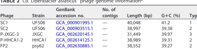
TABLE 2“Ca.Liberibacter asiaticus” phage genome informationa
Phage Strain
GenBank accession no.
No. of
contigs Length (bp) GⴙC (%) Type
SC1 UF506 GCA_000901995.1 — 40,048 41.2 1
SC2 UF506 GCA_000903515.1 — 38,997 39.38 2
P-JXGC-3 JXGC GCA_002620145.1 — 31,449 39.97 3
P-HHCA1-2 HHCA1 GCA_002614125.1 — 38,989 39.31 2
FP2 psy62 GCA_002630885.1 — 38,552 39.27 2
P-SGCA5-1 SGCA5 GCA_002945115.1 2 37,487 41.85 1
aCharacteristics of the 5 publicly available genomes of “Ca.Liberibacter asiaticus” phages are indicated as follows: phage name, associated “Ca.Liberibacter asiaticus” strain, GenBank accession number, number of contigs, sequence length, G⫹C content, and phage type. Dashes (—) in the contig column correspond to closed genomes.
on September 8, 2020 by guest
http://msphere.asm.org/
asiaticus” prophages were SC1 and SC2 (type 1 and 2 prophages, respectively). They were discovered when the sequencing of a “Ca. Liberibacter asiaticus” strain showed circular contigs rich in phage open reading frames (ORFs) (7). These phages were shown to integrate into the “Ca.Liberibacter asiaticus” genome (7). Moreover, a recent study identified P-JXGC-3 as the first type 3 prophage by using BLAST searches with phages SC1 and SC2 as query to locate phage-containing contigs within a “Ca.Liberibacter asiaticus” genome with incomplete read mapping to type 1 or type 2 prophage (13). The genomes of prophages of types 1, 2, and 3 can be divided into an early gene region and a late gene region (7, 13). The early gene regions of the three prophage types are highly similar and match about 50% of their genomes. In contrast, the late regions differ greatly in gene content between types (7, 13).
Current methods to identify prophage in “Ca.Liberibacter asiaticus” genomes rely on read mapping against known prophages (9, 12). As a consequence, failure of read mapping has been interpreted to mean that a genome is prophage free (e.g., as in the Ishi-1 strain [16]). More generally, we are unaware of any systematic analysis of prophage content in “Ca.Liberibacter asiaticus” genomes usingde novoprediction approaches. In this study, we examined the genome sequences of 15 “Ca.Liberibacter asiaticus” strains and extracted phage sequences usingde novoprediction tools, including Virsorter (18) and PHASTER (19). De novo prediction tools identify putative phage and prophage regions through detection of circular sequences and comparison of predicted proteins against complete phage databases (18, 19). In contrast to commonly used read-mapping methodologies, ade novoapproach can identify phage regions that do not necessarily resemble “Ca.Liberibacter asiaticus” prophages of type 1, 2, or 3. Via ade novo approach, we identified several potential prophage elements not available in databases, of which 5 belonged to type 1, 2, or 3 “Ca.Liberibacter asiaticus” phage elements that had not been previously reported. We also identified 12 phage-like sequences present in all strains that do not resemble any previously identified phage type. Subsequent analyses revealed that these 12 sequences do not match type 1, 2, or 3 prophages. We argue that, based on composition and evolutionary analysis, it is likely that these sequences belong to a different “Ca.Liberibacter asiaticus” prophage-like sequence, which we term “type 4.” Multiple lines of evidence suggest that type 4 prophage-like sequences are remnants of an integrated prophage. This study expanded the number of prophage-like elements from 16 to 33. The results will provide a genomic resource for future investigations into the role of prophage in shaping the ecology, evolution, and pathogenicity of “Ca.Liberibacter asiaticus.”
RESULTS
Identification of novel prophage-like sequences.A total of 35 putative prophage-like sequences were identified among the 15 “Ca.Liberibacter asiaticus” genomes by Virsorter and PHASTER. At least one sequence was predicted for each of the strains studied (Table 3) except for the genomes SGCA1 and SGpsy, where the absence of such a result might have been due to the high level of fragmentation of their assemblies (Table 1). Predicted sequences differ greatly in length, with the shortest sequence having⬃2,000 bp and the longest over 65,000 bp. The prophage-like sequences can be divided into two groups differing by the level of G⫹C content: those with around 40% (similar to the G⫹C content of type 1, 2, and 3 prophages) and those with around 36% (similar to the “Ca.Liberibacter asiaticus” value). We compared the predicted sequences to phage genomes available from NCBI (Table 2). Of the 35 predicted sequences, 6 belong to reported phage genomes— corresponding to 3 prophage sequences of HHCA1, 1 of JXGC, 1 of psy62, and 1 of SGCA5, belonging to phages P-HHCA1-2, P-JXGC-3, FP2, and P-SGCA5-1, respectively. Hence, we identified 29 prophage-like sequences that are not part of a publicly available prophage genome.
Classification of prophage-like predicted sequences.Of the 29 predicted prophage-like sequences, a total of 13 were successfully classified into type 1, 2, or 3 (see Materials and Methods and Table 4). As previously described, strains psy62, gxpsy, FL17, and YCPsy have sequences classified as type 1. Strains A4 and gxpsy have sequences
on September 8, 2020 by guest
http://msphere.asm.org/
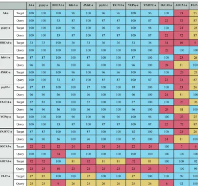
classified as type 2. Strain YNJS7C harbors a sequence that resembles a type 3 prophage (12, 15, 20). Novel prophage-like sequences of type 1 were found in strains TX1712 and TX2351, while type 2 sequences were also found in strain TX2351. In contrast to a previous report, no YNJS7C sequences were classified as type 2 (15); instead, a type 1 sequence was identified (see Addendum in Proof below). Although they seemed to resemble the representative phages, 4 sequences remained unclassified due to their small size. Finally, 12 sequences shared similar characteristics, but 10 had no apparent resemblance to the representative phages SC1, SC2, and P-JXGC-3, while a small subsequence of each of the other two (AHCA1-a and FL17-a) partially resembled the representative phages (Table 4).
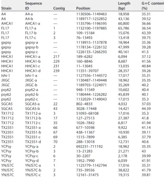
Identification of a new type of “Ca. Liberibacter asiaticus” prophage-like se-quences. We next set out to identify the “type” of prophage for the unclassified prophage-like sequences found via our de novo approach. Of the 12 unclassified sequences that do not resemble the three known phage types, 10 are highly similar to each other and belong to a candidate type 4 prophage-like sequence that includes the Ishi-1 prophage prediction (Table 5). Our evidence that these prophage-like sequences belong to a new type is as follows. First, there is no apparent resemblance to prophages of type 1, 2, or 3 at the nucleotide level or at the amino acid level. Second, the length of the type 4 sequences is about half the size of representative phages of types 1, 2, and 3 (Table 6). Third, in contrast to the previously known prophage types (with G⫹C TABLE 3Putative characteristics of prophage sequencesa
Strain
Sequence
ID Contig Position
Length (bp)
GⴙC content (%)
A4 A4-a — 1130506–1149463 18,958 35.31
A4 A4-b — 1189717–1232852 43,136 39.52
AHCA1 AHCA1-a — 1135796–1196595 60,800 36.66
FL17 FL17-a 1 1132100–1197885 65,786 36.54
FL17 FL17-b 2 109–15184 15,076 43.39
FL17 FL17-c 3 76–13493 13,418 39.75
gxpsy gxpsy-a — 1118915–1137878 18,964 35.36
gxpsy gxpsy-b — 1178134–1226132 47,999 39.28
gxpsy gxpsy-c — 1226133–1266293 40,161 41.5
HHCA1 HHCA1-a 217 189–6282 6,094 37.12
HHCA1 HHCA1-b 229 160–8846 8,687 41.56
HHCA1 HHCA1-c 231 11–13045 13,035 40.84
HHCA1 HHCA1-d 239 11351–39707 28,357 39.26
Ishi-1 Ishi-1-a — 1127556–1144572 17,017 35.31
JXGC JXGC-a — 1130487–1149448 18,962 35.35
JXGC JXGC-b — 1189703–1224971 35,269 40.2
psy62 psy62-a — 948–11549 10,602 40.4
psy62 psy62-b — 1180444–1226282 45,839 40.1
psy62 psy62-c — 1132029–1149043 17,015 35.3
SGCA5 SGCA5-a 22 802–4833 4,032 37.03
SGCA5 SGCA5-b 43 3028–17448 14,421 44.39
TX1712 TX1712-a 3 51093–68108 17,016 35.3
TX1712 TX1712-b 17 127–27513 27,387 41.8
TX1712 TX1712-c 35 1026–7842 6,817 41.94
TX2351 TX2351-a 66 677–10598 9,922 41.14
TX2351 TX2351-b 67 438–11367 10,930 39.11
TX2351 TX2351-c 69 1515–7899 6,385 37.79
TX2351 TX2351-d 70 288–13018 12,731 40.6
YCPsy YCPsy-a 2 692231–711192 18,962 35.35
YCPsy YCPsy-b 3 13–21283 21,271 43
YCPsy YCPsy-c 6 30–2207 2,178 39.99
YCPsy YCPsy-d 7 1952–7990 6,039 41.91
YNJS7C YNJS7C-a 1 1125779–1142794 17,016 35.31
YNJS7C YNJS7C-b 2 735–39556 38,822 41.79
YNJS7C YNJS7C-c 3 12161–31475 19,315 39.81
aCharacteristics of the sequences predicted by Virsorter and PHASTER are indicated as follows: “Ca. Liberibacter asiaticus” strain, sequence ID, the contig in which the sequence was found, the position in contig, the sequence length, and percent G⫹C content. Dashes (—) in the contig column indicate that the corresponding genome is fully assembled.
on September 8, 2020 by guest
http://msphere.asm.org/
TABLE 4Classification of novel prophage-like sequences into types 1, 2, and 3a
Type 1 Type 2 Type 3
SC1 SC2 P-JXGC-3
Target Query Target Query Target Query
Value (%) C I C I C I C I C I C I
SC1 100 100 100 100 52 98.49 52 98.49 42 97.89 53 97.89
SC2 52 98.49 52 98.49 100 100 100 100 47 97.43 58 97.43
P-JXGC-3 53 97.89 42 97.89 58 97.43 47 97.43 100 100 100 100
FL17-b 97 96.29 37 96.29 4 96.11 1 96.11 NA NA NA NA
FL17-c 98 98.62 34 98.62 62 96.35 22 96.35 63 98.15 27 98.15
gxpsy-c 89 97.83 92 97.83 44 96.38 45 96.38 41 98.47 52 98.47
psy62-b 71 99.8 81 99.8 29 98.07 33 98.07 19 98.8 28 98.8
TX1712-b 100 99.81 72 99.8 34 98.19 27 98.19 22 99.08 19 98.94
TX2351-a 100 99.92 25 99.92 89 99.54 23 99.54 89 98 28 98
YCPsy-b 93 97.88 52 97.88 17 99.63 11 99.63 8 99.94 5 99.94
YNJS7C-b 89 97.86 87 97.86 47 97.26 45 97.26 43 96.29 52 96.29
A4-b 46 96.01 48 96.01 90 98.28 98 98.28 43 94.99 58 95
gxpsy-b 50 98.22 53 98.22 87 98.64 97 98.64 44 99.49 61 99.49
TX2351-b 8 92.55 5 92.55 100 99.83 28 99.83 NA NA NA NA
TX2351-d 92 97.23 29 97.23 100 99.91 33 99.91 99 95.97 40 95.97
YNJS7C-c 85 95.91 42 95.91 94 96.03 47 96.03 100 98.11 61 98.11
TX1712-c 100 99.31 17 99.31 100 99.93 18 99.93 100 98.27 22 98.27
TX2351-c 79 98.07 13 98.07 100 99.83 17 99.83 81 91.83 16 91.83
YCPsy-c 100 97.8 6 97.8 100 97.8 6 97.8 99 99.8 7 99.8
YCPsy-d 99 97.73 15 97.73 99 98.11 16 98.11 99 99.42 20 99.42
AHCA1-a 2 93.87 4 93.87 4 96.18 6 96.18 4 98.02 7 98.02
FL17-a 6 95.72 13 95.72 5 92.5 11 92.51 3 94.84 6 94.85
A4-a NA NA NA NA NA NA NA NA NA NA NA NA
gxpsy-a NA NA NA NA NA NA NA NA NA NA NA NA
HHCA1-a NA NA NA NA NA NA NA NA NA NA NA NA
Ishi-1-a NA NA NA NA NA NA NA NA NA NA NA NA
JXGC-a NA NA NA NA NA NA NA NA NA NA NA NA
psy62-c NA NA NA NA NA NA NA NA NA NA NA NA
SGCA5-a NA NA NA NA NA NA NA NA NA NA NA NA
TX1712-a NA NA NA NA NA NA NA NA NA NA NA NA
YCPsy-a NA NA NA NA NA NA NA NA NA NA NA NA
YNJS7C-a NA NA NA NA NA NA NA NA NA NA NA NA
aThe novel predicted sequences were aligned against representative phages SC1, SC2, and P-JXGC-3 of types 1, 2, and 3. Coverage (C) values corresponding to two-way local alignments are presented in this table. Blue, mustard, and pink cells stand for high, intermediate, and low alignment values. Note that the threshold values used to classify coverage (C) vary according to sequence length and position in BLAST (either query or target) (see Materials and Methods). White cells with “NA” entries represent absence of significant hits. Sequences were categorized as type 1 (red), type 2 (blue), or type 3 (green) sequences; unclassified sequences with resemblance to the representative phages (black); or unclassified without resemblance (gray).
on September 8, 2020 by guest
http://msphere.asm.org/
content of⬃40%), type 4 sequences have G⫹C content of 35% to 37%, which is closer to that of the host, whose G⫹C content is⬃36% (Tables 1 and 6).
The other 2 predicted sequences, FL17-a and AHCA1-a, contain⬃80% of the type 4 sequence (Table 5) followed by bacterial genes and fragments highly resembling a part of a type 1 prophage genome and a part of a type 3 prophage genome, respectively (Fig. 1 and Table 4). Table S1 in the supplemental material shows alignment values for the 12 unclassified sequences using the type 4 region of AHCA1-a and FL17-a.
The prophage fragment at the end of FL17-a (end of contig 1) joined to type 1 sequences FL17-b (contig 2) and FL17-c (contig 3) reconstructs the sequence of a type 1 prophage (Fig. 1, top panel) with a sequence organization similar to that reported for the chromosomal integration of phage SC1 (7). Likewise, the last part of the AHCA1-a sequence and the following region found after a genome assembly gap reconstruct a type 3 prophage with the sequence organization predicted for P-JXGC-3 (13) (Fig. 1, bottom panel). Only a type 1 prophage has been reported in strain AHCA1 (9), but the sequence organization of the integrated prophage (Fig. 1) and the presence of a hypo-thetical protein (GenBank accession no.YP_007011137.1) found only in prophages of type 2 and 3 (Table S2) suggest that “Ca.Liberibacter asiaticus” strain AHCA1 harbors a type 3 prophage sequence. The truncation of the type 1 and type 3 prophage sequences and their closeness to the type 4 prophage-like sequence in strains FL17 and TABLE 5Resemblance between new type 4 sequencesa
aData represent results of comparisons of 12 predicted sequences that did not resemble any of the representative phages of types 1, 2, and 3. Coverage (C) values corresponding to two-way local alignments are presented in this table. Blue, mustard, and pink cells stand for high, intermediate, and low values, respectively (see Materials and Methods).
on September 8, 2020 by guest
http://msphere.asm.org/
AHCA1 explain the incorrect concatenation of these sequences into a single prediction by Virsorter and PHASTER. Table 6 shows all predicted prophage-like sequences with their assigned type and sequence characteristics.
Pan-genome analysis. In order to evaluate the robustness of the classification identified in prior sections, we analyzed the pan-genome content of prophages through a clustering approach (Fig. 2). Clustering of gene content in prophage reveals a separation between the 4 types of sequences. All type 1 sequences cluster together except for TX2351-a. Type 2 and type 3 sequences appear nearby in the clustering. The type 2 putative prophage for strain TX2351 clusters with type 3 sequences. The two mistaken assignments might result from the short size of sequences and the overall close relationship between prophage types 1, 2, and 3. All sequences of type 4 cluster together as the most distant group. The clustering of prophage-like sequences based on gene content predicts relationships that roughly agree with the classification.
Table 7 presents a summary of the associated prophage elements in the 15 “Ca. Liberibacter asiaticus” strains, including those previously reported and the ones intro-duced in this study. There are 5 newly reported elements that belong to prophages of type 1, 2, or 3 (see “Prophage-like sequence identification” section below) and 12 type TABLE 6Putative characteristics and classification of prophage sequencesa
Sequence
Length (bp)
GⴙC content (%)
Associated name
Prophage type
FL17-a/F2* 4,021 37.8 P-FL17-1 1
FL17-b 15,076 43.39 P-FL17-1 1
FL17-c 13,418 39.75 P-FL171-1 1
gxpsy-c 40,161 41.5 P-gxpsy-1 1
psy62-b 45,839 40.1 FP1 / P-psy62-1 1
SGCA5-b 14,421 44.39 P-SGCA5-1 1
TX1712-b 27,387 41.8 P-TX1712-1 1
TX2351-a 9,922 41.14 P-TX2351-1 1
YCPsy-b 21,271 43 P-YCPsy-1 1
YNJS7C-b 38,822 41.79 P-YNJS7C-1 1
A4-b 43,136 39.52 P-A4-2 2
gxpsy-b 47,999 39.28 P-gxpy-2 2
HHCA1-b 8,687 41.56 P-HHCA1-2 2
HHCA1-c 13,035 40.84 P-HHCA1-2 2
HHCA1-d 28,357 39.26 P-HHCA1-2 2
psy62-a 10,602 40.4 FP2 / P-psy62-2 2
TX2351-b 10,930 39.11 P-TX2351-2 2
TX2351-d 12,731 40.6 P-TX2351-2 2
AHCA1-a/F2* 2,334 39.3 P-AHCA1-3 3
JXGC-b 35,269 40.2 P-JXGC-3 3
YNJS7C-c 19,315 39.81 P-YNJS7C-3 3
A4-a 18,958 35.31 4
gxpsy-a 18,964 35.36 4
HHCA1-a 6,094 37.12 4
Ishi-1-a 17,017 35.31 4
JXGC-a 18,962 35.35 4
psy62-c 17,015 35.3 4
TX1712-a 17,016 35.3 4
YCPsy-a 18,962 35.35 4
YNJS7C-a 17,016 35.31 4
AHCA1-a/F1* 13,713 35.5 4
FL17-a/F1* 14,507 35.4 4
SGCA5-a 4,032 37.03 4
TX1712-c 6,817 41.94 Unknown
TX2351-c 6,385 37.79 Unknown
YCPsy-c 2,178 39.99 Unknown
YCPsy-d 6,039 41.91 Unknown
aCharacteristics of the predicted sequences are indicated as follows: sequence ID, length, G⫹C content, name (either previously assigned or assigned following the scheme proposed in reference 12), and prophage or prophage-like sequence type. Asterisks (*) mark sequences AHCA1-a/F1 and AHCA1-a/F2, which represent different fragments of sequence AHCA1-a, or sequences FL17-a/F1 and FL17-a/F2, which represent different fragments of sequence FL17-a.
on September 8, 2020 by guest
http://msphere.asm.org/
4 prophage-like sequences. Our findings expand the number of known prophage elements in “Ca.Liberibacter asiaticus” from 16 to 33.
Functional annotation of type 4 prophage-like sequences.In order to identify characteristic features of type 4 “Ca.Liberibacter asiaticus” prophage-like sequences, we functionally annotated the representative prophage-like sequence Ishi-1-a (Fig. 3). Annotation of Ishi-1-a revealed 26 putative coding DNA sequences (CDS). Of them, 14 ORFs corresponded to hypothetical proteins. Moreover, Ishi-1-a contains several ORFs that present premature stop codons. These ORFs code mainly for fragments of genes with phage structure and assembly functions. Table S3 contains the detailed list of the Ishi-1-a sequence annotation.
In Ishi-1, the sequence of a head protein found in “Candidatus Liberibacter so-lanacearum” is fragmented into ORF12 and ORF13. Three CDS (ORF15, ORF16, and ORF17) resemble different parts of a portal protein found also in “Ca. Liberibacter solanacearum.” ORF18 and ORF19 are remnants of a DNA packaging protein similar to one present in “Candidatus Liberibacter africanus.” There are three ORFs involved in DNA replication: a gene that codes for a transcriptional regulator of the XRE family (ORF22) and two fragments (ORF23 and ORF24) of a helicase coding gene. Furthermore, no attachment sites were found in the bacterial genome near the type 4 sequences.
FIG 1 Reconstruction of type 1 and 3 prophage-like sequences using AHCA1-a and FL17-a and nearby regions. (Top panel) Sequence FL17-a is formed by a type 4 sequence followed by bacterial genes and a region resembling a type 1 prophage. Joining sequences FL17-a, FL17-b, and FL17-c reconstructs a type 1 prophage sequence with the majority of its organization similar to that predicted for the integration of SC1 (7). (Bottom panel) AHCA-1 contains a type 4 sequence followed by bacterial genes and a region highly resembling a type 3 prophage. Using nearby regions in the AHCA1 genome, a type 3 prophage can be reconstructed with an organization similar to that predicted for the integration of P-JXGC-3 (13). Brown rectangles represent assembly gaps. Alignment visualizations were done using the R package genoPlotR (v0.8.9).
on September 8, 2020 by guest
http://msphere.asm.org/
The disruption of viral-particle-forming genes, along with the absence of att sites, the G⫹C content closely resembling that of the bacterial host, and the small sequence size, implies that type 4 sequences are remnants of a prophage integrated into the bacterial genome.
FIG 2 Gene composition in prophage-like sequences. A presence-absence heatmap of predicted pangenome proteins for the prophage-like sequences and phage available in public databases is shown. A clustering approach was used to obtain gene content relationships between sequences. Colors denote sequence classification as follows: red, type 1; blue, type 2; green, type 3; yellow, type 4. Sequences from the same strain that were classified as the same phage type, e.g., FL17 type 1 and TX2351 type 2, were used as a single phage for gene content analysis. Unclassified sequences were excluded from the analysis.
TABLE 7“Ca.Liberibacter asiaticus” genome information adding new prophage-like sequencesa
Strain Source
GenBank accession no.
No. of contigs
Length
(bp) GC (%)
Prophage type(s)
A4 Guangdong, China GCA_000590865.2 — 1,233,514 36.4 2,4
AHCA1 California, USA GCA_003143875.1 — 1,233,755 36.63 1,3,4
FL17 Florida, USA GCA_000820625.1 3 1,227,253 36.47 1,4
gxpsy Guangxi, China GCA_000346595.1 — 1,268,237 36.57 1, 2,4 HHCA1 California, USA GCA_000724755.2 239 1,150,620 36.55 2,4
Ishi-1 Okinawa, Japan GCA_000829355.1 — 1,190,853 36.32 4
JXGC Jiangxi, China GCA_002216815.1 — 1,225,162 36.4 3,4
psy62 Florida, USA GCA_000023765.2 — 1,227,328 36.47 1, 2,4
SGCA1 California, USA GCA_003149415.1 606 233,414 36.25 1
SGCA5 California, USA GCA_001430705.1 56 1,201,385 36.36 1,4 SGpsy California, USA GCA_003336865.1 1,402 769,888 36.32 1
TX1712 Texas, USA GCA_003160765.1 48 1,203,333 36.36 1,4
TX2351 Texas, USA GCA_001969535.1 71 1,252,002 36.52 1,2
YCPsy Guangdong, China GCA_001296945.1 9 1,233,647 36.48 1, 3,4 YNJS7C Yunnan, China GCA_003615235.1 3 1,258,986 36.58 1, 2, 3,4
aCharacteristics of the 15 publicly available “Ca.Liberibacter asiaticus” genomes are indicated as follows: strain, geographic origin, GenBank accession number, number of contigs, sequence length, G⫹C content, and associated prophage types. Prophage types identified in this study are indicated in bold and italics. Dashes (—) in the number of contigs column indicate complete (circularized) genomes.
on September 8, 2020 by guest
http://msphere.asm.org/
Evolutionary origins of prophage-like sequence type 4 in “Ca. Liberibacter asiaticus.”To assess the evolutionary origin of the type 4 prophage-like sequence and its time of insertion into the bacterial genome, we searched for type 4 sequences in “Ca. Liberibacter asiaticus”-related species. The uncultivated “Candidatus Liberibacter afri-canus” and “Candidatus Liberibacter americanus” species are closely related to “Ca. Liberibacter asiaticus” and have been identified as less-prevalent causative agents of HLB (1). According to nucleotide and protein alignments, the prophage-like sequence of type 4 is partially present in both “Ca. Liberibacter africanus” and “Ca.Liberibacter americanus” (Fig. 4). The presence of the type 4 sequence in these genomes could suggest that the integration of the type 4 prophage-like sequence in the Liberibacter genome occurred 309 million years (myr) ago or earlier (estimated time of lineage divergence for the “Ca. Liberibacter asiaticus”/“Ca. Liberibacter africanus” and the “Ca. Liberibacter americanus” clades [21]). To test this hypothesis, we compared the average nucleotide identity (ANI) and average amino acid identity (AAI) values determined for whole genomes to that of the prophage-like sequences. We observed high similarity in the ANI values determined for “Ca.Liberibacter africanus” and the Ishi-1 strain (81.60%) and in the ANI values determined for the corresponding predicted type 4 prophage-like sequences (81.69%). Similarly, the AAI values determined for “Ca.Liberibacter ameri-canus” and “Ca.Liberibacter africanus,” for “Ca.Liberibacter americanus” and Ishi-1, and for “Ca. Liberibacter africanus” and Ishi-1 (65.8%, 66.29%, and 76.79%, respectively) match the AAI values determined for the corresponding predicted type 4 prophage-like sequences (64.52%, 61.9%, and 77.83%, respectively). These findings are consistent with FIG 3 Annotation of representative sequence Ishi-1-a of type 4. The figure represents predicted CDS of Ishi-1-a, with ORFs color coded to predicted functions.
FIG 4 Presence of prophage-like sequence type 4 in related “CandidatusLiberibacter” species. The alignment shows a comparison of type 4 prophage-like sequence Ishi-1-a to genomes of “Ca.Liberibacter africanus” (GCA_001021085.1) and “Ca.Liberibacter americanus” (GCA_000496595.1).
on September 8, 2020 by guest
http://msphere.asm.org/
the integration of a type 4 prophage-like sequence prior to lineage divergence. The presence of a type 4 sequence in almost all evaluated genomes challenges the common view of “Ca.Liberibacter asiaticus” strains lacking prophage or prophage-like sequences.
DISCUSSION
SC1 and SC2 phages were first discovered in 2011 (7). Since then, the release of a new “Ca. Liberibacter asiaticus” genome has usually included analysis of prophage types (e.g., 10, 11, 15), whether of type 1, 2, or 3. These phage types can be used to compare evolutionary dynamics (9, 12) and to determine the origin of strains (9, 14, 17). Nonetheless, prior work has leveraged read-mapping-based approaches to identify new prophage-like sequences, albeit those that resemble type 1 or 2 phage. Indeed, even the discovery of a type 3 prophage (13) was due to an incomplete mapping of the known phages. This bias has led to categorization of multiple strains as lacking prophage (8, 13, 16) and to partial reduction of the importance of prophage sequences in analyses of the pathogenicity of “Ca.Liberibacter asiaticus” (8).
Here, we utilized ade novo-based search to identify new prophage sequences in “Ca. Liberibacter asiaticus” genomes. Our strategy allowed us to confirm the presence of publicly available phage genomes and of new prophages of types 1, 2, and 3, as well as of novel prophage-like sequences that did not resemble previously identified phage types. Subsequent classification analysis identified 12 sequences as part of new type 4 prophage-like sequences that are found in 12 of the 15 “Ca. Liberibacter asiaticus” strains. Sequence alignment and gene content analyses support the hypothesis that type 4 represents a new sequence unrelated to the previously identified prophages. Several characteristics suggest that type 4 sequences are remnants of a prophage integrated in the bacterial genome. These characteristics include the disruption of viral-particle-forming genes, the sequence size, the G⫹C content closely resembling that of the bacterial host, and the failure to find potential att sites. The presence of a type 4 prophage-like sequence in nearly all “Ca. Liberibacter asiaticus” genomes suggests that further studies of the relationship between prophage—whether active or remnant—and bacterial pathogenicity are warranted.
In summary, by adopting a de novo prediction approach, we have significantly expanded the diversity of prophage-like sequences that have been identified in “Ca. Liberibacter asiaticus.” Moving forward, combining reference-based andde novo ap-proaches is likely to contribute to understanding of the diversity, function, and evolu-tion of phage on “Ca.Liberibacter asiaticus” bacteria. As in other comparative studies of phage, many of the functions of genes in prophage and prophage-like sequences remain unknown. Investigation of the functions of the hypothetical proteins present in prophage-like sequences in bacterial genomes might further understanding of the mechanisms of “Ca.Liberibacter asiaticus” virulence and pathogenicity. We hope that the present report of “Ca. Liberibacter asiaticus” and associated prophage-like se-quences will help provide a new approach to identify both causes of and solutions to Huanglongbing disease.
MATERIALS AND METHODS
Genome sequences.A total of 15 genome sequences from different “Ca.Liberibacter asiaticus” strains were retrieved from the National Center for Biotechnology Information (NCBI) Nucleotide Data-base, under genome identifier (ID) 1750 (Table 1) (https://www.ncbi.nlm.nih.gov/genome/genomes/ 1750). All “Ca. Liberibacter asiaticus” prophages previously reported were also retrieved from NCBI (Table 2).
Prophage-like sequence identification.All “Ca.Liberibacter asiaticus” genomes were examined for prophage sequences using the phage identification tools Virsorter (18) and PHASTER (19). The genomes were imported into Virsorter (18) in the Discovery Environment (CyVerse) and evaluated against the available Refseq database. As PHASTER works only for sequences larger than 1,500 bp, contigs with a size smaller than the required were discarded. Only prophage-like sequences predicted by both tools were kept for further analysis.
Phage classification.Classification of putative prophages began with a comparison to phage genomes available in NCBI. Predicted prophages that matched publicly available phage were not subjected to further classification. In all other cases, SC1, SC2, and P-JXGC-3 phages were used as phages representative of
on September 8, 2020 by guest
http://msphere.asm.org/
type 1, type 2, and type 3. Two-way nucleotide BLAST analyses were performed with default parameters with the predicted sequence as query or target. Coverage and identity values were divided in high, intermediate, or low categories. Identity (I) was considered high with I valuesⱖ95, intermediate with I values⬍95 andⱖ85, and low with I values⬍85. Two sets of thresholds were used to evaluate the coverage. When the query sequence was shorter than the target, the coverage (C) was considered high with C valuesⱖ90, intermediate with C values⬍90 andⱖ75, and low with C values⬍75. When the query sequence was longer than the target, the coverage was considered high with C valuesⱖ33, intermediate with C values⬍33 andⱖ25, and low with C values⬍25. A sequence was classified as a particular phage type if none of its BLAST values were “low” and if it had the highest fraction of “high” scored values. If more than one phage type fulfilled the aforementioned conditions, the assigned phage type was associated with that with the highest identity value. The putative prophages that were not classified as type 1, 2, or 3 were aligned against each other as part of the process of identifying a potential (new) type 4 group.
CDS prediction, sequence clusterization, and gene annotation.For sequence annotation, open reading frames (ORFs) were identified with GeneMarkS-2 (22). Data representing homology between predicted proteins were obtained using get_homologues software (23) with default parameters for the bidirectional best hit. The presence or absence of genes in the pangenome was hierarchically clustered in R to group prophage-like sequences according to their shared CDS. Default parameters for the heatmap.2 function from the gplot package (version 3.0.1.1), including “average” linkage, were used. The protein predictions were annotated with BLAST against the nr database. The Tandem Repeats Finder (24) was used to find potential attachment sites 5,000 bp upstream and 5,000 bp downstream from the predicted prophage sequences. Additionally, average nucleotide identity (ANI) and average amino acid identity (AAI) were calculated using the enveomics collection (25).
Data availability.R (v3.3) and Bash were used to generate all figures. Code is available athttps:// github.com/WeitzGroup/CLas_prophageswith zenodo link athttps://doi.org/10.5281/zenodo.3405598.
SUPPLEMENTAL MATERIAL
Supplemental material for this article may be found at https://doi.org/10.1128/ mSphere.00409-19.
TABLE S1, DOCX file, 0.2 MB. TABLE S2, DOCX file, 0.02 MB. TABLE S3, DOCX file, 0.02 MB.
ACKNOWLEDGMENTS
We thank Burton H. Singer for discussions and Daniel Muratore for code review. We also thank Sarah R. Bordenstein, Brittany A. Leigh, Simon Roux, two anonymous reviewers, and the Weitz group for feedback on the manuscript.
ADDENDUM IN PROOF
At the proof stage, we learned that Chen et al. (15) had issued an erratum in which they corrected their classification and identified a type 1 (rather than type 2) sequence, which is consistent with our findings.
REFERENCES
1. Zheng Z, Chen J, Deng X. 2018. Historical perspectives, management, and current research of citrus HLB in Guangdong Province of China, where the disease has been endemic for over a hundred years. Phytopathology 108: 1224 –1236.https://doi.org/10.1094/PHYTO-07-18-0255-IA.
2. da Graça JV, Douhan GW, Halbert SE, Keremane ML, Lee RF, Vidalakis G, Zhao H. 2016. Huanglongbing: an overview of a complex pathosystem ravaging the world’s citrus. J Integr Plant Biol 58:373–387.https://doi .org/10.1111/jipb.12437.
3. Jain M, Fleites LA, Gabriel DW. 2015. Prophage-encoded peroxidase in ‘CandidatusLiberibacter asiaticus’ is a secreted effector that suppresses plant defenses. Mol Plant Microbe Interact 28:1330 –1337.https://doi .org/10.1094/MPMI-07-15-0145-R.
4. Clark K, Franco JY, Schwizer S, Pang Z, Hawara E, Liebrand TWH, Pagli-accia D, Zeng L, Gurung FB, Wang P, Shi J, Wang Y, Ancona V, van der Hoorn RAL, Wang N, Coaker G, Ma W. 2018. An effector from the Huanglongbing-associated pathogen targets citrus proteases. Nat Com-mun 9:1718.https://doi.org/10.1038/s41467-018-04140-9.
5. Duan Y, Zhou L, Hall DG, Li W, Doddapaneni H, Lin H, Liu L, Vahling CM, Gabriel DW, Williams KP, Dickerman A, Sun Y, Gottwald T. 2009. Com-plete genome sequence of citrus huanglongbing bacterium, ‘Candidatus
Liberibacter asiaticus’ obtained through metagenomics. Mol Plant Mi-crobe Interact 22:1011–1020.https://doi.org/10.1094/MPMI-22-8-1011.
6. Wang N, Pierson EA, Setubal JC, Xu J, Levy JG, Zhang Y, Li J, Rangel LT, Martins J. 2017. TheCandidatusLiberibacter– host interface: in-sights into pathogenesis mechanisms and disease control. Annu Rev Phytopathol 55:451– 482. https://doi.org/10.1146/annurev-phyto -080516-035513.
7. Zhang S, Flores-Cruz Z, Zhou L, Kang B-H, Fleites LA, Gooch MD, Wulff NA, Davis MJ, Duan Y-P, Gabriel DW. 2011. ‘Ca.Liberibacter asiaticus’ carries an excision plasmid prophage and a chromosomally integrated prophage that becomes lytic in plant infections. Mol Plant Microbe Interact 24:458 – 468.https://doi.org/10.1094/MPMI-11-10-0256. 8. Zheng Z, Bao M, Wu F, Chen J, Deng X. 2016. Predominance of single
prophage carrying a CRISPR/cas system in “CandidatusLiberibacter asi-aticus” strains in southern China. PLoS One 11:e0146422.https://doi.org/ 10.1371/journal.pone.0146422.
9. Dai Z, Wu F, Zheng Z, Yokomi RK, Kumagai L, Cai W, Rascoe J, Polek M, Deng X, Chen J. 2019. Prophage Diversity of ‘CandidatusLiberibacter asiaticus’ strains in California. Phytopathology 109:551–559.https://doi .org/10.1094/PHYTO-06-18-0185-R.
10. Zheng Z, Sun X, Deng X, Chen J. 2015. Whole-genome sequence of “Candidatus Liberibacter asiaticus” from a Huanglongbing-affected cit-rus tree in central Florida. Genome Announc 3:e00169-15.
11. Lin H, Han CS, Liu B, Lou B, Bai X, Deng C, Civerolo EL, Gupta G. 2013.
on September 8, 2020 by guest
http://msphere.asm.org/
Complete genome sequence of a Chinese strain of “Candidatus Liberib-acter asiaticus”. Genome Announc 1:e00184-13.
12. Zheng Z, Wu F, Kumagai L, Polek M, Deng X, Chen J. 2017. Two ‘CandidatusLiberibacter asiaticus’ strains recently found in California harbor different prophages. Phytopathology 107:662– 668.https://doi .org/10.1094/PHYTO-10-16-0385-R.
13. Zheng Z, Bao M, Wu F, Van Horn C, Chen J, Deng X. 2018. A type 3 prophage of ‘Candidatus Liberibacter asiaticus’ carrying a restriction-modification system. Phytopathology 108:454 – 461.https://doi.org/10 .1094/PHYTO-08-17-0282-R.
14. Zhou L, Powell CA, Hoffman MT, Li W, Fan G, Liu B, Lin H, Duan Y. 2011. Diversity and plasticity of the intracellular plant pathogen and insect symbiont “CandidatusLiberibacter asiaticus” as revealed by hypervari-able prophage genes with intragenic tandem repeats. Appl Environ Microbiol 77:6663– 6673.https://doi.org/10.1128/AEM.05111-11. 15. Chen Y, Li T, Zheng Z, Xu M, Deng X. 2019. Draft whole-genome
sequence of a “CandidatusLiberibacter asiaticus” strain from Yunnan. Microbiol Resour Announc 8:e01413-18.https://doi.org/10.1128/MRA .01413-18. (Erratum, 8:e00497-19,https://doi.org/10.1128/MRA.00497 -19.).
16. Katoh H, Miyata S-i, Inoue H, Iwanami T. 2014. Unique features of a Japanese ‘CandidatusLiberibacter asiaticus’ strain revealed by whole genome sequencing. PLoS One 9:e106109.https://doi.org/10.1371/ journal.pone.0106109.
17. Tomimura K, Miyata S-i, Furuya N, Kubota K, Okuda M, Subandiyah S, Hung T-H, Su H-J, Iwanami T. 2009. Evaluation of genetic diversity among ‘ Can-didatusLiberibacter asiaticus’ isolates collected in Southeast Asia. Phytopa-thology 99:1062–1069.https://doi.org/10.1094/PHYTO-99-9-1062.
18. Roux S, Enault F, Hurwitz B, Sullivan M. 2015. VirSorter: mining viral signal from microbial genomic data. PeerJ 3:e985.https://doi.org/10.7717/peerj .985.
19. Arndt D, Grant JR, Marcu A, Sajed T, Pon A, Liang Y, Wishart DS. 2016. PHASTER: a better, faster version of the PHAST phage search tool. Nucleic Acids Res 44:W16 –W21.https://doi.org/10.1093/nar/gkw387. 20. Zhou L, Powell CA, Li W, Irey M, Duan Y. 2013. Prophage-mediated
dynamics of ‘CandidatusLiberibacter asiaticus’ populations, the destruc-tive bacterial pathogens of citrus Huanglongbing. PLoS One 8:e82248.
https://doi.org/10.1371/journal.pone.0082248.
21. Teixeira D, Eveillard S, Sirand-Pugnet P, Wulff A, Saillard C, Ayres A, Bové J. 2008. The tufB–secE–nusG–rplKAJL–rpoB gene cluster of the liberibacters: sequence comparisons, phylogeny and speciation. Int J Syst Evol Microbiol 58:1414 –1421.https://doi.org/10.1099/ijs.0.65641-0. 22. Besemer J, Lomsadze A, Borodovsky M. 2001. GeneMarkS: a self-training method for prediction of gene starts in microbial genomes. Implications for finding sequence motifs in regulatory regions. Nucleic Acids Res 29:2607–2618.https://doi.org/10.1093/nar/29.12.2607.
23. Contreras-Moreira B, Vinuesa P. 2013. GET_HOMOLOGUES, a versatile soft-ware package for scalable and robust microbial pangenome analysis. Appl Environ Microbiol 79:7696 –7701.https://doi.org/10.1128/AEM.02411-13. 24. Benson G. 1999. Tandem repeats finder: a program to analyze DNA
se-quences. Nucleic Acids Res 27:573–580. https://doi.org/10.1093/nar/27.2 .573.
25. Rodriguez-R LM, Konstantinidis KT. 2016. The enveomics collection: a tool-box for specialized analyses of microbial genomes and metagenomes. PeerJ Preprints 4:e1900v1.