Aspects of the cell biology of
antigen processing for
M H C n
A thesis submitted for the degree of
Doctor of Philosophy
in the University of London, 1993
by
Dr. Timothy Paul Levine
Department of Biology
University College London
Gower Street
ProQuest Number: 10106541
All rights reserved
INFORMATION TO ALL USERS
The quality of this reproduction is dependent upon the quality of the copy submitted.
In the unlikely event that the author did not send a complete manuscript and there are missing pages, these will be noted. Also, if material had to be removed,
a note will indicate the deletion.
uest.
ProQuest 10106541
Published by ProQuest LLC(2016). Copyright of the Dissertation is held by the Author.
All rights reserved.
This work is protected against unauthorized copying under Title 17, United States Code. Microform Edition © ProQuest LLC.
ProQuest LLC
789 East Eisenhower Parkway P.O. Box 1346
ABSTRACT
Antigen processing for presentation by antigen presenting cells ( APCs) expressing class II major histocompatibility complex molecules (MHC II) involves partial digestion of
antigen, binding of antigen-derived peptides to MHC II, and intracellular trafficking of both antigen and MHC II. In this thesis, two major aspects of these intracellular events have been studied.
A flow cytometric assay was developed to measure fluid-phase endocytosis by dendritic cells, potent APCs which have been shown to have poor lysosomal function, and which
have been suggested to have a low level of endocytic activity. The assay used a two compartment model to measure the separate activities of early endosomes and late
endosomes. Comparison of dendritic cells with different B lymphocyte APCs showed that endocytic traffic through late endosomes, some of which are thought to be related to the site of peptide loading, was similar in dendritic cells and B cells. Thus, the low endocytic activity reported elsewhere was not detected in this project.
Organelles containing MHC II were studied in a cell-free system using membranes from disrupted APCs. These organelles were studied by density gradient centrifugation and separated from total membrane by immuno-isolation on magnetic beads. Density
gradient centrifugation demonstrated that organelles containing newly synthesised MHC n had a marginally higher density than early endosomes. Immuno-isolated MHC II
was found to co-isolate with endocytic markers, in particular markers of early endosomes. This indicates that MHC II is widely distributed throughout the endocytic pathway,
including early endosomes, even though it is not widely thought that antigen binds MHC II in early endosomes. In addition, immuno-isolated MHC II was enriched for
newly synthesised molecules, which indicates that the organelles where peptide loading
is thought to occur were immuno-isolated. In initial experiments, immuno-isolated newly
synthesised MHC II was used to perform some of the biochemical events of antigen processing.
ACKNOWLEDGMENTS
I would like to thank my supervisor, Dr. Benny Chain, for his great help over the last
four years. His enthusiasm for this project has been of great importance to me, and his
advice has been invaluable: I have learned a lot. I would also like to thank the many
people in the Medawar Building who have helped me throughout this project with
discussions, advice, and encouragement, including Karen Bennett, Huw Davies, Jo Ellis,
Liquan Gao, Gill Hastings, Karen Leyshon-Sorland, Lucienne Lopes, Ghislaine Poirier,
Louise Sealy, Christine Sinclair and Mike Siva-Jothy.
In addition, I would like to thank the following for their technical assistance: Prof Colin
Green, and Dr. Dave Becker (Dept, of Anatomy, U.C.L.) and Dr. Anthony Entwistle
CONTENTS page
Abstract iü
Acknowledgments iv
Contents v
List of figures , xiii
lis t of tables xiv
Abbreviations xv
CHAPTER 1. INTRODUCTION TO THE CELL BIOLOGY OF
ANTIGEN PROCESSING FOR PRESENTATION BY MHC II.
1
A. THE CONTEXT OF ANTIGEN PROCESSING. 2
(i) ANTIGEN PROCESSING AS A PART OF THE IMMUNE RESPONSE.
The cellular immune system: T cells recognise antigen in the context of
MHC. 2
The different functions of T cytotoxic and T helper cells are related to the
tissue distribution of MHC I and MHC II. 3
T cells recognise short peptides derived from protein antigens. 4
Peptide binds to a groove on the apical surface of MHC. 4
Theoretical minimum number of residues in T cell epitopes. 5
The definition of antigen processing. 6
Antigen processing of exogenous antigen is intracellular. 6
Different antigens are presented to T helper and T cytotoxic cells: there
are two intracellular pathways of antigen processing. 7
Only a small fraction of the MHC molecules of an APC express a single
epitope. 8
(ii) THE CELLS WHICH PROCESS AND PRESENT VIA MHC II.
Macrophages. 9
B cells. 9
Migratory dendritic cells. 11
Dendritic cells resident in primary lymphoid tissues, APCs without
Other cells. 13
Mechanism of antigen presentation. 14
MHC II expression and antigen processing capacity in an APC are related
to its role in antigen presentation. 15
B. THE BACKGROUND TO ANTIGEN PROCESSING FOR MHC II. 17
(i) ANTIGEN PROCESSING FOR MHC I.
MHC I binds 8-9mers. 17
The bound peptides have an MHC-dependent motif. 18
Production, translocation and binding to MHC I of 8-9mers. 18
Exogenous antigens are sometimes presented by MHC I. 20
(ii) THE ENDOCYTIC PATHWAY. 21
Uptake of antigen. 21
Early endosomes. 23
Late endosomes. 25
Lysosomes. 26
Connection between endocytic and exocytic pathways. 27
Maturation versus vesicular shuttle. 27
(iii) THE ENZYMES WHICH PROCESS EXOGENOUS ANTIGEN. 28
Processing determines which T cell epitopes are exposed. 28
Peptides are produced by a combination of endoproteinases,
exoproteinases, and other enzymes. 29
Endoproteinase inhibitors are blunt tools for the identification of
processing enzymes. 30
Positive identification of endosomal processing enzymes has been difficult. 31
The evidence for processing in early endosomes is weak. 32
Processing enzymes are anti-processing enzymes. 32
The role of extracellular degradative enzymes is unclear. 32
Future directions: How is partial degradation achieved? 33
C. THE MOLECULAR AND CELLULAR FUNCTIONS OF ANTIGEN
PROCESSING. 34
Structural comparison with MHC I. 34
MHC II binds 13-17mers derived mainly from molecules in the endosomal
membrane. 34
Conformational changes occur in MHC II. 35
The function of compact dimer formation is unclear. 36
(ii) THE CELL BIOLOGICAL INTERACTIONS OF MHC II.
MHC II is co-synthesised with invariant chain (Ii). 37
Ii assists folding of MHC II. 37
Ii controls peptide binding. 38
Invariant chain directs MHC II to endosomes. 38
Ii has an ER retention signal. 39
Ii dissociation follows Ii degradation in endosomes. 39
High level Ii expression creates large li*'^ vesicles. 40
Free Ii is found at the cell surface. 40
Ii has many modifications. 41
Overall role of Ii - a promoter of antigen processing. 41
The associations of MHC II are not limited to invariant chain. 42
Peptide binding protein (PBP72/74). 42
Phospholipids. 42
An important additional molecule remains undefined: chaperonin or
peptide supply factor (PSF)? 42
Recycling MHC II has a limited role in antigen processing. 43
D. UNRESOLVED ASPECTS AND FUTURE DIRECTIONS. 45
(i) PHENOMENOLOGY OF ANTIGEN PROCESSING. 45
Determinant selection. 45
Intermolecular competition. 46
Epitope hierarchy. 47
Epitope hierarchy and MHC-directed processing for MHC II. 49
(ii) THE SITE(S) OF ANTIGEN PROCESSING FOR MHC II. 51
The role of the plasma membrane is for antigen targeting. 51
Some antigens associate with MHC II in early endosomes. 51
Is there a specialised processing compartment? 53
Processing of some epitopes requires entry into lysosomes. 54
Summary: processing occurs throughout the endocytic pathway. 54
(iii) EXPERIMENTAL APPROACHES TO ANTIGEN PROCESSING.
Functional experiments are highly sensitive. 56
Antibodies which might reveal functional aspects of processing are powerful
tools for future experiments. 56
Histological experiments. 57
Biochemical experiments. 57
Cell-free systems. 58
(iv) THE AIMS OF THIS PROJECT. 60
CHAPTER 2. MATERIALS AND METHODS.
61
Chemicals, animals and cells. 61
Chapter 3. Flow cytometric assay for endocytosis of lucifer yellow (LY) by A20
cells. 61
Flow cytometric assay for exocytosis of LY by A20 cells. 62
Flow cytometric assay with rhodamine dextran (RD). 62
Use of Di.U'"® controls to calculate background for LY. 63
Conversion of flow cytometer units to absolute volume. 63
Comparison of the uptake of four fluid-phase markers. 64
Endocytosis by A20 cells across a range of LY concentrations. 64
[^"*C]-sucrose exocytosis after 6 and 60 minutes endocytosis. 64
Sequential pulses of LY and RD. 64
Adaptation of the flow cytometric assay for dendritic cells. 65
Confocal microscopy of dendritic cells. 65
Adaptation of the flow cytometric assay for B cells. 66
Chapter 4. Labelling of transferrin (Tf) with ^^I. 66
Labelling of Tf with horse radish peroxidase (HRP). 66
Disruption of A20 cells. 67
Labelling of plasma membrane and early endosomes with transferrin (Tf). 67
Labelling with fluid-phase HRP. 68
Measurement of bHx. 68
Measurement of galactosyltransferase. 69
Centrifugation of PNS into a 7.4ml percoll gradient. 69
Gel filtration of PNS used to determine the release of endocytic [^'*C]-sucrose. 70
Colorimetric assay for protein on percoll gradient samples. 70
incorporation assay for cellular protein. 71
Centrifugation of PNS into a 30ml percoll gradient. 71
Re-centrifugation of early endosomes. 71
Assessment of recycling of ^^I-Tf. 71
Raising antisera to peptide and FITC. 71
Enzyme linked immunosorbent assay (ELISA) to test antisera. 72
Preparation of solid matrix support for protein. 72
Affinity purification of anti-FITC and anti-a^yt antisera. 72
labelling of A20 cells. 73
Immunoprécipitation. 73
Sodium dodecyl sulphate polyacrylamide-gel electrophoresis (SDS-PAGE). 74
^S metabolic labelling for immunoprécipitation. 74
Pronase treatment of MHC II. 74
Precipitation of surface MHC II. 74
Precipitation of MHC II from percoll gradients. 75
Chapter 5. Identification of inverted vesicles containing MHC II on a percoll
gradient. 75
Immuno-isolation on magnetic immuno-adsorbent by anti-a^y^. 75
Di.I labelling of isolated vesicles for flow cytometry. 76
Inununo-isolation of early endosomes. 76
Immuno-isolation under different ionic conditions. 77
Direct inununo-isolation. 77
Effect of free anti-a^y^. 77
Acid stripping of vesicles in PNS. 77
Immuno-isolation of multiple intracellular compartments from a percoll
gradient. 77
Electron microscopy. 78
Precipitation of MHC II from isolated vesicles. 79
Chapter 6. Degradation of Ii. 79
Peptide loading of MHC II. 80
CHAPTER 3. FLUID-PHASE ENDOCYTOSIS BY DENDRITIC
CELLS.
83
Introduction. 83
Endocytic parameters can be obtained by a kinetic analysis of endocytosis and
exocytosis in a two compartment model. 84
Previous assays for endocytosis are not suitable for APCs; possible advantages
of a flow cytometric assay. 85
Flow cytometric assay for endocytosis applied to A20 cells. 86
Accurate controls are required to assess the slow phase of exocytosis. 86
Some fluid-phase markers are adsorbed onto plasma membrane. 87
[^"^C]-sucrose is concentrated within late endosomes and lysosomes. 88
The two compartment model using LY as a fluid-phase endocytic marker. 89
The flow cytometric assay can be adapted for dendritic cells. 90
Dendritic cells are endocytically heterogeneous. 91
Two compartment analysis for dendritic cells shows relatively high levels of late
endocytic traffic compared to other APCs. 91
CHAPTER 4. CELL-FREE STUDIES OF THE ENDOCYTIC AND
INTRACELLULAR MHC II PATHWAYS.
107
Introduction. 107
A. CHARACTERISATION OF ENDOCYTIC VESICLES FROM A20 CELLS. 107
Disruption of A20 cells. 108
Rapid separation of vesicles from 3 endocytic compartments by density gradient
centrifugation. 109
The integrity of endocytic vesicles is maintained. 110
HRP traffics through endocytic vesicles with increasing density. I l l
The profile of cell-derived protein on percoll gradients. 112
The overlap seen on percoll gradients between early endosomes and lysosomes
Early endosomes have sub-populations with distinct densities. 113
Transferrin recycles through vesicles in fractions 1-4. 113
Conclusions. 114
B. INTRACELLULAR PATHWAY OF MHC II IN A20 CELLS. 123
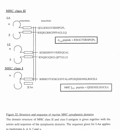
Recognition of I-Ace by antiserum to a peptide from its cytoplasmic domain. 123
Precipitation of Ii by the monoclonal antibody, In-1. 124
Post-synthetic modifications to MHC II in A20 cells. 125
MHC II is retained inside A20 cells for at least 2 hours. 127
At steady state MHC II resides mostly in vesicles of low density. 128
Newly synthesised MHC II resides in vesicles of intermediate density. 129
CHAPTER
5. A
CELL-FREE
SYSTEM
FOR
ANTIGEN
PROCESSING: IMMUNO-ISOLATION OF MHC II-POSITIVE
E N D O S O M E S
I N C L U D I N G
THE
P R O C E S S I N G
COMPARTMENT.
139
Introduction.
A. IMMUNO-ISOLATION OF MHC 11+^ ENDOCYTIC VESICLES.
A substantial minority of MHC IP ^ inverted vesicles have intermediate or high
density. 139
Anti-ttjyt immuno-isolates MHC IP'^ intracellular vesicles on magnetic
immuno-adsorbent. 140
Anti-a^ immuno-isolates MHC 11^'^ early endosomes. 141
B. METHODOLOGICAL ASPECTS OF THE IMMUNO-ISOLATION
TECHNIQUE. 143
Immuno-isolation requires an ionic environment. 143
Indirect isolation is better than direct isolation. 143
Anti-a^ is required in excess but free anti-a^yt is inhibitory to isolation. 144
Removal of cytosol. 144
c.
VESICLES ARE ISOLATED FROM ALL ELEMENTS OF THEINTRACELLULAR PATHWAY OF MHC II. 146
Right-side-out plasma membrane vesicles are not isolated by anti-a^yj. 146
Lysosomes, late endosomes and Golgi vesicles are immuno-isolated by anti-a^y^. 147
Electron microscopy of vesicles isolated by anti-a^^. 148
D. ISOLATION BY ANTI-a^^ ENRICHES FOR RECENTLY SYNTHESISED
MHC II 149
Early endosomes contain Ii. 149
Anti-ttcyt efficiently isolates vesicles containing recently synthesised a/B/Ii. 150
Conclusion. 151
CHAPTER 6. A PRELIMINARY STUDY OF CELL-FREE
ANTIGEN PROCESSING FUNCTION USING MATERIAL
OBTAINED BY IMMUNO-ISOLATION,
166
Introduction. 166
Ii is degraded by immuno-isolated proteinases. 166
Compact dimers do not form after peptide-loading of I-A^. 167
Future directions for the cell-free assay of antigen processing. 169
REFERENCES
172
LIST OF FIGURES
1. The range of density of percoll gradients used in this study 82
2. The two compartment model for endocytosis into early and late endosomes 94
3. Endocytosis and exocytosis of LY by A20 cells 95
4. Background adjustment for exocytosis of RD 96
5. Flow cytometric separation of mixed populations of A20 cells 97
6. Comparison of endocytosis of four different fluid-phase markers by A20 cells
over a 0, 2, 6, 20 and 60 minute pulse 98
7. ^^C-sucrose exocytosis from A20 cells 99
8. Separation of early and late endosomal contents by analysis of exocytosis 100
9. Two compartment analysis of endocytosis by A20 cells over 60 minutes 101
10. Detection of fast and slow emptying/filling in the same cell at the same time 102
11. Flow cytometric analysis of dendritic cells 103
12. Dendritic cells labelled with endocytosed RD 104
13. Volume of LY in early and late endosomal compartments of four different
APCs 105
14. Separation of cytosol and membranes on a percoll gradient 115
15. Profiles of endocytic markers on a percoll gradient 116
16. Distributions of HRP and Tf-HRP after pulse with or without chase 117
17. Position of a fluid-phase marker according to duration of endocytosis 118
18. Profiles of membrane-associated protein, measured by two different
techniques 119
19. Overlap between early endosomal transferrin and B-hexosaminidase as a
function of gradient size 120
20. The range of densities displayed by early endosomes 121
21. Assessment of recycling of ^^I-Tf through dense vesicles 122
22. Structure and sequence of murine MHC cytoplasmic domains 131
23. Precipitation of MHC II by anti-a^y^ 132
24. Precipitation and of Ii by In-1 133
25. Post-synthetic modifications to MHC II in A20 cells 134
26. Proteolysis of Ii 135
27. Export of newly synthesised MHC II to the cell surface 136
29. Precipitation of newly synthesised MHC II from fractions of a percoll
gradient 138
30. Density gradient of vesicles identified by anti-a^^ compared to plasma
membrane 153
31. Isolation of MHC^'^ vesicles by anti-a^^ 154
32. Direct visualisation of immuno-isolated MHC 11^^ membranes 155
33. Specific immuno-isolation of early endosomal marker by anti-a^y^ 156
34. Immuno-isolation in varying ionic environments 157
35. Comparison of indirect and direct immuno-isolation 158
36. Immuno-isolation of membranes during repeated isolation with sub-optimal
amounts of ImAd 159
37. Immuno-isolation of markers from a percoll gradient 160 and 161
38. Electron micrography of isolated vesicles from a light fraction of the
percoll gradient 162
39. Electron micrography of isolated vesicles from a dense fraction of the
percoll gradient 163
40. Inununo-isolation of early endosomal marker by In-1 164
41. Isolation of newly synthesised MHC II by anti-ûj^ 165
42. Degradation of Ii after inununo-isolation 170
43. Incubation of I-A** with peptide to form compact dimers 171
LIST OF TABLES
1. Calibration of sucrose refractometer 81
a/ 6 MHC II molecule containing a IL interleukin
and 6 chains ImAd immuno-adsorbent
a /6/Ii MHC n molecule containing KLH keyhole limpet haemocyanin
a, 6 and Ii chains LE late endosomes
^cyt cytoplasmic domain of LMP lo w m o l e c u l a r m a s s
I - A a ^ / W f / w polypeptide
AMTR anti-mouse transferrin receptor LY lucifer yellow
APC antigen presenting cell MHC m ajor histocom patibility
^ x y z absorbance at xyz nm complex
62m 62-microglobulin M H CI^t cytoplasmic domain of
BSA bovine serum albumin H-2L‘*
Di.I l ,l ’-dioctadecyl-3,3,3’,3’-tetra- mig membrane immunoglobulin
methylindocarbocyanine MPR mannose-6-phosphate receptor
perchlorate MVB multi-vesicular body
Di.O l,r-d ih ex ad ecy lo x a carb o - OPD O-phenylene-diamine
cyanine perchlorate PBP peptide-binding protein
DMEM Dulbecco’s modified Eagle’s PBS phosphate-buffered saline
medium PNS post-nuclear supernatant
DTT dithiothreitol PSF peptide supply factor
E.M. electron microscopy PTA phosphotungstic acid
EE early endosomes RD rhodamine dextran
EOF epidermal growth factor SDS-PAGE sodium dodecyl
sulphate-ELISA enzyme-linked immunosorbent polyacrylam ide gel
assay electrophoresis
ER endoplasmic reticulum SPDP 3-(2-pyridyldithio)propionic
FcR Fc receptor acid N-hydroxysuccinimide
FCS fetal calf serum ester
FITC fluorescein isothiocyanate SRBC sheep red blood cells
HB homogenisation buffer Tf transferrin
HPLC high performance liquid TfR transferrin receptor
chromatography TON rrwzj-Golgi network
HRP horse radish peroxidase TLC thin layer chromatography
IFN interferon t t tetanus toxoid
Ig immunoglobulin WB wash buffer
CHAPTER 1, INTRODUCTION TO THE CELL BIOLOGY OF ANTIGEN
PROCESSING FOR PRESENTATION BY MHC II
The cellular immune system has been the focus of much research since the discoveries
of lymphocyte recirculation, clonal selection, and acquired tolerance over 30 years ago.
Subsequent discoveries have revealed that antigen presenting cells convert antigen into
a specific form for recognition by the cellular immune system. This conversion is termed
antigen processing. Antigen processing is a complex series of intracellular events which
make use of previously unknown pathways. Therefore, antigen processing is as much in
the realm of cell biology as immunology. This review of antigen processing for
presentation by MHC II deals with the subject as follows:
A. antigen processing is placed in context: (i) within the overall immune response, (ii)
by contrasting different cells which process for presentation by MHC II;
B. the background for antigen processing for MHC II is described: (i) antigen processing
for MHC I; (ii) the endocytic pathway, (iii) the enzymes which degrade exogenous
antigens;
C. the molecular and cellular functions for antigen processing for MHC II are
summarised: (i) the interactions of MHC II and peptide, (ii) the cell biological
interactions of MHC II;
D. in the light of current knowledge of antigen processing, unresolved aspects and future
directions are discussed: (i) the phenomena which indicate a role for processing in
immune regulation, (ii) the intracellular sites where antigen is processed, (iii) the
experimental approaches for studying the cell biology of MHC II, including cell-free
A. THE CONTEXT OF ANTIGEN PROCESSING
fn ANTIGEN PROCESSING AS A PART OF THE IMMUNE RESPONSE
The cellular immune system: T cells recognise antigen in the context of MHC. The
immune system is capable of recognising and responding to non-self using primitive, non-
adaptive mechanisms. However, on their own these responses are inadequate for defence
against microbial colonisation. In phylogenetically more advanced animals, the immune
system has evolved an adaptive arm, the importance of which has recently been
publicised by the appearance of Acquired Immune Deficiency Syndrome. The adaptive
arm of the immune system also recognises and responds to non-self, but differs from the
non-adaptive arm by its capacity for amplification and memory. The adaptive immune
system has two divisions: humoral and cellular, which communicate with each other
closely. The humoral immune system recognises antigen with antibodies synthesised by
B cells. The antigens are intact, i.e. in the native state. Thus, foreign structures can be
detected in solution or on the surface of either invading micro-organisms or host cells.
The cellular immune system functions in the limited context of intracellular interactions.
The effector cells are termed T cells, and the cell recognised by a T cell is an antigen
presenting cell (APC). The ligand on APCs for T cells was determined to be the product
of major histocompatibility complex class I and class II genes (MHC I and MHC II)
(Zinkemagel and Doherty, 1974; Shevach and Rosenthal, 1973; Rosenthal, 1978). There
is more than one locus within each MHC class, the products of the different loci being
termed isotypes. Within each locus, there is a large amount of polymorphism, each allele
being termed an allotype. The differences between allotypes are smaller than the
differences between isotypes.
MHC II was also shown to control the level of response of in-bred laboratory animals
to foreign antigens, being called the "Immune response", or Ir genes (McDevitt et al,
1972; Benacerraf and McDevitt, 1972). In addition, the response of an individual animal’s
T cells to antigen was shown to occur only in the context of antigen presenting cells
expressing the same MHC as that animal, this phenomenon being termed MHC
restriction (Zinkemagel and Doherty, 1974). Therefore, antigen recognition by T cells
was shown to require the co-recognition of both MHC and antigen, and theoretically
shown that there is a single T cell receptor, structurally similar to an immunoglobulin
Fab, for both MHC and antigen (Haskins et oL, 1983; Chien et oL, 1984).
The different functions of T cytotoxic and T helper cells are related to the tissue
distribution of MHC I and MHC II. Two major T cells subsets were identified by the
mutually exclusive expression of CD4 and CD8 surface markers. The CD4^'^ subset,
termed T helper cells, is responsible for delayed-type hypersensitivity, and amplification
of antibody and cytolytic responses. The CD8^'^ subset, termed T cytotoxic cells, is
responsible for killing virally infected and tumour cells. The functional division of these
subsets is correlated with the requirement of T helper cells for APC which are
MHC II whereas T cytotoxic cells require APC to express MHC I. The molecular
basis for this has been demonstrated recently from the binding of CD4 to MHC II and
CD8 to MHC I. Without this extra binding, the activation of T cells is diminished (Bierer
et oL, 1989). In addition, CD4 and CD8 have signal transduction functions which are
important for T cell signalling (Janeway, 1992a). Therefore, CD4 and CD8 act as co
receptors for the interaction of T helper and cytotoxic cells with APCs bearing MHC II
and MHC I respectively.
The tissue distribution of the two classes of MHC differ widely. MHC I is expressed by
almost all nucleated cells. In contrast, MHC II is expressed primarily by cells in the
immune system (B cells, dendritic cells, activated macrophages, activated T cells and
thymic epithelium) as well as some cells outside the immune system. Therefore, most
MHC I^'"® cells are MHC 11'^. The term APC would lose its currency if applied to
antigen presentation by MHC I, and has traditionally been reserved for cells expressing
MHC II. The distribution of MHC I and MHC II indicates the differing roles of the T
cell subsets. T cytotoxic cells scan all nucleated cells, killing those with abnormal
expression of virus- or tumour-associated genes. T helper cells scan APCs within the
immune system, controlling the level and nature of responses to antigens the APCs have
acquired from microorganisms or other cells. A co-ordinated immune response is largely
the result of T helper function, for example at the level of cytokine secretion - different
T helper cell subsets secrete specific patterns of cytokines (Mosmann and Coffman,
T cells recognise short peptides derived from proteii antigens. The form of antigen
recognised by T cell receptor in the context of co-reccgnition of MHC is not the same
as the form recognised by antibody. Denatured antigem can be recognised by T cells but
fail to be detected by antibody. The antigen recognised by antibodies (termed B cell
epitopes) are generally the 3-dimensional configuration of tertiary structure (Atassi,
1975), not necessarily protein. In contrast, the antigens recognised by T cell receptors (T
cell epitopes) are the primary structures of proteins.
The earliest evidence was that antigen recognised by T cells was in the form of fragments
intimately associated with MHC (Benacerraf, 1978). Subsequently it was shown that short
peptides derived from internal sequences within an ant%en could substitute for epitopes
(Shimonkevitz et al, 1983 and 1984). The importance of antigen-derived peptides was
confirmed by the demonstration of binding with moderate affinity between short peptides
containing T cell epitopes and the MHC II molecules for which these epitopes were
restricted (Babbitt et al, 1985; Buus et al, 1986). Antigens which elicit antibody
responses but cannot elicit T cell responses are either very small (termed haptens) or
large, non-protein polymers (termed T-independent antigens); in both cases the antigen
cannot form short peptides.
Peptide binds to a groove on the apical surface of MHC. MHC molecules consist of 4
extracellular domains with 2-fold rotational symmetry. Two membrane-proximal domains
are part of the immuno-globulin superfamily and form a V-region-like heterodimer. Two
membrane-distal domains are similar to each other and form a closely linked
heterodimer, but are dissimilar to any other known polypeptide. MHC I is formed by a
heavy chain («44 kDa), which contains both the membrane-distal domains (a^, and
one membrane-proximal domain (a^). The fourth domain of MHC I is provided by Bg-
microglobulin (Bjm, 12 kDa), encoded outside the MHC. MHC II is formed from two
chains, a («33 kDa) and 6 («28 kDa), each with one membrane-distal domain (aj and
Bj) and one membrane-proximal domain («2 and B2).
The precise mechanism of antigen recognition emerged from the structure of an MHC I
molecule which was solved by X-ray crystallography (Bjorkman et al, 1987a). This
interaction of MHC I with T cell receptor (Bjorkman et oL, 1987b). The membrane-distal domains together form a longitudinal groove on the apical surface of the molecule. The
floor of this groove is constructed from a B-pleated sheet, and the two walls of the
groove consist of a-helices. In the groove, material was detected which was consistent
with a mixture of peptides. This is the only site with such clear peptide binding capacity.
In addition, the residues which are most responsible for polymorphism between allotypes
form the walls and floor of the groove. This indicates that the groove is indeed the
antigen binding site. More recent studies have shown that a characteristic set of peptides
is bound to each allotype in its apical groove.
The manner in which T cell receptors recognise MHC and peptide has been hypothesised
to be via direct contacts between the germ-line-encoded CDRl and CDR2 regions of the
T cell receptor and the a-helices of MHC, while antigen is recognised by CDR3, which
is extremely variable due to V(D)J joining and random nucleotide addition (Davis and
Bjorkman, 1988). In confirmation of this, specific interactions, such as acid-base pairs,
have been demonstrated between residues of CDR3 and MHC-bound peptide (Jorgensen
et aLy 1992). The conformation of an epitope is largely determined by polymorphic residues in the MHC groove to which it binds. Thus, the basis of alloreactivity across
small allelic differences is the appearance as non-self of self peptides in altered
conformations (Nathenson et aly 1986; Lechler et aly 1990; Cotner et aLy 1991). The solution of the structure of crystals of the MHC/peptide/T cell receptor complex is
awaited.
Theoretical minimum number of residues in T cell epitopes. The repertoire of T cell
receptors created by rearrangement has been suggested to have in the order of 10^
specificities (Kronenberg et aly 86). On this basis the T cell receptor must contact at least 5 residues (20^ « 3 x 10^). MHC binding must be less specific than T cell binding
to allow a wide range of pathogen-derived molecules to be presented. Assuming that the
smallest pathogens such as viruses, which may encode 1000 residues or less, might be
expected to yield at least one epitope, MHC binding might impose specificity on 2
residues (20^ = 400). Therefore, the shortest peptide which can fit the theoretical
The definition of antigen processing. Protein antigens are recognised in the form of a
short peptide bound to an MHC molecule. The series of events for both antigen and
MHC which bring this about is termed antigen processing. The subsequent interaction
with a T cell is termed antigen presentation.
Antigen processing of exogenous antigen is intracellular. The minimal molecular
requirements for antigen processing are the mechanism to expose T cell epitopes, and
the expression of MHC II which binds the epitopes. Antigen processing also involves
intracellular traffic of antigen and MHC II. The need for internalisation of antigen was
suggested by the finding that processing was sensitive to lysosomotropic agents such as
NH^"*" and chloroquine which neutralise lysosomal acidic pH (Ziegler and Unanue, 1982).
This indicated that antigen processing might occur in lysosomes. Mild fixation of APCs
abolishes antigen processing, while not affecting presentation either of antigen processed
prior to fixation, or of peptides (Shimonkevitz et al, 1983). In addition, processing was
shown to require cellular energy expenditure (Ziegler and Unanue, 1981; Werdelin and
Buus, 1983). Therefore, a complex intracellular event is required to process antigen.
Processed antigen is not usually released from inside one APC back to the extracellular
medium for other APCs to present (Harding et al, 1990; Steinman, 1991). This shows
that either epitope loading is internal or that epitopes are always kept associated with
membrane and thus their loss is prevented. Prevention of loss of soluble epitopes to the
extracellular medium has the advantage of preventing binding to MHC II on the surface
of nearby B cells, with the induction of incorrect B-T pairing (Mitchison, 1971).
Internalisation of antigen prior to degradation circumvents this difficulty by exporting
only those T cell epitopes which are bound to MHC II.
It has been proposed that dendritic cells are unable to process antigen but acquire
processed fragments from macrophages (Inaba et al, 1981). Such synergistic processing
has not been found subsequently (Inaba et al, 1990; Pure et al, 1990). Therefore, all
available evidence suggests that antigen processing occurs inside the presenting APC.
There are cell surface proteinases which might be capable of procesing antigen (Kenny,
1977; Buus and Werdelin, 1986a), but their role remains unclear. The plasma membrane
the likely site where synthetic peptides bind, with the therapeutic possibility to modulate
specific immune responses, either for stimulation (Milich et al, 1987; Berzofsky et al,
1991) or for inhibition (Wraith et al, 1989).
Different antigens are presented to T helper and T cytotoxic cells: there are two
intracellular pathways of antigen processing. The separation of function between T
cytotoxic and T helper cells requires that a different set of antigens must be presented
to each subset of T cells. For example, virally infected cells must be recognised by T
cytotoxic cells, while an APC must present a virus to T helper cells without itself being
infected with the virus. This is achieved by the presentation of endogenous antigens
synthesised in the cytosol of the cell by MHC I, and of exogenous antigens acquired from
outside the cell by MHC II.
MHC I presents cytosolic molecules (Townsend et al, 1986). Processing is insensitive to
disrupting lysosomal function (Morrison et al, 1986), but is dependent on protein export
(Yewdell and Bennink, 1989; Nuchtern et al, 1989). MHC I does not enter the endocytic
pathway (Neefjes et al, 1990). In contrast, MHC II presents exogenous molecules
(Germain, 1986; Yewdell and Bennink, 1990; Long and Jacobson, 1989). Processing is
less dependent on protein export than MHC I (Harding and Unanue, 1989; Adorini et
al, 1990; St.Pierre and Watts, 1990), and MHC II intersects the endocytic pathway
(Neefjes et al, 1990; Neefjes and Ploegh, 1992a). The general rule separating these two
pathways has notable exceptions (Carbone and Bevan, 1990; Jaraquemada et al, 1990;
Rock et al, 1990).
With two classes of MHC, and a corresponding division in the source of antigens, there
are two pathways of antigen processing. For MHC I the pathway is: (1) synthesis in
cytosol, (2) degradation, (3) translocation into ER, (4) binding to MHC I, (5) export to
plasma membrane. For MHC II the pathway is: (1) uptake by APC, (2) traffic into
MHC lU'^ endosome, (3) degradation, (4) binding to MHC II, (5) export to plasma
membrane. The pathways are kept separate by the absence of peptide binding to MHC II
in the ER, and the targeting of MHC II to endocytic compartments. Otherwise, the basic
schemes of the pathways differ only in the order of degradation and delivery to the MHC
of MHC is demonstrated by those peptides which bind to MHC of both classes (Choppin
et aly 1990; Hickling et oL, 1990; Perkins et aL, 1989). The dissimilarity is therefore largely a product of the differing cell biological roles of MHC I and n.
Only a small fraction of the MHC molecules of an APC express a single epitope. It has
been shown by analysis of peptide/MHC complexes extracted from APCs that the copy
number of a particular complex required to stimulate a T cell is surprisingly low.
Estimates using T hybridomas as responding cells indicate between 50 and 300 complexes
per APC are sufficient for T cell responses, i.e. 0.01% to 0.1% of the total MHC
(Demotz et al, 1990; Harding and Unanue, 1990a; Christinck et al, 1991). There are a
number of peptides represented at much higher levels than this, for example in one
MHC II species, 6 peptides occupy 50%-75% of the binding sites (Rudensky et al,
1991a). It has been suggested that T cell clones are more sensitive than T hybridomas,
and that in vivo responses may be to as few as 10-20 complexes (Germain, 1991). This
A.nn THE CELLS WHICH PROCESS AND PRESENT VIA MHC II
Macrophages. Macrophages consist of a heterogenous population of bone-marrow
derived, phagocytic cells found in many tissues, for example Kupffer cells lining hepatic
sinusoids. Unstimulated macrophages are MHC Expression of MHC II and the
second signal are both induced by bacterial cell wall components and inflammatory
cytokines such as gamma-IFN and IL-1 (Weaver and Unanue, 1990; Janeway, 1992b).
Antigen uptake can occur via several routes. Fluid-phase endocytic traffic is the least
efficient, but is at a higher rate than in other cell types (Besterman et oL, 1981). Receptor-mediated endocytosis of antigens opsonised with IgG or complement can occur
via receptors for Fc, C3b and C4b receptors (Gammon et al, 1987; Arvieux et al, 1988;
Gosselin et al, 1992). In addition, scavenger receptors with a broad, ill-defined specificity
recognise and enhance uptake of desialylated molecules and polyanions (Rohrer et al,
1990; Naito et al, 1991). Finally, phagocytosis delivers particulate antigens, such as whole
micro-organisms, into an MHC lU'^ compartment with many properties of late
endosomes (Harding and Geuze, 1992).
The highly active degradative pathway of macrophages does not imply a greater capacity
for processing of all antigens than less active cells (Chain et al., 1986). In contrast to dendritic cells, macrophages may act as APCs in the periphery, since they respond to
activation by inhibition of migration, and secretion of centrally and locally acting
cytokines, including IL-6, IL-1 and TNF, which can amplify the immune response.
B cells. Resting B cells process and present specific antigen, and activated B cells can
respond to several antigens (Chesnut and Grey, 1981; Chesnut et al, 1982). MHC II
expression is constitutive on B cells. Expression of the co-stimulatory molecule B7/BB1
is induced by bacterial and other inflammatory molecules (Liu and Janeway, 1991). Co
operation between B and T lymphocytes specific for the same antigen is an important
part of the adaptive immune response (Mitchison, 1971; Lanzavecchia, 1988). Initial
models were of simultaneous recognition by B and T cells, but since T cell epitopes are
the result of antigen processing within B cells, recognition must be sequential
(Lanzavecchia, 1988). B cells take up and processes antigen; epitopes are presented to
T cells; antigen-specific T cells in turn stimulate the presenting B cell. Therefore, to
be from an antigen which the B cell’s membrane immunoglobulin (mig) binds. This is
teological evidence for the importance of mIg in antigen presentation by B cells.
The importance of mig has been directly demonstrated by the presentation of antigen
by specific B cells at antigen concentrations as low as upto 10,000x less than by
irrelevant B cells (Pierce et cd., 1988; Lanzavecchia, 1985 and 1990). Once a response to
infection is underway, B cells are especially important when the antigen concentration
is low, when other APCs cannot take up sufficient antigen. As the infection is overcome,
antigen levels fall still further to a point where secreted antibody is in excess of antigen,
so there is no free antigen to be taken up. This mechanism for terminating successful
immune responses explains the prophylactic effect of passive immunisation with anti-D
in Rhesus disease of the newborn.
In the absence of ligand, mig (approximately 10^ per cell) recycles between pools of
roughly equal size on the plasma membrane and in early endosomes, with an average
cycling time of 15 minutes (Davidson et al, 1990). After antigen binding, mig is removed
from the cell surface. Polyvalent antigen/mlg complexes traffic into late endosomes and
lysosomes (Tony et al, 1985), the same route as other cross-linked receptors (for example
transferrin receptor, TfR) (Ekblom et al, 1983). For monovalent antigen/mlg, endocytic
routing is also altered away from recycling (Davidson et al, 1990). The signals for mig
routing are in its transmembrane and short cytoplasmic domains, and in the molecules
associated with mig (Reth et al, 1991) which are regulated by phosphorylation.
Antigen processing by specific B cells is extremely efficient (Lanzavecchia, 1985). EBV-
transformed B cells specific for Tetanus-toxoid (TT) presented antigen after a 48 hour
incubation at antigen concentrations at which 0.05% of mig was occupied with antigen
(Lanzavecchia, 1990), equivalent to 50 tetanus toxoid molecules per cell. Assuming
clearance of surface mIg-TT complexes every 15 minutes (Davidson et al, 1990), the
uptake of approximately 10,000 surface bound TT molecules was sufficient for
presentation. Assuming delivery of all these complexes for processing, and that T cells
recognise 50-300 specific MHC/peptide complexes, processing by these APC yielded a
single epitope from 0.5%-3% of antigen. The mechanisms which lead to this efficiency
(2) mig-bound antigen at a high concentration in the plane of the membrane; (3)
protection from further processing by mig of T cell epitopes within partially degraded
antigen.
Antigen is delivered, by mechanisms (1) and (2), into a compartment where processing
is efficient. There is ample evidence that mechanism (3) leads to the control of
processing by the fine specificity of the B cell’s mig (Ruud et aL, 1986; Davidson and
Watts, 1989; Davidson et aL, 1990). The B cell epitope is protected from degradation,
although other portions of antigen are lost and mig is cleaved into Fab/antigen remnant
+ Fc. Similar antibody-directed processing is seen for uptake via FcR. In a study using
macrophages as APCs, a panel of monoclonal antibodies specific for a single antigen
were used to enhance responses by a panel of T cell clones specific for the same antigen.
However, some combinations of antibody and T cell clones were not enhancing.
Antibody-directed processing, with the protection of T cell epitopes near to B cell
epitopes may explain the proximity of B and T cell epitopes on some antigens (Milich
et aL, 1986).
Migratoiy dendritic cells. Dendritic cells are the most potent in vitro APC, in particular
for primary responses. The first dendritic cells to be described were those in the
epidermis, which have taken the name of their discoverer in 1868, Paul Langerhans.
Dendritic cells are now known to form a small population in almost all organs. The
precursors of tissue dendritic cells are a small population of MHC'^ peripheral blood
mononuclear cells which differentiate into tissue dendritic cells in response to GM-CSF
(Inaba et aL, 1992) or in response to the combination of TNFa and GM-CSF (Caux et
aL, 92). The induction of tissue dendritic cells may be a way in which these cytokines
amplify local immune responses. After differentiation these cells become MHC 11^'^,
with multiple cytoplasmic extensions which contact a large number of nearby
parenchymal cells. Langerhans’ cells contain unique, membrane bound, acidified
organelles, named Birbeck Granules, which are in contact with the endocytic pathway
(Stossel et aL, 1990). Dendritic cells are non-phagocytic (Girolomoni et aL, 1990), do not
have many electron-dense lysosomes (Stossel et aL, 1990), and are correspondingly poor
at terminally degrading foreign material (Chain et aL, 1986). The poor lysosomal function
The tissue stage of dendritic cells is only an intermediate stage prior to migration and
maturation. This is enhanced by non-specific tissue injury either by direct signalling from
injured parenchymal cells, or via cytokines, possibly TNFa. Upon activation, dendritic
cells migrate into lymphatics, and draining secondary lymphoid organs (Steinman, 1991),
where they reside as interdigitating dendritic cells in T cell areas for several days before
dying (Macatonia et aL, 1987; Larsen et al, 1990). The maturation of tissue dendritic
cells has been studied by in vitro culture of Langerhans' cells (Schuler and Steinman,
1985; Romani et aL, 1989; Aiba and Katz, 1991). Cell biological aspects of the
maturation include the cessation of synthesis of E and MHC II, with undiminished
expression of cell surface MHC II (Pure et al, 1990; Kampgen et aL, 1991). In addition,
a reduction in the ability to process antigens upon maturation has been deduced from
the loss of acidification of endocytic organelles and Birbeck granules (Stossel et aL,
1990). Therefore, antigen acquired and processed at the early stages of activation in the
periphery is retained for presentation after migration. Thus, mature dendritic cells are
less able to process antigen encountered after leaving the periphery.
Dendritic cells' potency as APCs derives from high level expression of MHC I, MHC II,
B7/BB1 and the adhesion molecules CD 11a, CD 18 and CD54 (LFA-1, ICAM-1 and
LFA-3), and from low levels of cell surface glycoprotein sialylation (King and Katz, 1990;
Young et aL, 1992). Intracellular MHC II has been found throughout the endocytic
pathway, as well as in non-endocytic vesicles (Arkema et al, 1991). The reduced surface
sialylation may enhance (1) uptake of cationic antigens (Apple et aL, 1988), (2) specific
immune recognition of MHC/peptide (Boog et al, 1989), (3) clustering with T cells
which occurs prior to engagement of MHC II by T cell receptor and CD4 (King and
Katz, 1990). Dendritic cells present antigen to and stimulate T cytotoxic cells without
ensuing lysis, and may therefore provide an important site for T helper cells to control
T cytotoxic cell responses in three cell clusters (Mitchison and O'Malley, 1987). In
support of this role for dendritic cells, dendritic cells have been suggested to be the
APCs capable of presenting exogenous antigens by MHC I (Carbone and Bevan, 1990;
Rock et aL, 1990), although this activity has not been detected in all systems (Lopes and
Chain, 1992).
uptake. Although some dendritic cells in primary lymphoid tissues are post-migratory
following inflammatory responses, interdigitating dendritic cells are detected at all times
in lymphoid tissue, not only after inunune stimulation (Kamperdijk et aL, 1985). This
non-stimulated population is likely to include the cells which acquire antigens from the
circulation (Crowley et aL, 1990). Further evidence in favour of additional subsets of
splenic dendritic cells comes from the heterogeneity of dendritic cell markers in the
spleen (Aiba and Katz, 1990; Agger et aL, 1992). Unlike macrophages, splenic dendritic
cells have very low levels of cell surface receptors for antigen: i.e. opsonin or scavenger
receptors (Steinman, 1991; Naito et aL, 1991). This implies that non-specific means of
uptake including fluid-phase endocytosis may be an important mechanism of antigen
entry for splenic dendritic cells (Janeway, 1992b).
However, splenic dendritic cells have been shown to have poor fluid-phase endocytic
activity, especially in comparison with macrophages (Kapsenberg et al, 1986; Inaba et al,
1990). Although evidence for co-operation between macrophages and dendritic cells was
obtained some years ago, and taken to imply the transfer of pre-processed antigen (Inaba
et aL, 1981), this finding has not been repeated (Inaba et al, 1990; Pure et aL, 1990). Therefore, how might non-migratory splenic dendritic cells acquire antigen? It is possible
that the claim of endocytic inactivity is inaccurate, since these studies measured uptake
after long pulses of a fluid-phase marker. From kinetic studies of endocytosis in
macrophages and fibroblasts, a pulse of several hours or more leads to saturation of
endosomes and uptake into lysosomes (Besterman et aL, 1981; Swanson et aL, 1985).
Relatively poor lysosomal function clearly does not inhibit processing of all antigens by
dendritic cells. Therefore, it is important to measure the traffic of fluid-phase markers
through pre-lysosomal compartments.
In Chapter 3, a technique to assess fluid-phase endosomal traffic in dendritic cells is
described. Fluid-phase traffic through early and late endosomes was shown to be similar
to several B cell APCs. This confirms that splenic dendritic cells can take up antigen by
fluid-phase endocytosis.
Other cells. The expression of MHC II and antigen processing in the thymus (thymic
from non-self (Robertson er a/., 1991). In addition, the expression of MHC II can be
induced on many cell types. Within the immune system, activated human, but not rodent
T cells express MHC II and are able to process and present antigen (Hewitt and
Feldmann, 1989), although the physiological importance of this is not known. On cells
outside the immune system, for example pancreatic islet cells, expression of MHC II is
induced as part of a non-specific inflammatory response, and may be responsible for
auto-immune responses to (normally) sequestered antigens (Todd et oL, 1985; Bottazzo
et aLy 1985). MHC II is also expressed on activated endothelial cells exposed to gamma- IFN, where it may be involved in delayed-type hypersensitivity responses (Pober and
Cotran, 1990). MHC II is found at all times on gut epithelium, although its function is
not known.
A wide range of cell types express MHC II under some circumstances. Transfection of
other cell types also demonstrates that often the only additional requirement for a cell to
process and present antigen is the expression of MHC II and li. Therefore, the other
molecules involved are constitutively expressed in the large majority of cells, implying
that antigen processing uses many facets of the pre-existing exocytic, endocytic and
degradation pathways.
Mechanism of antigen presentation. The binding between MHC/peptide and T cell
receptor together with CD4/8 is usually insufficient to activate T cells (Springer et aL,
1987). There are a number of adhesion molecules expressed on T cells with a paired,
complementary protein on the target cells. Examples of such pairs are CD2/LFA-3,
LFA-l/ICAM-1. These multiple interactions occur prior to engagement of T cell
receptor by MHC, and are thought to be a means of focusing T cells to their targets.
Other interactions may also determine the outcome of the encounter between T cell and
target. There is a second signal which is required for presentation to lead to T cell
activation rather than T cell anergy. This signal is delivered by B7/BB1 on APCs, the
ligand for which is CD28 on T cells (June et al., 1990).
The strength of the antigen-non-specific interaction determines the amount of antigen-
specific interaction which must occur. For T cell hybridomas and other secondary T cell
for naive cells because of greater antigen-non-specific interaction (Cerottini and
MacDonald, 1989). Therefore, secondary responses require less antigen processing before
successful presentation.
MHC II expression and antigen processing capacity in an APC are related to its role in
antigen presentation. The immune response is partly regulated by control of APC
functions: MHC II expression; second signal expression; and, in some cases, the antigen
processing machinery. The main APC types have distinctive roles for which they have
adapted different patterns of regulation of APC function.
Macrophages are particularly suited to process particulate antigens. Thus, they are likely
to be the predominant APC for bacteria. To avoid presentation of self cellular
components, for example apoptotic bodies, the expression of both MHC II and second
signal is only induced in the presence of pathogen-related molecules.
The important aspect of B cells as APCs is to recruit T cell help for the production of
Ig by focusing T helper cells onto the very same antigens that are recognised by the B
cells’ mig. The constitutive expression of MHC II, compared to macrophages, may allow
initiation of secondary immune responses after antigen uptake by resting memory B cells.
It has remained uncertain whether resting B cells can elicit primary T helper responses.
However, a recent study shows that this interaction can occur, mediated via the induction
of B7/BB1 (Koulova et al, 1991).
The role of migratory dendritic cells is to scan parenchymal cells of non-lymphoid organs
for abnormal protein synthesis, and present antigens in primary lymphoid tissues.
Although the second signal is constitutively expressed, these cells do not contact large
numbers of T cells until inflammatory signals lead to migration. The maturation process
is the best-documented example of down-regulation of the antigen-processing machinery,
in this case to preserve a cohort of previously processed antigen.
The only APCs in primary lymphoid organs which constitutively express the second signal
are the resident dendritic cells, which also are the APCs with the most limited means of
non-specific recognition, possibly detecting pathogens which do not express molecules
such as lipopolysaccharide, mannan and dsRNA. In addition, these cells may be
particularly involved in responses to viruses, the pathogens which are the least able to
B. THE BACKGROUND TO ANTIGEN PROCESSING FOR MHC II
(\) ANTIGEN PROCESSING FOR MHC I
MHC I binds 8-9mers. The crystal structure of MHC I was shown to contain peptide in
the apical binding groove (Bjorkman et aL, 1987 a and b). Since then, it has been shown
that MHC I retains low molecular weight material during immuno-precipitation.
Separation on reverse-phase HPLC reveals a large number of peptides (for example:
Van Bleek and Nathenson, 1990; Rotzschke et aL, 1990), although even more peptides
which bind with low affinity may be lost during the precipitation. Some MHC I molecules
yield mostly 9mers; others yield Smers. These peptides are much shorter than was
previously expected (Townsend et aL, 1986). This length of the processed epitope is very
similar to the theoretical minimum of 7 residues which could contain sufficient
information for specific immune recognition by both T cell receptor and MHC (see
above), demonstrating the efficiency of the tertiary complex of MHC-peptide-T cell
receptor. One or two residues have been found to be fairly invariant for all peptides
binding to a particular MHC I species, and have been termed anchor residues (see
below).
The peptide is in an extended confirmation (Madden et aL, 1991), not an a-helix as was
once postulated (DeLisi and Berzofsky 85, Rothbard and Taylor, 88). The peptide
backbone has key interactions at its N- and C- termini, which fix the peptide into
position. Peptide side-chains of certain residues, particularly the anchor residues, fit into
well-defined side-pockets in the peptide-binding groove (Garrett et aL, 1989). An
individual side chain is selected by the lining of the side-pocket, which is partly made up
of the polymorphic residues within MHC I. In this way different MHC molecules are
able to select different groups of peptides.
The structures of several MHC I-peptide complexes have now been solved (Fremont et
aL, 1992; Matsumura et al., 1992; Zhang et aL, 1992; Silver et aL, 1992; Guo et aL, 1992;
Madden et aL, 1992). These structures demonstrate that peptide can form a bulge away
from the floor of the binding groove 4-5 residues from the N-terminus, this being the site
in the peptide which accommodates the single residue variation in length between
different complexes for the same MHC I molecule. An important, but subtle feature of
groove is affected by peptide binding (Fremont et al, 1992). This may reflect the trapping of bound peptide, and thus account for the slow dissociation of peptide (Buus
et aL, 1986; Elliott et aL, 1991).
The bound peptides have an MHC-dependent motif. In a single species of MHC I, the
eluted peptides have been sequenced as a pool. An overall pattern, or motif, is apparent,
which is specific for an individual MHC I species (Falk et al, 1991). Anchor residues are
represented by a single amino acid, or a limited set of related amino acids. Less strict
requirements are imposed on other positions, at which the amino residues are thought
to interact less specifically with MHC. A third group of positions contains a large range
of amino acids, these positions being proposed to interact not with MHC but with T cell
receptor.
More recently, HPLC has been combined with mass spectroscopy to achieve better
resolution of peptides eluted from MHC I, and to sequence the major species. The
results confirm that peptides are mainly derived from cytosolic proteins, such as the
human nuclear protein p68 (Jardetzky et aL, 1991; Hunt et aL, 1992a). In addition,
peptides derived from signal sequences have been detected (Hunt et al, 1992a;
Henderson et aL, 1992).
A different method of determining the range of peptides bound by MHC I has been to
synthesise a wide range of 8-9mers which are all related to a suggested motif
(Schumacher et aL, 1992). The peptides which bind can be eluted from MHC and
visualised in 2 dimensions (HPLC and TLC) to produce a "fingerprint" for that MHC
molecule. Point mutations in the MHC molecule alter peptide binding by loss and gain
of certain specificities.
Production, translocation and binding to MHC I of 8-9mers. There is very little data on
the cytosolic enzymes which process antigen. It has been suggested that the ubiquitin
pathway, a common cytosolic degradation system, can lead to MHC I processing
(Townsend, 1987; Monaco, 1992a). However, multiple routes are possible since non-
ubiquitinated antigens introduced into the cytosol are also presented (Moore et aL, 1988).
molecular mass polypeptide (LMP), a cytosolic multi-chain proteolytic complex (Monaco,
1992a) which might deliver peptides from the ubiquitin degradative pathway to the
translocation step. Despite the candidacy of these molecules for a role in MHC I
processing, their importance has recently been thrown into doubt (Arnold et aL, 1992),
and their role is unclear.
Some mutant cell lines selected for low MHC I expression present peptides but not
endogenous antigens (Townsend et aL, 1989; Hosken and Bevan, 1990), due to lack of
peptide supply for MHC I binding (Schumacher et aL, 1990). The genetic defect in the
mutant cell lines has been detected as a deletion within the MHC II locus (Deverson et
aL, 1990), which contains 2 largely homologous ATP-binding cassette proteins (Trowsdale
et aL, 1990; Spies and DeMars, 1991). It has been postulated, but not proven, that these proteins are, like other members of this family, membrane transporters. The dimer
formed by the two polypeptides, termed peptide supply factor (PSF), may translocate 8-
9mers from the cytosol into the ER (Spies et aL, 1992). In the absence of PSF, MHC I
epitopes are not recovered from a total cell lysate (Falk et aL, 1990). Therefore, these short peptides are either not created or rapidly degraded in the absence of MHC I.
Signal sequence-derived peptides tend to be longer than 8-9 residues, indicating that the
usual length of epitopes is imposed by pre-ER processing. In the rat, which has less
polymorphic MHC I genes than mouse and human, allelic variants have been detected
in the PSF genes (Monaco, 1992b). The PSF alleles have been shown to lead to loading
of MHC I with different sets of peptides, thus enhancing overall MHC polymorphism
(Powis et aL, 1992).
Increasing the length of peptides from 8-9mers leads to rapid loss of affinity. For
example, an impurely synthesised peptide of 12 or 16 residues bound only weakly to
MHC I, while a minor contaminant of 9 residues, which was not formed by degradation
after MHC binding, was recovered from MHC I (Schumacher et aL, 1991). Therefore,
longer peptides have a much reduced affinity for the binding site. This is likely to be
caused by the failure of longer peptides to be bound by MHC I at both termini of the
peptide back-bone. A longer peptide may bind, but one or both termini are incompletely
It has been shown that MHC I expression can be stabilised on PSP*““ cells by peptide,
B2I11, or low temperature. The mechanism underlying this stabilisation is the acquisition
of a temperature-stable conformation after binding both Bgm and peptide (Ljunggren et
al, 1990; Schumacher et al, 1990; Townsend et al, 1990; Elliott et al, 1991), which shows that peptide has a structural role. As discussed below, peptide plays a similar, though
lesser role, in determining MHC II structure.
Presentation of endogenous antigens by MHC I is inhibited by treatment of cells with
Brefeldin A, a fungal product which has pleiotropic actions on several compartments in
the cell, including preventing exit of newly synthesised proteins from the Golgi (Nuchtern
et al, 1989; Yewdell and Bennink, 1989; Cox et al, 1990). This shows that only newly synthesised MHC I binds peptide. The most favoured site for this binding is the ER.
Exogenous antigens are sometimes presented by MHC I. It has generally been found that
cytosolic loading is required for presentation of antigen to CD8'^'* T cells via MHC I (for
example: Lopes and Chain, 1992; Collins et al, 1992). However, exceptions have been
reported. For instance,anMHC 11^'^ sub-population of splenocytes does not need cytosolic
loading of antigen (Rock et al, 1990). A further exception to the rule has been described
for cell-associated antigens, which are processed in vivo for presentation by MHC I
(Carbone and Bevan, 1990). This indicates a pathway involving phagocytosis of cells, and
subsequent presentation of their contents by MHC I. Although the APCs involved in
either of these exceptions have not been identified, dendritic cells have been suggested