R E S E A R C H
Open Access
Comparison of the types of candidate
reference samples for quality control of
human epidermal growth factor receptor 2
status detection
Yulong Li
1,2,3, Rui Zhang
1,3, Yanxi Han
1,3, Tian Lu
1,2,3, Jiansheng Ding
1,2,3, Kuo Zhang
1,3, Guigao Lin
1,3,
Jiehong Xie
1,3and Jinming Li
1,2,3*Abstract
Background:Human epidermal growth factor receptor 2 (HER2) is as a target gene for trastuzumab in patients with breast cancer. Accurate determination of HER2 status and strict quality control are necessary to ensure reproducibility and accuracy of the techniques used for the determination of HER2 status.
Methods:We used three different types of samples: formalin-fixed and paraffin-embedded (FFPE) samples prepared from cell lines, agarose gel samples using cell lines, and xenograft tumor samples. One cell line for FFPE or
xenografts did not overexpress HER2, while the others showed different levels of HER2 overexpression. We compared the morphology,HER2gene amplification status, and HER2 protein expression status of these samples with those of clinical specimens.
Results:We successfully produced three kinds of samples for quality control. Cells from the cell line-sample sections were dispersed while those from the agarose gel-sample sections and xenograft tumor sample sections (prepared from the both cell lines) were concentrated in one area. The FISH results for all three kinds of samples were as expected. The IHC results of the cell line samples and xenograft tumor samples were as expected, but the staining level of the agarose gel samples, using HER2-overexpressed cell lines was weak which might be regarded as a false negative result.
Conclusions:Xenograft tumor samples might be used as an additional option for quality control in FISH and IHC. However, it might not replace the clinical specimen quality controls directly.
Keywords:Quality control, Cell lines, Agarose gel, Xenograft, Clinical specimens, FISH, IHC
Abbreviations:ASCO, American Society of Clinical Oncology; CAP, College of American Pathologists;
DAB, Diaminobenzidine; DAPI, 4′,6-Diamidine-2′-phenylindole dihydrochloride; EQA, External quality assurance; ER, Estrogen receptor; FDA, Food and Drug Administration; FFPE, Formalin-fixed and paraffin-embedded;
FISH, Fluorescence in situ hybridization; HE, Hematoxylin-eosin; HER2, Human epidermal growth factor receptor 2; IHC, Immunohistochemistry; IQC, Internal quality control; PBS, Phosphate buffered saline; PR, Progesterone receptor; SCID, Severe combined immunodeficient; TMA, Tissue microarray; US, The United States
* Correspondence:[email protected]
1National Center for Clinical Laboratories, Beijing Hospital, National Center of
Gerontology, No1 Dahua Road, Dongdan, Beijing 100730, People’s Republic of China
2Graduate School, Peking Union Medical College, Chinese Academy of
Medical Sciences, Beijing, People’s Republic of China
Full list of author information is available at the end of the article
Background
Approximately 25 % of patients with invasive breast can-cer overexpress human epidermal growth factor receptor 2 (HER2) [1]. HER2 overexpression, mainly because of
HER2amplification, is significantly associated with aggres-sive disease and poor prognosis [2]. Trastuzumab can bind the extracellular domain of HER2, and has a remarkable impact on the treatment of patients with HER2-positive breast by several pathways [3–5]. Therefore, trastuzumab was approved by the United States (US) Food and Drug Administration (FDA) for the treatment of breast cancer. Hence, accurate determination of the HER2 status in pa-tients with breast cancer is crucial [6].
Immunohistochemistry (IHC), for measuring protein overexpression, and fluorescence in situ hybridization (FISH), for measuring HER2 amplification, are the most commonly applied methods for the assessment of HER2 status [7]. Although both these methods can be applied to evaluate protein overexpression and gene amplifica-tion in relaamplifica-tion to the morphological features of tumors, there is large inter-laboratory variation in the procedure, staff, and interpretation, which affects the analytical sen-sitivity and specificity of these assays, especially in the case of indeterminate samples [8].
Ensuring the accuracy and reproducibility of HER2 de-tection by IHC and FISH testing is a fundamental pre-requisite for targeted therapy. Recommendations for HER2 testing were developed by the American Society of Clinical Oncology (ASCO) and the College of Ameri-can Pathologists (CAP) in 2007 and were updated in 2013 [9, 10] to improve the performance of these tests. HER2 detection requires precisely characterized and uni-versally available reference controls [11]. Adequate qual-ity control is necessary to diminish inter-laboratory variation and external quality assurance.
Recently, a consensus was reached to create clinical specimen or cell line controls as reference materials for HER2 testing [12]. Quality control of clinical specimens facilitates the assessment of variation in methodologies and laboratories. However, some disadvantages have lim-ited the development of these quality controls, such as production and ethics. Therefore, we developed new xenograft tumor controls and checked their suitability for internal quality control (IQC) and external quality assurance (EQA). In this study, we compared three qual-ity control samples with traditional clinical specimens. The aim of our study was to identify the most appropri-ate quality control that would ensure maximum accur-acy and reproducibility of IHC and FISH.
Methods Cell lines
The breast cancer cell lines MCF-7, MDA-MB-453, BT474, and SKBR-3 were obtained from the Cell Based
Medical Center of Basic Medicine of Peking Union Med-ical College (Beijing, China). Cells were cultured as rec-ommended by the suppliers. The HER2 status of the cell lines has been reported in previous studies [1, 13]. MDA-MB-453, BT474, and SKBR-3 expressed HER2, while MCF-7 did not. All four cell lines were applied for the preparation of three quality-control samples as de-scribed below.
Samples
Two surgical specimen cases of invasive breast cancer with of known HER2 status were selected and obtained from the Pathology Department of Beijing Hospital, Beijing, China. Our protocol and specimen were ap-proved by the Ethics Committee of the National Center for Clinical Laboratories. One of the samples tested negative for HER2 (A2; FISH result: negative; IHC score: 0), whereas the other sample tested positive (A12; FISH result: positive; IHC score: 3+). The HER2 status of these two samples was evaluated together with our pre-pared samples.
Preparation of formalin-fixed and paraffin-embedded (FFPE) cell line samples
Approximately 1 × 10⁷–2 × 10⁷ cells were obtained and fixed in 10 % neutral-buffered formalin for approxi-mately 2 h at room temperature. Subsequently, cell pellets were processed with gradient ethanol for dehy-dration and with xylene for transparency. Cell pellets were treated with melted liquid paraffin in small Eppendorf tubes and waxed thoroughly for 10 min; liquid paraffin was mixed with the cell pellets, using pipette tips. The so-lution was then chilled to achieve solidification, and the resulting blocks were embedded in paraffin, using a stand-ard histochemical apparatus. Hematoxylin-eosin (HE) staining was performed to preserve the representative morphology of each sample and the number of cells to as-sess the uniformity of cellular distribution for each sec-tion. Each section was 5 μm in thickness. The sections were mounted onto slides and dried in an oven at 70 °C for approximately 3 h to ensure maximum adhesion.
Preparation of FFPE agarose gel samples
was embedded in prewarmed and equilibrated 3 % PBS-buffered agarose gel in 5-mm diameter × 5-mm deep iron molds from which small agarose-cell mixture cylinders were extruded after the agarose was sufficiently chilled. The agarose gel cylinders were then placed in a standard tissue-processing cassette and processed with paraffin wax. HE staining was performed; each section was 5 μm in thickness.
Preparation of FFPE xenograft tumor samples
Female severe combined immunodeficient (SCID) mice between 21 and 28 days of age (Vital River, Beijing, China) were used; the mice were quarantined for at least 3 days prior to the study. The mice were anesthetized with vaporized isoflurane, and suspensions of cells (all the four cell lines, approximately 1 × 10⁷ cells) mixed with Matrigel™ basement membrane matrix (Corning, New York, USA) were injected subcutaneously under sterile conditions into the backs of mice, using a 5-gauge needle. Once the size of the xenograft tumor had pro-gressed to approximately 500 mm3, the mice were eu-thanized via cervical dislocation, and all xenograft tumors belonging to different cell lines were removed surgically. All animal experiments were performed in compliance with the guidelines specified by the Institute for Experimental Animals, Beijing Hospital. Following surgical removal of xenograft tumors from the SCID mice, the tumors were cut into multiple pieces of ap-proximately 250 mm3, placed into appropriately labeled disposable plastic tissue cassettes, and then immediately fixed in 10 % neutral-buffered formalin at 4 °C overnight. Subsequently, automated tissue embedding was per-formed using a Renaissance Tissue Processor (Ventana Medical Systems, US). This included washing in a graded ethanol series for dehydration followed by xylene washes for transparency, and heated paraffin washes prior to em-bedding in paraffin. HE staining was performed to ensure that the representative morphology of each sample was maintained. Each section was cut to a thickness of 5μm.
Evaluation ofHER2status by FISH
FISH assays were performed following the manufacturers’ instructions (PathVysion HER-2 DNA Probe Kit, Abbott, Chicago, IL, USA). In brief, deparaffinization, heat pre-treatment, and protease treatment were performed se-quentially for all the FFPE samples. After pretreatment, the sample sections and probes were co-denatured and hybridized. A post-hybridization wash was performed for all sample sections in the dark using the wash buffer sup-plied by the manufacturer. 4′,6-Diamidine-2′ -phenylin-dole dihydrochloride (DAPI) counter-stain was applied to the target area of the sections, and fluorescence microscopy was performed to view areas of positive
hybridization. The results were evaluated qualitatively based on the kit recommendations.
Evaluation of HER2 expression by IHC analysis
The HER2 protein expression status of the sample slides was assessed using a commercial primary antibody (PATHWAY® anti-HER-2/neu (4B5) Rabbit Monoclonal Primary Antibody; Ventana, Tucson, AZ, USA), as per manufacturer’s instructions. Briefly, all the sample slides were processed according to a standard tissue-processing process, including deparaffinization, rehydration and anti-gen retrieval. After cooling for 15 min, IHC reactions were manually performed manually using the primary mono-clonal antibody, 2-step plus® Poly-HRP Anti Mouse/Rabbit IgG Detection System (ZSGB-BIO, Beijing, China) and di-aminobenzidine (DAB) staining. HER2 protein expressed on the membrane of tumor cells was scored as 0+, 1+, 2+, or 3+ as recommended [10].
Results
Characteristics of the three types of samples for quality control
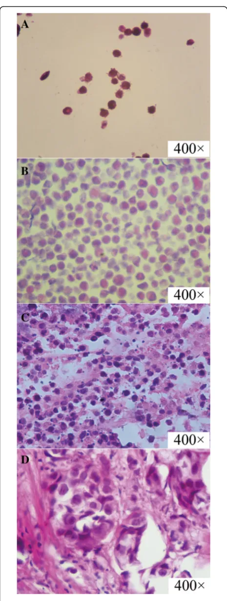
In this study, we successfully developed three types of samples that may be used for quality control in the de-termination of HER2 status. The morphological features of these samples in comparison with those of clinical specimens are shown in Fig. 1. In brief, the target cells in the cell line samples were well dispersed, but cells embedded in the agarose gel samples were concentrated in one area. However, the xenograft tumor samples were similar to tumor tissue samples derived from patients with breast cancer. Nests of carcinoma were found in both the xenograft tumor samples and clinical speci-mens. However, some distinctions were also found in the samples. In the clinical specimens, a sizeable number of normal cells was found, and the tissue structure was complicated. Apart from the nests of carcinoma, breast catheter and breast glands were observed. Inflammatory cell infiltration was evident around the nests of carcin-oma. In the xenograft tumor samples, almost all the cells were tumor cells, and the tissue structure was simple. Apart from the target tumor cells, only red blood cells, fibroblast cells, and vascular endothelial cells were de-tected. The other characteristics of the samples, as well as the technology used and costs are shown in Table 1.
Evaluation ofHER2status by FISH
positive for HER2 amplification. Together, these results suggested that all the samples could be applied for qual-ity control in FISH assays.
Evaluation of HER2 expression by IHC analysis
A comparison of HER2 expression between the samples developed by us and the clinical specimens is shown in Table 2 and Fig. 3. Cell line samples and xenograft tumor samples prepared from SKBR-3, BT474, and MDA-MB-453 showed overexpression of HER2 protein localized to the cell membrane (IHC score: 3+) relative to samples prepared using MCF-7 (lower expression; IHC score: 0) and the negative clinical sample (A2). In addition, cell line samples and xenograft tumor samples using SKBR-3 and BT474 expressed higher levels of HER2 than samples prepared using MDA-MB-453 and the positive clinical sample (A12). There was no differ-ence of staining between the cell line samples and their corresponding xenograft tumor samples. However, agar-ose gel samples prepared using MCF-7, MDA-MB-453, and SKBR-3 showed negative results, and the HER2 ex-pression in agarose gel samples prepared using BT474 was lower than that in the cell line samples and xeno-graft tumor samples prepared using BT474. The de-tailed IHC staining scores are shown in Table 2 and Additional file 2.
Discussion
Quality controls are required in HER2 status detection to ensure the accuracy and reproducibility of testing [2, 10] and hence, choosing a suitable quality control is ne-cessary for FISH and IHC assays. Currently, the best quality controls for HER2 testing are tissue-based con-trols from clinical specimens exhibiting both HER2 over-expression and HER2 amplification that have been confirmed by IHC and FISH [2]. Some studies have also reported that cell line samples can be used for EQA as additional quality controls instead of traditional quality controls derived from patient tumor tissue [13, 14]. In this study, we have developed three quality controls and compared them with cell line samples and clinical speci-mens to identify a suitable quality control for FISH and IHC assays.
Different quality controls possess characteristic advan-tages and disadvanadvan-tages. At present, tissue microarray (TMA)-based or FFPE-based quality controls prepared from clinical specimens are widely used for EQA or IQC in IHC or FISH testing [15, 16]. The protocol utilizing
Fig. 1The HE staining results of three kinds of samples compared
clinical specimen quality controls is robust, cost-effective, and rapid. Moreover, it allows continuous monitoring of the performance of FISH and IHC testing [16]. However, some potential limitations of clinical spe-cimen quality controls should be considered. Since, EQA requires a larger number of quality controls, a collection of large clinical specimens from different patients with breast cancer is needed. Previous studies have reported that the properties of the samples being tested may in-fluence the results and create bias in the comparison [17]. Thus, heterogeneity among clinical specimens from different patients may affect the reproducibility of assays. Moreover, mass production of clinical specimens is diffi-cult, and ethical problems should be taken into consider-ation. As additional quality controls, cell line quality controls are currently applied for IQC and EQA instead of traditional clinical specimens. Compared with trad-itional paraffin blocks of tumor tissues, large amounts of such controls can be obtained easily, and the reproducibil-ity allos for sensitive detection and decreases analytical
errors [11]. In our study, we found that cell line samples can be applied for both IHC and FISH, as reported by pre-vious studies [11, 18], and that the protein and gene status of different cell lines can be determined accurately.
However, we found two main disadvantages in the use of cell line quality controls. First, the tumor cells showed diffuse distribution in the entire section, and the diffused cells were difficult to observe and count. The diffused cells were also prone to drop off for lack of tissue struc-ture. Second, cultured cells were prone to polysomy, which is uncommon in patient tumor tissue. This polys-omy might affect the reproducibility of cell line quality controls [19].
To resolve these issues, agarose gel samples prepared using different cell lines were produced to optimize the basic cell line samples. Embedding in agarose gel helped gather the cells and increase the viscosity of the speci-men. However, the morphological features of actual hu-man tumors could not be simulated. Furthermore, we found that the agarose gel samples could only be applied
Table 1The characteristic of three samples for quality control
Quality control samples Cell lines samples Agarose gel samples Xenograft tumor samples Clinical specimens
Morphological feature Diffused distribution without tissue structure
Concentrated distribution without tissue structure
Concentrated distribution with simple tissue structure
Concentrated distribution with complicated tissue structure
Target cells count in each section >104 >103 Countless >102
Adhesion characteristic Bad Good Good Bad
Production process Easy Easy Difficult Difficult
Production period 1 month 1 month 3–4 months About 2–4 months [15]
Production cost (£/sample on one section)
2.03 2.07 2.22 About 10 [15]
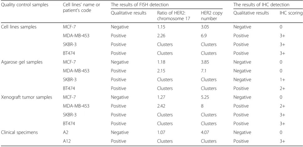
Table 2The results of FISH and IHC method to evaluate three samples made of different cell lines for quality control
Quality control samples Cell lines’name or patient’s code
The results of FISH detection The results of IHC detection
Qualitative results Ratio of HER2: chromosome 17
HER2 copy number
Qualitative results IHC scoring
Cell lines samples MCF-7 Negative 1.15 3.05 Negative 0
MDA-MB-453 Positive 2.26 6.9 Positive 3+
SKBR-3 Positive Clusters Clusters Positive 3+
BT474 Positive Clusters Clusters Positive 3+
Agarose gel samples MCF-7 Negative 1.18 3.85 Negative 0
MDA-MB-453 Positive 2.15 7.1 Negative 0
SKBR-3 Positive Clusters Clusters Negative 1+
BT474 Positive Clusters Clusters Positive 2+
Xenograft tumor samples MCF-7 Negative 1.27 5.25 Negative 0
MDA-MB-453 Positive 2.42 8 Positive 2+
SKBR-3 Positive Clusters Clusters Positive 3+
BT474 Positive Clusters Clusters Positive 3+
Clinical specimens A2 Negative 1.07 4.07 Negative 0
for FISH. The use of an overheated agarose gel led to changes in the morphology of cells. Additionally, the use of agarose gel affected the positive results because of weak or faded staining. The probable reason for this is the effect of fixation or storage on the agarose gel, which has been previously reported to decrease the staining in IHC [20]. Thus, agarose gel samples were not suitable as quality controls.
On the basis of the results of this study, we suggest xenograft tumor samples as suitable additional quality controls instead of cell line quality controls. Our obser-vations indicate that xenograft tumor samples are similar to patient tumor specimens, providing evidence that they can be implemented for quality control. A previous study, which applied xenograft tumor samples as the quality control in the IHC assessment of estrogen
receptor (ER) and progesterone receptor (PR), reported that the use of xenograft tumor samples as quality con-trols provides advantages in relation to reproducibility of biomarker, homogeneity of tissue histology, specimen processing, availability, and cost compared with archived tumor quality controls and cell line quality controls [21]. Similarly, our study confirmed that xenograft tumor samples could be applied for quality control in the de-tection of HER2 status. Furthermore, our study proved that xenograft tumor quality controls could be applied for the detection of both protein status (IHC) and gene status (FISH). Regarding the economic factor, the cost of producing xenograft tumor samples is similar to that in-volved in producing cell line samples but lesser than that involved in preparing clinical specimen tissue TMA or FFPE-based quality controls (Table 1) [15].
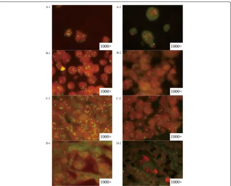
Fig. 2The results of HER2 gene amplification status evaluated by FISH assay. A-1 was section of cell lines sample without HER2 gene amplification
However, some potential disadvantages need to be considered. The characteristic features of cell lines for establishing xenografts should be monitored during long-term culture to ensure reliability. In theory, al-though the target tumor cells in xenografts are human-derived [22], they might contain vessels or fibrocytes derived from mice. This indicates that the non-specificity of the secondary antibody might lead to some background staining in IHC. However, in our study, we found that the influence of this background staining was negligible. Nevertheless, poor tumorigenic potential and slow growth of HER2-positive xenograft tumors might impede its application [23]. In our study, we chose SCID mice and Matrigel basement membrane matrix to im-prove the tumorigenic potential [24]. Moreover, some distinctions were found between the xenograft tumor
samples and clinical specimens in histological analysis. In the clinical specimens, a larger number of normal cells was observed and the tissue structure was more complicated. Breast catheter, breast gland, and inflam-matory cell infiltration was evident. In the xenograft tumor samples, almost all the cells were tumor cells, and the tissue structure was simple. As a result, the evalu-ation of clinical specimens was more difficult than in the case of xenograft tumor samples. Some performance characteristics, especially the interpretation by observers in the routine work, might be neglected to be evaluated in EQA.
Conclusions
In this study, we compared three types of candidate refer-ence samples with clinical specimens for quality control of
Fig. 3The results of HER2 protein expression status evaluated by IHC assay. A-1 was section of cell lines sample without HER2 overexpression
HER2 status detection. The study indicated that xenograft tumor samples might be more suitable for FISH and IHC as additional alternative quality controls than cell line quality controls. However, xenograft tumor samples cannot replace the traditional clinical specimen quality controls until issues such as tumorigenic potential and distinctions in histology are resolved.
Additional files
Additional file 1:The detailed results of HER2 gene amplification status
evaluated by FISH assay. A-1 to A-4 were section of cell lines samples which were derived from MCF-7, MDA-MB-453, SKBR-3, BT474, respectively. B-1 to B-4 were section of agarose gel within cell lines samples which were derived from MCF-7, MDA-MB-453, SKBR-3, BT474, respectively. C-1 to C-4 were section of xenograft tumor samples which were derived from MCF-7, MDA-MB-453, SKBR-3, BT474, respectively. The magnification was 1000 power. (PDF 17214 kb)
Additional file 2:The detailed results of HER2 protein expression status
evaluated by IHC assay. A-1 to A-4 were section of cell lines samples which were derived from MCF-7, MDA-MB-453, SKBR-3, BT474, respectively. B-1 to B-4 were section of agarose gel within cell lines samples which were derived from MCF-7, MDA-MB-453, SKBR-3, BT474, respectively. C-1 to C-4 were section of xenograft tumor samples which were derived from MCF-7, MDA-MB-453, SKBR-3, BT474, respectively. The magnification was 400 power. (PDF 17387 kb)
Acknowledgements
This work was edited by professional editors at Editage.
Funding
This work was supported by a grant from the Special Fund for Health Scientific Research in the Public Interest from National Population and Family Planning Commission of the PR China (No. 201402018).
Availability of data materials
All data generated or analysed during this study are included in this published article and its supplementary information files.
Authors’contributors
Conception, design, analysis and interpretation of the work: YL and JL. Writing of the manuscript and Check: YL, RZ and JL. Experiment operation: YL, RZ, YH and JD. Data acquisition: TL, KZ, GL, JX. All authors read and approved the final manuscript.
Competing interests
The authors declare that they have no competing interests.
Consent for publication
No individual person’s data was included.
Ethics approval and consent to participate
Our protocol and specimen were approved by the Ethics Committee of the National Center for Clinical Laboratories (Reference Number: NCCL-2014-0912). Surgical specimens were obtained from patients that had given general consent at the time of surgery for future use of tissue for research. All animal experiments were performed in compliance with the guidelines specified by the Institute for Experimental Animals, Beijing Hospital.
Author details
1National Center for Clinical Laboratories, Beijing Hospital, National Center of
Gerontology, No1 Dahua Road, Dongdan, Beijing 100730, People’s Republic of China.2Graduate School, Peking Union Medical College, Chinese Academy of Medical Sciences, Beijing, People’s Republic of China.3Beijing Engineering
Research Center of Laboratory Medicine, Beijing Hospital, National Center of Gerontology, Beijing 100730, People’s Republic of China.
Received: 7 April 2016 Accepted: 2 September 2016
References
1. Liu W, Xu J, Liu Y, Yu X, Tang X, Wang Z, et al. Anthocyanins potentiate the activity of trastuzumab in human epidermal growth factor receptor 2-positive breast cancer cells in vitro and in vivo. Mol Med Rep. 2014;10(4): 1921–6. doi:10.3892/mmr.2014.2414.
2. Rakha EA, Pinder SE, Bartlett JM, Ibrahim M, Starczynski J, Carder PJ, et al. Updated UK Recommendations for HER2 assessment in breast cancer. J Clin Pathol. 2015;68(2):93–9. doi:10.1136/jclinpath-2014-202571.
3. Gianni L, Dafni U, Gelber RD, Azambuja E, Muehlbauer S, Goldhirsch A, et al. Treatment with trastuzumab for 1 year after adjuvant chemotherapy in patients with HER2-positive early breast cancer: a 4-year follow-up of a randomised controlled trial. Lancet Oncol. 2011;12(3):236–44. doi:10.1016/ S1470-2045(11)70033-X.
4. Gianni L, Eiermann W, Semiglazov V, Manikhas A, Lluch A, Tjulandin S, et al. Neoadjuvant chemotherapy with trastuzumab followed by adjuvant trastuzumab versus neoadjuvant chemotherapy alone, in patients with HER2-positive locally advanced breast cancer (the NOAH trial): a randomised controlled superiority trial with a parallel HER2-negative cohort. Lancet. 2010;375(9712):377–84. doi:10.1016/S0140-6736(09)61964-4.
5. Baselga J, Bradbury I, Eidtmann H, Di Cosimo S, de Azambuja E, Aura C, et al. Lapatinib with trastuzumab for HER2-positive early breast cancer (NeoALTTO): a randomised, open-label, multicentre, phase 3 trial. Lancet. 2012;379(9816):633–40. doi:10.1016/S0140-6736(11)61847-3.
6. Hanna WM, Ruschoff J, Bilous M, Coudry RA, Dowsett M, Osamura RY, et al. HER2 in situ hybridization in breast cancer: clinical implications of polysomy 17 and genetic heterogeneity. Mod Pathol. 2014;27(1):4–18. doi:10.1038/ modpathol.2013.103.
7. Jimenez RE, Wallis T, Tabasczka P, Visscher DW. Determination of Her-2/Neu status in breast carcinoma: comparative analysis of immunohistochemistry and fluorescent in situ hybridization. Mod Pathol. 2000;13(1):37–45. doi:10. 1038/modpathol.3880007.
8. Perez EA, Suman VJ, Davidson NE, Martino S, Kaufman PA, Lingle WL, et al. HER2 testing by local, central, and reference laboratories in specimens from the North Central Cancer Treatment Group N9831 intergroup adjuvant trial. J Clin Oncol. 2006;24(19):3032–8. doi:10.1200/JCO.2005.03.4744.
9. Wolff AC, Hammond ME, Schwartz JN, Hagerty KL, Allred DC, Cote RJ, et al. American Society of Clinical Oncology/College of American Pathologists guideline recommendations for human epidermal growth factor receptor 2 testing in breast cancer. Arch Pathol Lab Med. 2007;131(1):18–43. doi:10. 1043/1543-2165(2007)131[18:ASOCCO]2.0.CO;2.
10. Wolff AC, Hammond ME, Hicks DG, Dowsett M, McShane LM, Allison KH, et al. Recommendations for human epidermal growth factor receptor 2 testing in breast cancer: American Society of Clinical Oncology/College of American Pathologists clinical practice guideline update. J Clin Oncol. 2013;31(31):3997–4013. doi:10.1200/JCO.2013.50.9984.
11. Xiao Y, Gao X, Maragh S, Telford WG, Tona A. Cell lines as candidate reference materials for quality control of ERBB2 amplification and expression assays in breast cancer. Clin Chem. 2009;55(7):1307–15. doi:10.1373/ clinchem.2008.120576.
12. Hammond ME, Barker P, Taube S, Gutman S. Standard reference material for Her2 testing: report of a National Institute of Standards and Technology-sponsored Consensus Workshop. Appl Immunohistochem Mol Morphol. 2003;11(2):103–6.
13. Bartlett JM, Ibrahim M, Jasani B, Morgan JM, Ellis I, Kay E, et al. External quality assurance of HER2 fluorescence in situ hybridisation testing: results of a UK NEQAS pilot scheme. J Clin Pathol. 2007;60(7):816–9. doi:10.1136/ jcp.2006.040840.
14. Zhang R, Han Y, Huang J, Ma L, Li Y, Li J. Results of first proficiency test for KRAS testing with formalin-fixed, paraffin-embedded cell lines in China. Clin Chem Lab Med. 2014;52(12):1851–7. doi:10.1515/cclm-2014-0227. 15. Hung T, Wolber R, Garratt J, Kalloger S, Gilks CB. Improved breast cancer
biomarker detection through a simple, high frequency, low cost external proficiency testing program. Pathology. 2010;42(7):637–42. doi:10.3109/ 00313025.2010.520306.
17. Vyberg M, Nielsen S, Roge R, Sheppard B, Ranger-Moore J, Walk E, et al. Immunohistochemical expression of HER2 in breast cancer: socioeconomic impact of inaccurate tests. BMC Health Serv Res. 2015;15:352. doi:10.1186/ s12913-015-1018-6.
18. Rhodes A, Jasani B, Couturier J, McKinley MJ, Morgan JM, Dodson AR, et al. A formalin-fixed, paraffin-processed cell line standard for quality control of immunohistochemical assay of HER-2/neu expression in breast cancer. Am J Clin Pathol. 2002;117(1):81–9. doi:10.1309/4NCM-QJ9W-QM0J-6QJE. 19. Vanden Bempt I, Van Loo P, Drijkoningen M, Neven P, Smeets A, Christiaens
MR, et al. Polysomy 17 in breast cancer: clinicopathologic significance and impact on HER-2 testing. J Clin Oncol. 2008;26(30):4869–74. doi:10.1200/JCO. 2007.13.4296.
20. Wester K, Andersson AC, Ranefall P, Bengtsson E, Malmstrom PU, Busch C. Cultured human fibroblasts in agarose gel as a multi-functional control for immunohistochemistry. Standardization Of Ki67 (MIB1) assessment in routinely processed urinary bladder carcinoma tissue. J Pathol. 2000;190(4):503–11. doi:10.1002/(SICI)1096-9896(200003)190:4<503::AID-PATH537>3.0.CO;2-E. 21. Hasan T, Carter B, Denic N, Gai L, Power J, Voisey K, et al. Evaluation of
cell-line-derived xenograft tumours as controls for immunohistochemical testing for ER and PR. J Clin Pathol. 2015;68(9):746–51. doi:10.1136/jclinpath-2015-203066. 22. Taetle R, Jones OW, Honeysett JM, Abramson I, Bradshaw C, Reid S. Use of
nude mouse xenografts as preclinical screens. Characterization of xenograft-derived melanoma cell lines. Cancer. 1987;60(8):1836–41.
23. Holliday DL, Speirs V. Choosing the right cell line for breast cancer research. Breast Cancer Res. 2011;13(4):215. doi:10.1186/bcr2889.
24. Czerski L, Majors A, Ng TC, Vijayakumar S, Weichselbaum R. Growth and magnetic resonance characteristics of human squamous cell carcinoma xenografts implanted with cells suspended in Matrigel. NMR Biomed. 1993;6(5):297–301.
• We accept pre-submission inquiries
• Our selector tool helps you to find the most relevant journal • We provide round the clock customer support
• Convenient online submission • Thorough peer review
• Inclusion in PubMed and all major indexing services • Maximum visibility for your research
Submit your manuscript at www.biomedcentral.com/submit