Imaging and mapping of mouse bone using MALDI-imaging
mass spectrometry
Yoko Fujino
a,1, Tomoko Minamizaki
b,1, Hirotaka Yoshioka
b, Mitsugi Okada
c, Yuji Yoshiko
b,⁎
aDepartment of Special Care Dentistry, Hiroshima University Graduate School of Biomedical and Health Sciences, Hiroshima University, Hiroshima, Japan b
Department of Calcified Tissue Biology, Hiroshima University Institute of Biomedical and Health Sciences, Hiroshima University, Hiroshima, Japan c
Special Care Dentistry, Hiroshima University Hospital, Hiroshima, Japan
a b s t r a c t
a r t i c l e i n f o
Article history:
Received 15 July 2016
Received in revised form 23 September 2016 Accepted 28 September 2016
Available online 29 September 2016
Matrix-assisted laser desorption/ionization-imaging mass spectrometry (MALDI-IMS) is an advanced method used globally to analyze the distribution of biomolecules on tissue cryosections without any probes. In bones, however, hydroxyapatite crystals make it difficult to determine the distribution of biomolecules using MALDI-IMS. Addition-ally, there is limited information regarding the use of this method to analyze bone tissues. To determine whether MALDI-IMS analysis of bone tissues can facilitate comprehensive mapping of biomolecules in mouse bone, we first dissected femurs and tibiae from 8-week-old male mice and characterized the quality of multiplefixation and decalcification methods for preparation of the samples. Cryosections were mounted on indium tin oxide-coated glass slides, dried, and then a matrix solution was sprayed on the tissue surface. Images were acquired using an iMScope at a mass-to-charge range of 100–1000. Hematoxylin-eosin, Alcian blue, Azan, and periodic acid-Schiff staining of adjacent sections was used to evaluate histological and histochemical features. Among the variousfi xa-tion and decalcification conditions, sections from trichloroacetic acid-treated samples were most suitable to exam-ine both histology and comprehensive MS images. However, histotypic MS signals were detected in all sections. In addition to the MS images, phosphocholine was identified as a candidate metabolite. These results indicate success-ful detection of biomolecules in bone using MALDI-IMS. Although analytical procedures and compositional adjust-ment regarding the performance of the device still require further developadjust-ment, IMS appears to be a powerful tool to determine the distribution of biomolecules in bone tissues.
© 2016 The Authors. Published by Elsevier Inc. This is an open access article under the CC BY-NC-ND license (http://creativecommons.org/licenses/by-nc-nd/4.0/). Keywords:
Matrix-assisted laser desorption/ionization-imaging mass spectrometry
Tissue cryosection Bone
Fixation Decalcification
1. Introduction
Recent advances in omics approaches allow for the identification of whole molecules constituting an organism, which can provide insight into pathogenic processes and serve as biomarker candidates. Informa-tion obtained from omics research, such as proteomics and metabolo-mics, has become particularly important for elucidating etiology and for diagnosis of diseases. However, conventional imaging techniques, such as immunohistochemistry, require labeling and have limited use for revealing new pathological molecules in tissue.
Mass spectrometry (MS) is a commonly used technology to detect analytes in proteomics and metabolomics research, and can directly
de-fine individual molecular species in complex samples. Among MS methods, matrix-assisted laser desorption/ionization-imaging mass spectrometry (MALDI-IMS) enables analysis of the molecule distribu-tion without any disrupdistribu-tion of the morphology or architecture. In
contrast, liquid chromatography-MS or gas chromatography-MS analy-ses require the use of tissue homogenates, which retains no tissue local-ization information.
Although cellular and molecular analyses are useful for many types of tissues, they are difficult to use with calcified tissues such as bones and teeth. This limits investigation of the function of cells such as oste-ocytes and cementoste-ocytes, or the distribution of organic matter such as proteins and peptides. For example, osteocytes, which terminally differ-entiate from osteoblasts and are embedded in the bone matrix, play an important role in the maintenance of homeostasis in the network be-tween osteoblasts and osteoclasts (Bonewald, 2011). To examine the physiological functions of osteocytes, it is preferable to retain the origi-nal distribution of biomolecules. However, MALDI-IMS has certain lim-itations for quantitative and qualitative uncertainty analyses. Additionally, there are few studies that have applied MALDI-IMS to bone tissues to identify molecules because of the lack of appropriate methods to prepare sections for ionization (Hirano et al., 2014; Cillero-Pastor et al., 2015; Seeley et al., 2014). Hirano et al. (Hirano et al., 2014) reported MALDI-IMS for tooth cryosections prepared by the
Kawamoto method using adhesivefilm without any pretreatment,
such asfixation or decalcification. However, the signals obtained from the enamel and dentin were not listed in the metabolomics database. ⁎ Corresponding author at: Department of Calcified Tissue Biology, Hiroshima
University Institute of Biomedical & Health Sciences, 1-2-3, Kasumi, Minami-ku, Hiroshima 734-8553, Japan.
E-mail address:[email protected](Y. Yoshiko).
1
Yoko Fujino and Tomoko Minamizaki contributed equally to this work.
http://dx.doi.org/10.1016/j.bonr.2016.09.004
2352-1872/© 2016 The Authors. Published by Elsevier Inc. This is an open access article under the CC BY-NC-ND license (http://creativecommons.org/licenses/by-nc-nd/4.0/).
Contents lists available atScienceDirect
Bone Reports
They concluded that almost all of these signals were mineral which can interrupt ionization of large components.
Therefore, in this study we established a protocol tofix and decalcify samples derived from bone to detect MS using MALDI-IMS and provide a comprehensive map of proteins and peptides.
2. Materials and methods 2.1. Materials
Conductive indium tin oxide (ITO)-coated glass slides (8–12Ω) were purchased from Sigma-Aldrich Co. (St. Louis, MO).α -Cyano-4-hydroxycinnamic acid (CHCA) matrices were purchased from Bruker Daltonics (Bremen, Germany). Carboxymethylcellulose (CMC; 2%) was
purchased from Leica Microsystems (Wetzlar, Germany).
Trifluoroacetic acid (TFA), 2,5-dihydroxy-benzoic acid (DHB), and all other chemicals, unless specified otherwise, were purchased from Sigma-Aldrich Co.
2.2. Animals
C57BL/6J mice were purchased from CLEA Inc. (Osaka, Japan). The mice were housed and handled to minimize pain and discomfort ac-cording to protocols approved by the Institutional Animal Care and Use Committee at the Central Institute for Experimental Animals and the Committee of Animal Experimentation at Hiroshima University. 2.3. Specimen preparation
Femurs and/or tibiae from 8-week-old male mice werefixed and decalcified in various combinations offixatives [4% paraformaldehyde (PFA), Carnoyfluid or trichloroacetic acid (TCA)] and decalcification so-lutions (formic acid, EDTA-NH4, or TCA) (Table 1). Fresh samples, with or without decalcification, were also prepared. The samples were then embedded in a stainless steel containerfilled with 2% CMC and placed in dry ice-cooled hexane to prepare frozen CMC blocks. Each frozen block was stored at−80 °C until sectioning. Tissues were sectioned (5μm for staining and MALDI-IMS of fresh samples without any
pre-treatment by the Kawamoto method (Kawamoto, 2003), and 10μm
for MALDI-IMS of samples with pretreatment) with a CM 3050 S cryo-stat (Leica Microsystems). For staining, the sections were placed on nor-mal glass slides and washed with 100% ethanol. For MALDI-IMS, the sections were placed on ITO-coated glass slides with electrically conducting double-adhesive tape for samples without pretreatment, followed by washing with 70% and then 100% ethanol, and dried. 2.4. Staining
Adjacent sections were stained with hematoxylin-eosin (H-E), Alcian blue, Azan, and Periodic Acid-Schiff (PAS) to evaluate histological and histochemical features. Sections that did not undergo previousfi x-ation werefixed with 4% PFA before staining.
2.5. MALDI-IMS and MS-MS
After drying at room temperature, cryosections were coated with a DHB or CHCA matrix vapor deposition using an iMlayer (Shimadzu Cor-poration, Kyoto, Japan) at a thickness of 1.5 or 0.7μm, respectively. MALDI images were acquired using an iMScope (Shimadzu
Corporation) in positive or negative ion modes in the mass-to-charge ratio (m/z) range of 100–1000, at a laser frequency of 1000 Hz,
Table 1
Combinations offixation and decalcification solutions.
A B C D E F G H
Fixation – TCA, overnight PFA, overnight Carnoy, overnight PFA, overnight Carnoy, overnight – –
Decalcification – EDTA, 7 days EDTA, 7 days Formic acid, 2 days Formic acid, 2 days Formic acid, 2 days EDTA, 4 days
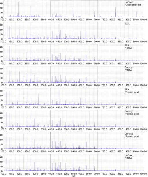
Fig. 1.Histological observations of femurs and tibiaefixed and decalcified with each solution by hematoxylin-eosin (H-E), Alcian blue, Azan, and Periodic Acid-Schiff (PAS) staining. (A) Sections of unfixed/undecalcified tibiae. (B) Sections of trichloroacetic acid (TCA)-treated femurs. (C–H) Sections of femurs treated with 4% paraformaldehyde (PFA)/EDTA (C), Carnoy/EDTA (D), PFA/formic acid (E), or Carnoy/formic acid (F). (G, H), Unfixed sections of femurs decalcified with formic acid (G) or EDTA (H). Representative images are shown; n = 2–5.
accumulating 50 laser shots. Detector and sample voltages were 1.7– 1.9 kV and 3–3.5 kV, respectively. Spatial resolution was 10μm and laser intensity ranged from 23 to 45. To prevent any influence offixation and decalcification solutions as well as matrices, 1μL of each solution and matrix was placed onto a stainless steel plate and applied to the iMScope after drying. Mass spectra obtained from this mixture were omitted from spectra of the samples. Principal component analysis (PCA) was used to extract the peak matrix from the mass spectrum and search for principal components as characteristic patterns in the images (Shao et al., 2012).
To identify the metabolites in bones, the TCA-treated sections from mouse femurs were applied to iMScope in positive ion mode with the DHB matrix. MS-MS data were evaluated using the Human Metabolome
Database search engine (version 3.6, The Metabolomics Innovaton Cen-tre, Edmonton, Canada) for metabolite identification.
Phosphocholine inferred by MS-MS data was mixed with DHB and applied to the iMScope. The MS-MS spectrum was compared with that detected in sections.
3. Results
3.1. Histological and histochemical features of bones with or without pretreatment
To evaluate the influence offixation and decalcification solutions on the histological and histochemical features of femurs and tibiae,
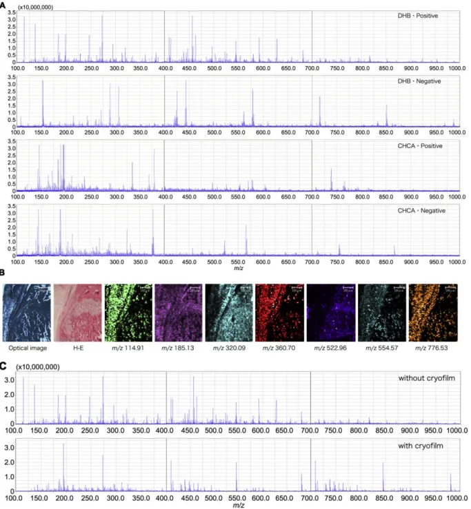
Fig. 2.Mass spectra (maximum intensity) and MALDI-IMS analysis of TCA-treated samples in the range ofm/z100–1000 under various conditions. (A) Analysis with a 2,5-dihydroxy-benzoic acid (DHB) orα-cyano-4-hydroxycinnamic acid (CHCA) matrix in positive or negative ion modes in the range ofm/z100–1000. (B) Imaging of the histotypic distribution of several mass peaks from the sections. Left panels show optical images and H-E staining. (C) Mass spectra detected in sections with or without cryofilm in the range ofm/z100–1000. Representative images and data are shown. nN3.
cryosections were stained with H-E, Alcian blue, Azan, and PAS. Among the variousfixation and decalcification conditions, sections from TCA-treated samples were the most suitable to examine both histology and comprehensive MS images (Fig. 1B), followed by samples decalcified with EDTA afterfixation (Fig. 1C, D). Bone marrow peeled from trabec-ular bone surfaces in samples decalcified with formic acid (Fig. 1E, F). Cartilage in the growth plate and bone marrow were unable to keep their structure in decalcified samples withoutfixation (Fig. 1G, H). There was no difference between samples with Azan or PAS staining. However, with Alcian Blue staining, bone marrow in unfixed and Carnoy/EDTA-treated samples turned dark blue (Fig. 1A, D, G, and H). 3.2. Comparison of MALDI-IMS between bones with or without pretreatment
In MALDI-IMS analysis, an appropriate matrix is required to obtain successful images (Setou and Kurabe, 2011). To establish the measure-ment conditions of MALDI-IMS, all samples were applied to the iMScope in positive or negative ion modes with DHB or CHCA matrices. Among these conditions, the positive mode with DHB enabled detection of many mass spectra in a wide range ofm/z(Fig. 2A, samples treated
with TCA). Imaging by MALDI-IMS showed tissue-specific distribution of MS with DHB (Fig. 2B). Because the Kawamoto method requires cryofilm, MALDI-IMS of TCA-treated sections mounted on ITO-coated glass slides with or without cryofilm were analyzed to measure the in-terference of cryofilm. With cryofilm, the number of mass spectra was much less than without cryofilm (Fig. 2C).
A comparison of mass spectra detected in positive ion mode with
DHB showed that the undecalcified sample with the Kawamoto
method exhibited few peaks in the range ofm/z100–700 (Fig. 3). Each section had its own characteristic features of the appearance of peaks, but there was no significant difference in the obtained number of mass spectra between samples that were decalcified,
fixed, or both (Fig. 3).
In MALDI-IMS, histotypic MS signals were detected in all sections. The signals of the molecule atm/z554.57 were located mainly in bone marrow in all sections except undecalcified sections. The molecule at m/z185.13 was located mainly in cortical bones, trabecular bones, and cartilage in all sections except the Carnoy/formic acid sample (Fig. 4). In the undecalcified section, there were a few signals atm/z554.57, and the signals atm/z185.13 were localized diffusely in the Carnoy/ formic acid sample (Fig. 4).
Fig. 3.Mass spectra (maximum intensity) of each treatment with MALDI-IMS in the range of 100–1000 and the positive ion mode with DHB. Sections of unfixed/undecalcified tibiae, femurs treated with TCA, PFA/EDTA, Carnoy/EDTA, PFA/formic acid, or Carnoy/formic acid, and femurs decalcified with formic acid or EDTA in the range ofm/z100–1000. Representative data are shown. nN3.
PCA, a statistical method to extract thefirst principal component in variance between samples, revealed that thefirst principal component was the same in samples except undecalcified, Carnoy/EDTA, and PFA/ formic acid samples in the range ofm/z100–700 (Table 2).
3.3. Metabolomics of mouse femurs
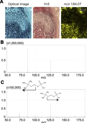
To identify metabolites in bone, TCA-treated sections from mouse fe-murs were applied to the iMScope in positive ion mode with the DHB matrix. Several MS-MS data sets were subjected to the human metabo-lome database, and some high-scoring candidates were recovered. Of these, the molecule atm/z184.07 was identified as phosphocholine (Fig. 5B). For confirmation, an authentic sample of phosphocholine was applied to the iMScope. The MS-MS spectra were in agreement with those obtained from the tissue sections (Fig. 5C).
4. Discussion
In this study, we comprehensively determined the metabolomics of mouse bones with localization information using MALDI-IMS. We also characterized differentfixation and decalcification techniques and established an effective method for preparation of mouse bone tissue.
When preparing cryosections of hard tissues without decalcification, the Kawamoto method requires cryofilm that can attach to the cutting surface under freezing conditions (Kawamoto, 2003). Furthermore, for ionization, an electrically conducting double-adhesive tape is needed to affix sections on the ITO-glass slide (Hirano et al., 2014). Comparison of mass spectra between sections with or without tape revealed that many more peaks were obtained from the section without tape. A meth-od has been reported to remove the tape before application to MALDI-IMS (Seeley et al., 2014), but a high degree of technical skill is required to do so.
For MALDI-IMS, fresh frozen sections (withoutfixation) are usually used for analysis. This study showed thatfixation and decalcification of bones facilitates the preparation of sections, and detection of MS from organic components is possible because of mineral removal. Be-cause the utility of MALDI-IMS has been reported for formalin-fixed,
paraffin-embedded (FFPE) tissues (Lemaire et al., 2007), researchers have focused on analysis of FFPE samples by MALDI-IMS (Ronci et al., 2008; Djidja et al., 2009; Stauber et al., 2010; Cole et al., 2013; Powers et al., 2014; Gravius et al., 2015; O'Rourke et al., 2015). Formalinfixation can be used to avoid degradation and spoilage of samples. However, the cross-linked molecules between formalin and primary amines are un-able to be ionized. Therefore, the number of identified proteins in FFPE sections is less than that in cryosections, and several steps are required to remove formalin, break crosslinks, and cleave proteins to peptides (Ronci et al., 2008; O'Rourke et al., 2015; Fowler et al., 2013). For re-search targeting nucleotides, lipids, or peptides without primary amines, of which formalin does not form crosslinks, it is possible to apply the samples without such steps. Organic solvents such as ethanol can alsofix tissues by coagulation and precipitation of proteins. Fixation with organic solvents has less impact on MALDI-IMS analysis, but it dis-solves lipids from tissues. Some of thefirst principal components detect-ed by PCA were the same among the samples, but some were not. These
findings suggest that researchers should select afixation solution ac-cording to the object used for analysis. The selection of the appropriate matrix is also crucial; DHB and CHCA are suitable for imaging small or-ganic compounds, such as lipids and peptides, respectively (Setou and Kurabe, 2011).
Decalcification with EDTA or TCA was better than with formic acid for maintaining the tissue structures. Bone marrow had peeled from tra-becular bone surfaces using formic acid decalcification, which can occur during the preparation of sections (Prasad and Donoghue, 2013). Treat-ment with TCA is useful because of rapid one-stepfixation and
decalci-fication that can preserve antigens and tissue morphology (Athanasou et al., 1987). In this study, sections from TCA-treated samples were the most suitable to examine both histology and comprehensive MS images.
By performing metabolomics of TCA-treated samples from mouse bone with MALDI-IMS, we were able to identify biomolecules, including phosphocholine, an important biomolecule for bone mineralization (Roberts et al., 2007; Yadav et al., 2011; Stewart et al., 2006; Kvam et al., 1992; Stern and Vance, 1987). Phosphocholine can be cleaved by PHOSPHO1, a soluble phosphatase that is responsible for initiating
Table 2
First principal component with or without pretreatment.
Unfixed/undecalcified TCA PFA/EDTA Carnoy/EDTA PFA/formic acid Carnoy/formic acid Unfixed/formic acid Unfixed/EDTA
m/z100–400 165.07 114.91 114.91 114.91 184.08 114.91 114.91 114.91
m/z400–700 456.11 462.73 462.73 412.71 462.73 462.73 462.73 462.73 Fig. 4.MALDI-IMS atm/z554.57 andm/z185.13 of each treatment. Optical images (upper). MALDI-IMS atm/z554.57 (middle) and atm/z185.13 (lower) with indicated treatment. Representative images and data are shown. nN3.
hydroxyapatite crystal formation inside osteoblast-derived matrix vesi-cles (Roberts et al., 2007; Stewart et al., 2006). PHOSPHO1-R74X null mutant mice display skeletal and growth plate abnormalities and a de-crease in growth rate (Yadav et al., 2011). Phosphocholine is one of the abundant phosphomonoesters in cartilage (Kvam et al., 1992), and low accumulation of phosphocholine in calvarial tissue compared with liver is consistent with the upregulation of PHOSPHO1 activity in calvaria, whose function reduces the levels of phosphocholine in chondrocytes and osteoblasts (Stern and Vance, 1987).
This study successfully detected biomolecules in bones using MALDI-IMS. Although analytical procedures and compositional adjust-ment on device performance require further developadjust-ment, MALDI-IMS appears to be a powerful tool to search for biomolecules in bones. Conflicts of interest
None.
Acknowledgements
We thank Prof. Mitsutoshi Setou from Hamamatsu University School of Medicine for his excellent suggestions. We are deeply grateful to the members of Center of Oral Clinical Examination, Hiroshima University Hospital, for their support.
This research did not receive any specific grant from funding agen-cies in the public, commercial, or not-for-profit sectors.
References
Athanasou, N.A., Quinn, J., Heryet, A., Woods, C.G., McGee, J.O., 1987.Effect of decalcifi ca-tion agents on immunoreactivity of cellular antigens. J. Clin. Pathol. 40, 874–878.
Bonewald, L.F., 2011.The amazing osteocyte. J. Bone Miner. Res. 26, 229–238.
Cillero-Pastor, B., Eijkel, G.B., Blanco, F.J., Heeren, R.M., 2015.Protein classification and dis-tribution in osteoarthritic human synovial tissue by matrix-assisted laser desorption ionization mass spectrometry imaging. Anal. Bioanal. Chem. 407, 2213–2222.
Cole, L.M., Mahmoud, K., Haywood-Small, S., Tozer, G.M., Smith, D.P., Clench, M.R., 2013.
Recombinant“IMS TAG”proteins - a new method for validating bottom-up matrix-assisted laser desorption/ionisation ion mobility separation mass spectrometry imag-ing. Rapid Commun. Mass Spectrom. 27, 2355–2362.
Djidja, M.C., Claude, E., Snel, M.F., Scriven, P., Francese, S., Carolan, V., Clench, M.R., 2009.
MALDI-ion mobility separation-mass spectrometry imaging of glucose-regulated protein 78 kDa (Grp78) in human formalin-fixed, paraffin-embedded pancreatic ad-enocarcinoma tissue sections. J. Proteome Res. 8, 4876–4884.
Fowler, C.B., O'Leary, T.J., Mason, J.T., 2013.Toward improving the proteomic analysis of formalin-fixed, paraffin-embedded tissue. Expert Rev. Proteomics 10, 389–400.
Gravius, S., Randau, T.M., Casadonte, R., Kriegsmann, M., Friedrich, M.J., Kriegsmann, J., 2015.Investigation of neutrophilic peptides in periprosthetic tissue by matrix-assisted laser desorption ionisation time-of-flight imaging mass spectrometry. Int. Orthop. 39, 559–567.
Hirano, H., Masaki, N., Hayasaka, T., Watanabe, Y., Masumoto, K., Nagata, T., Katou, F., Setou, M., 2014.Matrix-assisted laser desorption/ionization imaging mass spectrom-etry revealed traces of dental problem associated with dental structure. Anal. Bioanal. Chem. 406, 1355–1363.
Kawamoto, T., 2003.Use of a new adhesivefilm for the preparation of multi-purpose fresh-frozen sections from hard tissues, whole-animals, insects and plants. Arch. Histol. Cytol. 66, 123–143.
Kvam, B.J., Pollesello, P., Vittur, F., Paoletti, S., 1992.31
P NMR studies of resting zone carti-lage from growth plate. Magn. Reson. Med. 25, 355–361.
Lemaire, R., Desmons, A., Tabet, J.C., Day, R., Salzet, M., Fournier, I., 2007.Direct analysis and MALDI imaging of formalin-fixed, paraffin-embedded tissue sections. J. Proteome Res. 6, 1295–1305.
O'Rourke, M.B., Djordjevic, S.P., Padula, M.P., 2015.A non-instrument-based method for the analysis of formalin-fixed paraffin-embedded human spinal cord via matrix-assisted laser desorption/ionisation imaging mass spectrometry. Rapid Commun. Mass Spectrom. 29, 1836–1840.
Powers, T.W., Neely, B.A., Shao, Y., Tang, H., Troyer, D.A., Mehta, A.S., Haab, B.B., Drake, R.R., 2014.MALDI imaging mass spectrometry profiling ofN-glycans in formalin-fixed paraffin embedded clinical tissue blocks and tissue microarrays. PLoS One 9, e106255.
Prasad, P., Donoghue, M., 2013.A comparative study of various decalcification techniques. Indian J. Dent. Res. 24, 302–308.
Roberts, S., Narisawa, S., Harmey, D., Millán, J.L., Farquharson, C., 2007.Functional involve-ment of PHOSPHO1 in matrix vesicle-mediated skeletal mineralization. J. Bone Miner. Res. 22, 617–627.
Ronci, M., Bonanno, E., Colantoni, A., Pieroni, L., Di Ilio, C., Spagnoli, L.G., Federici, G., Urbani, A., 2008.Protein unlocking procedures of formalin-fixed paraffin-embedded tissues: application to MALDI-TOF imaging MS investigations. Proteomics 8, 3702–3714.
Seeley, E.H., Wilson, K.J., Yankeelov, T.E., Johnson, R.W., Gore, J.C., Caprioli, R.M., Matrisian, L.M., Sterling, J.A., 2014.Co-registration of multi-modality imaging allows for com-prehensive analysis of tumor-induced bone disease. Bone 61, 208–216.
Setou, M., Kurabe, N., 2011.Mass microscopy: high-resolution imaging mass spectrome-try. J. Electron Microsc. 60, 47–56.
Shao, C., Tian, Y., Dong, Z., Gao, J., Gao, Y., Jia, X., Guo, G., Wen, X., Jiang, C., Zhang, X., 2012.
The use of principal component analysis in MALDI-TOF MS: a powerful tool for estab-lishing a mini-optimized proteomic profile. Am. J. Biomed. Sci. 4, 85–101.
Stauber, J., MacAleese, L., Franck, J., Claude, E., Snel, M., Kaletas, B.K., Wiel, I.M., Wisztorski, M., Fournier, I., Heeren, R.M., 2010.On-tissue protein identification and imaging by MALDI-ion mobility mass spectrometry. J. Am. Soc. Mass Spectrom. 21, 338–347.
Stern, P.H., Vance, D.E., 1987.Phosphatidylcholine metabolism in neonatal mouse calvaria. Biochem. J. 244, 409–415.
Stewart, A.J., Roberts, S.J., Seawright, E., Davey, M.G., Fleming, R.H., Farquharson, C., 2006.
The presence of PHOSPHO1 in matrix vesicles and its developmental expression prior to skeletal mineralization. Bone 39, 1000–1007.
Yadav, M.C., Simão, A.M., Narisawa, S., Huesa, C., McKee, M.D., Farquharson, C., Millán, J.L., 2011.Loss of skeletal mineralization by the simultaneous ablation of PHOSPHO1 and alkaline phosphatase function: a unified model of the mechanisms of initiation of skeletal calcification. J. Bone Miner. Res. 26, 286–297.
Fig. 5.MS-MS data atm/z184.07 in sections and an authentic sample of phosphocholine. (A) An optical image (left), H-E staining (middle), and MS image atm/z184.07 (right). (B) MS-MS spectra of an authentic sample of phosphocholine. (C) Product ions predicted a part of the structure as indicated by the arrow. n = 3.