Assay for Diagnosis of
Clostridium difficile
Infection: Performance,
Cost, and Correlation with Disease
Marie L. Landry,a,b,cDavid Ferguson,cJeffrey Topalb ‹Departments of Laboratory Medicinea
and Internal Medicine,b
Yale University School of Medicine, and Clinical Virology Laboratory, Yale-New Haven Hospital,c New Haven, Connecticut, USA
SimplexaClostridium difficileuniversal direct PCR, a real-time PCR assay for the detection of theC. difficiletoxin B (tcdB) gene using the 3M integrated cycler, was compared with a two-step algorithm which includes the C. Diff Chek-60 glutamate dehydro-genase (GDH) antigen assay followed by cytotoxin neutralization. Three hundred forty-two liquid or semisolid stools submitted for diagnosticC. difficiletesting, 171 GDH antigen positive and 171 GDH antigen negative, were selected for the study. All sam-ples were tested by the C. Diff Chek-60 GDH antigen assay, cytotoxin neutralization, and Simplexa direct PCR. Of 171 GDH-positive samples, 4 were excluded (from patients on therapy or from whom duplicate samples were obtained) and 88 were deter-mined to be true positives for toxigenicC. difficile. Of the 88, 67 (76.1%) were positive by the two-step method and 86 (97.7%) were positive by PCR. Seventy-nine were positive by the GDH antigen assay only. Of 171 GDH antigen-negative samples, none were positive by PCR. One antigen-negative sample positive by the cytotoxin assay only was deemed a false positive based on chart review. SimplexaC. difficileuniversal direct PCR was significantly more sensitive for detecting toxigenicC. difficile bacte-ria than cytotoxin neutralization (Pⴝ0.0002). However, most PCR-positive/cytotoxin-negative patients did not have clearC. difficiledisease. The estimated cost avoidance provided by a more rapid molecular diagnosis was outweighed by the cost of iso-lating and treating PCR-positive/cytotoxin-negative patients. The costs, clinical consequences, and impact on nosocomial trans-mission of treating and/or isolating patients positive for toxigenicC. difficileby PCR but negative forin vivotoxin production merit further study.
C
lostridium difficileinfection (CDI) is a major cause of nosocomial diarrhea, leading to morbidity and mortality in hospitalized pa-tients (1). Anaerobic culture and bacterial glutamate dehydrogenase (GDH) antigen tests detect the presence of both nontoxigenic and toxigenic strains ofC. difficile. Toxigenic culture and PCR for toxin genes detect only the presence of bacteria capable of making toxin; these tests cannot separate active disease from colonization. Tests for toxin productionin vivo, e.g., enzyme-linked immunosorbent assay (ELISA) to detect toxins in stool specimens or cell culture to determine cytotoxicity, have been considered insensitive com-pared to molecular methods (2). In a previous report, we com-pared our two-stepC. difficilealgorithm testing for GDH antigen and cytotoxin to one of the first commercially available PCR tests (3). While PCR was significantly more sensitive than toxin ELISA, its results were not statistically different from those of the two-step algorithm, it failed to detect nine cytotoxin-positive stools, and it was more expensive. Furthermore, patients positive by PCR but not for cytotoxin did not appear to haveC. difficiledisease.The objective of this study was to compare the performance of the newly available SimplexaC. difficileuniversal direct PCR (Fo-cus Diagnostics) for detection of theC. difficiletoxin B gene with that of our standard two-step method, namely, the C. Diff Chek-60 GDH antigen assay followed by cytotoxin neutralization. In addition, the costs and savings of the projected change from cytotoxin assay to PCR with regard to reagents, labor, contact isolation (CI), and antibiotics were estimated.
MATERIALS AND METHODS
Clinical samples.Stool samples submitted forC. difficiletesting from
patients at Yale-New Haven Hospital were entered into the study from
August 2012 to October 2012. All samples were stored at 4°C and tested within 24 h of receipt using the C. Diff Chek-60 GDH antigen ELISA as part of the hospital’s standard two-step diagnostic routine. AllC. difficile
GDH antigen-positive samples with sufficient stool available and an equivalent number of GDH antigen-negative stools were selected on each study day. All study samples were tested within 24 h of receipt by cytotoxin neutralization. Aliquots were labeled with study numbers and then tested by SimplexaC. difficileuniversal direct PCR. PCR was performed by study personnel blind to the results of the two-step method. Only semisolid or liquid stools, one stool per patient per day and no more than two stools per patient in a 7-day period, were included. Formed stools, repeat sam-ples sent on the same day, and samsam-ples obtained from patients onC. difficiletherapy were excluded.
Two-step method using C. Diff Chek-60 and a cytotoxicity assay.
The C. Diff Chek-60 (TechLab, Blacksburg, VA) was performed according to the manufacturer’s instructions. Briefly, 0.1 ml of specimen was added to 0.4 ml of specimen diluent, vortexed, and then centrifuged at 5,000⫻g
for 10 min. Next 0.05 ml of conjugate solution was added to the test microwells, followed by 0.1 ml of centrifuged specimen. The wells were covered and incubated for 50 min at 37°C and washed with 0.35 ml wash solution (7 cycles). Then 0.1 ml of substrate was added, and wells were incubated at room temperature for 10 min. After addition of 0.05 ml of
Received2 October 2013Returned for modification3 November 2013 Accepted7 November 2013
Published ahead of print13 November 2013
Editor:P. H. Gilligan
Address correspondence to Marie L. Landry, [email protected].
Copyright © 2014, American Society for Microbiology. All Rights Reserved.
doi:10.1128/JCM.02545-13
on May 16, 2020 by guest
http://jcm.asm.org/
stop solution, optical density was measured on a microplate reader. A positive result had an optical density ofⱖ0.080, and a negative result had an optical density of⬍0.080 using the spectrophotometric dual wave-length 450/620 nm. All antigen results were reported clinically.
All study samples were stored at 4°C and tested by cytotoxicity assay within 24 h of receipt. Stool samples (0.5 ml) were added to 0.5 ml of phosphate-buffered saline with antibiotics (vancomycin, gentamicin, and amphotericin B) and then vortexed, and the toxin was allowed to elute for 5 min. After centrifugation for 10 min in a microcentrifuge, the superna-tant was removed and passed through a 0.45-m-pore-size filter. Then, 20
l of filtrate was inoculated in duplicate onto foreskin fibroblast mono-layers (MRHF cells; BioWhittaker, Walkersville, MD) in 96-well plates prepared weekly in the laboratory. To assess cytotoxicity, serial 10-fold dilutions (1:10 to 1:10,000) were made without antitoxin. For neutraliza-tion,C. difficileantitoxin (20l; TechLab, Inc., Blacksburg, VA) was added to two wells inoculated with 1:10 and 1:100 dilutions of sample. Thus, after addition of antitoxin, the final dilution in the first culture well was 1:20. Monolayers were read at 4, 24, and 48 h after inoculation using an inverted microscope. A known positive control, run with each assay, was required to show cytotoxicity in the expected range. A positive result consisted of cytotoxicity that was neutralized byC. difficileantitoxin. Re-sults were recorded as the highest dilution showing specific cytotoxicity. Cytotoxin results were reported for GDH antigen-positive samples only, as per the routine clinical protocol.
SimplexaC. difficileuniversal direct PCR.The SimplexaC. difficile
universal direct PCR (Focus Diagnostics, San Diego, CA) was performed directly on stool specimens according to the manufacturer’s instructions. It utilizes real-time PCR to amplify theC. difficiletoxin B (tcdB) gene, an internal control, and bifunctional fluorescent probes/primers for the identification of amplified target DNA. The amplification, detection, and interpretation of the assay are done using the 3M integrated cycler instru-ment. Results were positive, negative, or invalid. Invalid results were due to technical errors or inhibitors in the sample lysate. According to the manufacturer’s instructions, lysates of unresolved samples were frozen at
⫺70°C, thawed, and then retested. The entire procedure requires about 2 h, depending on the number of samples in the run. All samples not tested within 24 h were stored at⫺20°C and tested within 5 days. PCR testing was done without knowledge of two-step test results, and PCR results were not reported clinically.
Discrepant analysis.Samples positive only for GDH antigen were
considered to represent colonization with nontoxigenic strains ofC. dif-ficile. Samples positive by two or more tests were accepted as true posi-tives. Discrepant samples were defined as those positive by either the cytotoxicity assay or PCR but not both. Discrepant samples from patients who were on treatment for CDI at the time of sample collection were excluded from analysis. Chart reviews for inpatients with discrepant re-sults were conducted independently by two of the authors to determine antibiotic therapy, use of contact isolation (CI), frequency and persistence of diarrhea, and subsequentC. difficiletesting results and disease.
CDI.CDI was defined as three or more liquid or semisolid stools in less than 24 h, without other explanation; it resolves on therapy, persists if not treated, or shows a characteristic appearance on colonoscopy or by pathological exam (1).
Statistical analysis. Statistical analysis was performed using
Mc-Nemar’s test and the unpairedttest.
IRB review.The work was considered routine clinical practice and was
deemed exempt from Institutional Review Board (IRB) review.
RESULTS
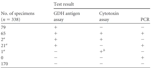
Performance of the Simplexa PCR.A total of 342 samples (171 GDH antigen-positive and 171 GDH-negative samples) were ini-tially tested by the Simplexa PCR. Of 342 samples tested by PCR, only 1 (0.3%) had an invalid internal-control result, and its DNA was successfully amplified on repeat testing. Four GDH-positive samples were excluded from the final analysis: two samples be-cause the patients were on treatment for CDI and two bebe-cause the number of samples exceeded one per day or two in a 7-day period. In the final analysis, 338 stool samples from 300 patients were included. Results are shown inTable 1. Overall, 79 samples were positive by the GDH antigen assay only, 23 were positive by the GDH antigen assay and positive either by the cytotoxin assay or by PCR, and 65 were positive by all three tests. When threshold cycle (CT) values for samples that were PCR positive/cytotoxin positive (meanCT, 30.784⫾3.199) were compared toCTvalues for sam-ples that were PCR positive/cytotoxin negative (mean CT, 34.936⫾3.100), the difference between the two groups was sig-nificant (P⬍0.0001, unpairedttest). However, substantial over-lap inCTvalues was evident (range, 23.5 to 39.8 versus 28.9 to 39.4, respectively).
[image:2.585.39.288.78.190.2]Of 171 GDH-negative samples, 1 was positive by the cytotoxin assay only and 170 were negative for all three tests. Particularly notable was the fact that no samples were positive by PCR only. Of the 88 true-positive samples, 67 (76.1%) were positive by the two-step method and 86 (97.7%) were positive by the Simplexa PCR assay. There was a 92.9% concordance of PCR results with the two-step method. Simplexa PCR was significantly more sensitive than cytotoxin neutralization for detection of toxigenicC. difficile bacteria (P⫽0.0002, McNemar’s test) (Table 2). However, it was not clear that PCR-positive/cytotoxin-negative patients had CDI. Clinical correlation by chart review.Twenty-four of the sam-ples were considered discrepant, and 19 of these were from inpatients (16 GDH-positive/PCR-positive/cytotoxin-negative specimens, 2
TABLE 1Results for all three methods
No. of specimens (n⫽338)
Test result
GDH antigen assay
Cytotoxin
assay PCR
79 ⫹ ⫺ ⫺
65 ⫹ ⫹ ⫹
2a ⫹ ⫹ ⫺
21a ⫹ ⫺ ⫹
1a ⫺ ⫹b ⫺
0 ⫺ ⫺ ⫹
170 ⫺ ⫺ ⫺
aChart reviews were performed for 19 inpatients. b
[image:2.585.298.546.97.198.2]Falsely positive for cytotoxin.
TABLE 2Comparison of SimplexaC. difficileuniversal direct PCR assay with the two-step GDH antigen/cytotoxin algorithm for detection of toxigenicC. difficile
Simplexa direct PCR result
No. of specimens with indicated two-step GDH antigen/cytotoxin algorithm resulta
Total Positiveb Negative
Positive 65 21 86
Negative 2 250 252
Total 67 271 338
aThe difference in sensitivity between PCR and the two-step algorithm was significant
(P⫽0.0002, McNemar’s test).
bOne GDH antigen-negative/cytotoxin-positive specimen was considered a false
positive and was not reported clinically.
on May 16, 2020 by guest
http://jcm.asm.org/
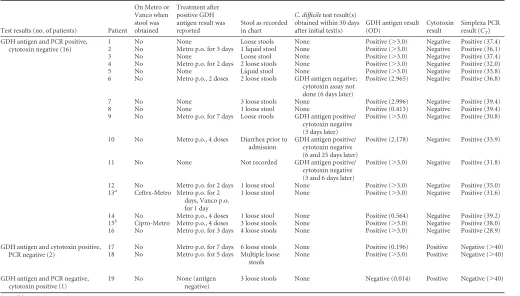
GDH-positive/PCR-negative/cytotoxin-positive specimens, and 1 GDH-negative/PCR-negative/cytotoxin-positive specimen). Charts for the 19 inpatients were reviewed to assess diarrhea, antibiotic ther-apy, subsequentC. difficiletesting, and CDI (Table 3). Since the Simplexa assay was not validated for clinical use and PCR results were not reported, clinical decisions were based solely on the stan-dard two-step algorithm. All GDH antigen results were reported, followed by cytotoxin results if GDH was positive, as per standard practice.
Of the 16 GDH-positive/PCR-positive but ultimately cytotox-in-negative inpatients, 6 were not treated, 9 were treated for up to 3 days until the cytotoxin results returned negative, and 1 was treated for 7 days. Of the 16, only 3 met the requirement of 3 diarrheal stools within a 24-h period. Only four had repeat testing within 30 days, and none were cytotoxin positive. One (6.3%) had symptomaticC. difficileinfection 6 weeks later, with positive GDH antigen and cytotoxin results and diarrhea.
Two patients positive by the two-step algorithm but negative by PCR had 3 or 4 loose stools per 24 h and were treated according to clinical guidelines. No further samples were sent in 60 days of follow-up. One patient was both GDH antigen and PCR negative but had a low positive cytotoxin result. Only the negative GDH result was reported (not the positive cytotoxin result) as per stan-dard clinical protocol; in a nonstudy situation, the cytotoxin assay would not have been done. The patient was lactose intolerant and had been given milk in error. This was considered a false-positive cytotoxin result.
Since most patients with stools positive by PCR but not for cytotoxin did not meet the criteria of 3 loose stools in 24 h and symptoms resolved with minimal or no therapy, it is highly likely that most if not all were carriers of toxigenicC. difficile.
Cost per reportable result.The average turnaround times, re-agents, materials, and labor costs per test in our laboratory are given inTable 4. If used as the sole test, PCR is significantly more expensive than the two-step algorithm. Costs can be reduced sig-nificantly if the GDH antigen ELISA is used as the screening test and PCR is performed only on GDH antigen-positive samples.
[image:3.585.40.560.77.373.2]Impact of PCR on infection control and antibiotic costs. Though the cost of laboratory testing is higher for PCR, potential savings by avoidance of 2 days of contact isolation (CI) and anti-biotic therapy were investigated for antigen-positive patients who were ultimately found to be cytotoxin negative.
TABLE 3Chart review of inpatients with discrepant cytotoxin and PCR resultsc
Test results (no. of patients) Patient
On Metro or Vanco when stool was obtained
Treatment after positive GDH antigen result was reported
Stool as recorded in chart
C. difficiletest result(s) obtained within 30 days after initial test(s)
GDH antigen result (OD)
Cytotoxin result
Simplexa PCR result (CT)
GDH antigen and PCR positive, cytotoxin negative (16)
1 No None Loose stools None Positive (⬎3.0) Negative Positive (37.4) 2 No Metro p.o. for 3 days 1 liquid stool None Positive (⬎3.0) Negative Positive (36.1) 3 No None Loose stool None Positive (⬎3.0) Negative Positive (37.4) 4 No Metro p.o. for 2 days 2 loose stools None Positive (⬎3.0) Negative Positive (32.0) 5 No None Liquid stool None Positive (⬎3.0) Negative Positive (33.8) 6 No Metro p.o., 2 doses 2 loose stools GDH antigen negative;
cytotoxin assay not done (6 days later)
Positive (2.965) Negative Positive (36.8)
7 No None 3 loose stools None Positive (2.996) Negative Positive (39.4) 8 No None 1 loose stool None Positive (0.413) Negative Positive (39.4) 9 No Metro p.o. for 7 days Loose stools GDH antigen positive/
cytotoxin negative (3 days later)
Positive (⬎3.0) Negative Positive (30.8) 10 No Metro p.o., 4 doses Diarrhea prior to
admission
GDH antigen positive/ cytotoxin negative (6 and 25 days later)
Positive (2.178) Negative Positive (33.9)
11 No None Not recorded GDH antigen positive/ cytotoxin negative (3 and 6 days later)
Positive (⬎3.0) Negative Positive (31.8) 12 No Metro p.o. for 2 days 1 loose stool None Positive (⬎3.0) Negative Positive (35.0) 13a Ceftrx-Metro Metro p.o. for 2
days, Vanco p.o. for 1 day
1 loose stool None Positive (⬎3.0) Negative Positive (31.6) 14 No Metro p.o., 4 doses 1 loose stool None Positive (0.564) Negative Positive (39.2) 15b
Cipro-Metro Metro p.o., 4 doses 3 loose stools None Positive (⬎3.0) Negative Positive (38.0) 16 No Metro p.o. for 3 days 4 loose stools None Positive (⬎3.0) Negative Positive (28.9) GDH antigen and cytotoxin positive,
PCR negative (2)
17 No Metro p.o. for 7 days 6 loose stools None Positive (0.196) Positive Negative (⬎40) 18 No Metro p.o. for 5 days Multiple loose
stools
None Positive (⬎3.0) Positive Negative (⬎40) GDH antigen and PCR negative,
cytotoxin positive (1)
19 No None (antigen negative)
3 loose stools None Negative (0.014) Positive Negative (⬎40)
aSmall bowel obstruction. bSurgical-site infection.
cMetro, metronidazole; Ceftrx, ceftriaxone; Cipro, ciprofloxacin; Vanco, vancomycin;C
T, cycle threshold; OD, optical density reading (ⱖ0.080 is positive); p.o.,per os.
TABLE 4Time to result and estimated costs for different methods
Test
Turnaround time (h)a Cost of
materials ($) Cost of
labor ($) Total ($) GDH antigen screen 2–24 4.73 2.50 7.23 Cytotoxin neutralizationb,c
6–48 11.76 6.46 18.22 Simplexa direct PCRb
2–24 36.05 6.25 42.30
aBased on one PCR or antigen assay run per day. b
Four samples per run; kit pricing is volume dependent.
cWith the 2-step method, GDH antigen-negative results are reported within 2 to 24 h.
GDH-positive results are then tested for cytotoxin, and cytotoxin results are reported after 4, 24, and 48 h of incubation. Negative results require 48 h.
on May 16, 2020 by guest
http://jcm.asm.org/
[image:3.585.298.544.631.679.2]Analysis of GDH antigen-positive specimens revealed 137 unique inpatients, and of these, 76 were GDH antigen positive but ultimately cytotoxin negative. Of these 76, 38 (50%) were already on CI prior toC. difficiletesting due to multidrug-resistant organ-isms; the remaining 38 (50%) were put on contact precautions as a consequence of the positive GDH antigen result. Empirical an-tibiotic therapy was given to only 41 of 76 patients (54%) for a median of 2 days and a mean of 3 days (range, 1 to 18 days). Metronidazole (Flagyl) was given to 33 (80%) and oral vancomy-cin to 8 (20%) patients. If PCR was reported at the same time as the GDH antigen-positive result, these costs could be avoided for patients who were ultimately found to be negative. The cost of CI was estimated at $61.44 per person per day, and the average cost of antibiotic therapy was $20.96 per patient per day. However, only 60 of 76 cytotoxin-negative patients were PCR negative, and 16 were PCR positive. Thus, if same-day PCR results replaced the 2-day cytotoxin assay results, the cost savings for 2 days of CI and antibiotic therapy for PCR-negative patients would be out-weighed by the costs incurred for 10 days of CI and antibiotic therapy for the additional PCR-positive patients (Table 5). Length of stay would also likely increase.
DISCUSSION
Ours is the first study comparing the performance characteristics of the commercial Focus Diagnostics SimplexaC. difficile univer-sal direct PCR and the two-step GDH antigen ELISA/cytotoxin neutralization protocol. Simplexa PCR was more sensitive than the two-step algorithm in detecting toxigenic bacteria (97.7% ver-sus 76.1%, respectively;P⫽0.0002). The concordance of the Sim-plexa PCR assay with cytotoxicity results (92.9%) is similar to that of other commercial PCR assays (3–5). The Simplexa direct assay produced results in approximately 2 h, compared to 6 to 48 h for cytotoxin neutralization, was simple to perform, and was done outside a molecular laboratory.
The C. Diff Chek-60 assay for detection of GDH antigen did not miss any PCR-positive samples, thus confirming its value as an initial screening test in a two-step protocol, using a toxin assay or PCR as the second step (2,6–8). In a microplate ELISA format, it is a relatively inexpensive test that takes 1 h to complete. Further-more, since it is read in a spectrophotometer, it may be more sensitive than GDH antigen lateral-flow tests read by eye, but this has not been studied.
Twenty-one PCR-positive/cytotoxin-negative samples were detected, and of these, 16 were from inpatients whose charts were available for review. Since PCR results were not reported to clini-cians, these samples were reported as GDH positive and ultimately cytotoxin negative. Investigation revealed that 10 patients re-ceived brief treatment while their cytotoxin results were pending. While unlikely, it is possible that clinical outcomes were affected. Six received no treatment for CDI, with no apparent adverse
con-sequences. According to the nurses’ notes, only 3 of the 16 had diarrhea that met the criteria for testing at the time that the sam-ples were sent. In a follow-up period of 30 days, only 4 of 16 patients had repeatC. difficiletesting and all remained cytotoxin negative. Our previous study of the BD GeneOhm PCR kit also found that the 18 patients positive by PCR only did not have CDI and did well (3).
Two patients were PCR negative/cytotoxin positive. These two patients met the criteria of 3 or more diarrheal stools per day and were treated. In our institution, cytotoxin testing is performed on site by experienced virology laboratory personnel, using cell culture plates freshly prepared on site, and samples are tested starting at a low dilu-tion (i.e., 1:20). Thus, our cytotoxicity results are likely more sensitive than those obtained using higher starting dilutions of 1:50 (5) or 1:100 (4), commercially prepared cell culture, or samples shipped a distance, during which toxin can degrade. Of note, Simplexa PCR missed fewer cytotoxin-positive samples (n ⫽ 2) than the BD GeneOhm test (n⫽9) in our previous study (3). Falsely negative PCR results can be due to low levels of bacteria, inhibitors, or genetic vari-ance leading to primer or probe mismatch (3,9,10).
Our study contributes to the growing concerns regarding tests that target bacteria rather than toxin. Both toxigenic bacterial cul-ture and toxin gene PCR detect toxigenic bacteria, while toxin assays detectin vivotoxin production. More positive results are detected by toxigenic culture and PCR than by toxin assays, and PCR can be associated with a⬎50% increase in the rate of inci-dence (11). However, the reported greater sensitivity of PCR may be misleading, as colonized patients are also detected. Further-more, toxigenic bacteria can remain detectable in stool for weeks after treatment (12).
The positive predictive value of a PCR result for CDI depends on having significant diarrhea (13). Too often, samples are sent after only one or two loose stools. In addition, carriers can have diarrhea due to other causes, such as norovirus gastroenteritis, tube feedings, laxatives, antibiotics, or other causes, and toxigenic C. difficileis merely a bystander (14,15). Carriers have been creasingly recognized in asymptomatic individuals of all ages, in-cluding 10% or more of hospitalized patients and up to 51% of patients in a long-term-care facility (16–21). Attempting to erad-icate carriage by treating with antibiotics may have adverse effects on the host-pathogen balance, increasing the risk of subsequent diarrhea, and is not currently recommended (1,16,22,23). Thus, separating colonized patients from those with true CDI is essential to avoid treating patients unnecessarily. To do this, a toxin assay is essential.
[image:4.585.40.557.78.135.2]Importantly, several recent studies have found that only toxin-positive stools, and not PCR-toxin-positive/toxin-negative stools, are associated with high morbidity and mortality as well as longer hospital stays (11,24,25). A large multicenter study found that cytotoxin positivity, and not GeneXpert PCR or toxigenic culture
TABLE 5Estimated impact of same-day PCR result on CI and antibiotic usage for inpatients
PCR result for GDH antigen-positive/ cytotoxin-negative
inpatients Impact
Total no. of patients
No. (%) of patients requiring CI
Days of CI required
Total CI days
CI cost per day ($)a
No. (%) of patients treated for CDI
No. of antibiotic treatment days per patient
Total antibiotic treatment days
Antibiotic cost ($)b
Negative (same day) Cost avoidance 60 30 (50) 2 60 3,686.40 32 (54) 2 64 1,341.44 Positive Cost incurred 16 8 (50) 10 80 4,825.20 16 10 160 3,353.60
aCI cost per day, $61.44. b
Antibiotic cost per day, $20.96 (80% were treated with metronidazole for $1.20; 20% were treated with oral vancomycin for $100).
on May 16, 2020 by guest
http://jcm.asm.org/
positivity, correlated with clinical outcome and thus best defined true cases of CDI (24). Consequently, they recommended against using PCR alone to diagnose CDI due to its low positive predictive value. Instead, they recommend a new diagnostic category ofC. difficileexcretor for carriers whose diarrhea is probably not due to CDI but who can potentially transmit infection.
It has been postulated that even asymptomatic carriers are a cause of nosocomial transmission and should be placed in isola-tion (21,26,27). A recent study used genotyping to investigate the role of asymptomatic carriers in transmission in a ward and con-cluded that carriers played an important role (27). However, it is not yet known whether carrier identification and isolation will reduce transmission and improve patient outcomes.
Commercial PCR test kits are more expensive than toxin assays and may require purchase of expensive equipment (3). Thus, compensatory savings from adoption of PCR were sought for the wards. It was anticipated that cost avoidance from a same-day diagnosis would outweigh the costs of PCR reagents. However, by our estimation, isolation and treatment of the additional patients positive by PCR lead to a net increase in costs. Unfortunately, we were not able to assess whether implementing PCR for toxigenic C. difficilewill reduce nosocomial transmission and disease and thus generate long-term savings. Such studies are urgently needed. As in our previous report (3), inappropriate stool sample sub-mission forC. difficiletesting, including multiple samples from the same patient on the same day, was common. Thirteen percent of samples were solid stools, which were rejected. Chart review revealed submission of samples from patients with minimal diar-rhea and from patients who were already on treatment. Though same-day samples and samples from patients on therapy were excluded from the study, these problems illustrate the difficulty in both enforcing testing guidelines and interpreting a positive PCR result. Nurses observe the stool and collect the specimens, but physicians receive the test results and act on them. To avoid un-necessary treatment and reduce costs of PCR testing and CI, cli-nicians need to limitC. difficiletesting and treatment to patients with a reasonable probability of having disease, such as those pa-tients having 3 or more loose stools per day for 1 to 2 days (1,28). Accomplishing this is a challenge.
There were a number of limitations of this study. All GDH antigen-positive samples submitted on study days that met study criteria were included, but only a subset of GDH antigen-negative samples were included. Cytotoxin and PCR results were not con-firmed by toxigenic culture but were accepted as true if GDH was also positive. C. difficileribotypes were not determined, which may influence clinical disease determination as well as perfor-mance of the GDH antigen assay. The number of stools as re-corded in the chart may have been underestimated. Colonoscopy was not performed to establish the presence or absence ofC. dif-ficiledisease. Rather we relied on cytotoxin to separate CDI from colonization.
In conclusion, Simplexa PCR detected significantly more pos-itive samples than a two-step method for diagnosis of toxigenicC. difficile. The integrated cycler has a small footprint and can be obtained by reagent rental rather than capital purchase. Addition-ally, the test can be performed outside a molecular laboratory. However, as in our prior study of a different toxin gene PCR kit, BD GeneOhm, samples positive by PCR and negative by cytotox-icity assay did not clearly indicate disease, yet their detection by PCR will lead to both contact isolation and treatment. The costs
and consequences of identifying, isolating, and treatingC. difficile carriers, either with or without significant diarrhea, should be a high priority for further study.
ACKNOWLEDGMENT
We thank the Clinical Virology Laboratory staff at YNHH for their work.
REFERENCES
1.Cohen SH, Gerding DN, Johnson S, Kelly CP, Loo VG, McDonald LC, Pepin J, Wilcox MH, Society for Healthcare Epidemiology of America, Infectious Diseases Society of America.2010. Clinical practice guidelines for Clostridium difficile infection in adults: 2010 update by the Society for Healthcare Epidemiology of America (SHEA) and the Infectious Diseases Society of America (IDSA). Infect. Control Hosp. Epidemiol.31:431– 455. http://dx.doi.org/10.1086/651706.
2.Burnham CA, Carroll KC.2013. Diagnosis ofClostridium difficile infec-tion: an ongoing conundrum for clinicians and for clinical laboratories. Clin. Microbiol. Rev.26:604 – 630.http://dx.doi.org/10.1128/CMR.00016 -13.
3.Kvach EJ, Ferguson D, Riska PF, Landry ML.2010. Comparison of BD GeneOhm Cdiff real-time PCR assay with a two-step algorithm and a toxin A/B enzyme-linked immunosorbent assay for diagnosis of toxigenic
Clostridium difficileinfection. J. Clin. Microbiol.48:109 –114.http://dx .doi.org/10.1128/JCM.01630-09.
4.Barbut F, Braun M, Burghoffer B, Lalande V, Eckert C.2009. Rapid detection of toxigenic strains ofClostridium difficilein diarrheal stools by real-time PCR. J. Clin. Microbiol. 47:1276 –1277.http://dx.doi.org/10 .1128/JCM.00309-09.
5.Stamper PD, Alcabasa R, Aird D, Babiker W, Wehrlin J, Ikpeama I, Carroll KC.2009. Comparison of a commercial real-time PCR assay for
tcdBdetection to a cell culture cytotoxicity assay and toxigenic culture for direct detection of toxin-producingClostridium difficilein clinical sam-ples. J. Clin. Microbiol. 47:373–378. http://dx.doi.org/10.1128/JCM .01613-08.
6.Wilcox MH.2012. Overcoming barriers to effective recognition and di-agnosis of Clostridium difficile infection. Clin. Microbiol. Infect.
18(Suppl. 6):13–20.http://dx.doi.org/10.1111/1469-0691.12057. 7.Shetty N, Wren MW, Coen PG.2011. The role of glutamate
dehydroge-nase for the detection of Clostridium difficile in faecal samples: a meta-analysis. J. Hosp. Infect.77:1– 6.http://dx.doi.org/10.1016/j.jhin.2010.07 .024.
8.Carman RJ, Wickham KN, Chen L, Lawrence AM, Boone JH, Wilkins TD, Kerkering TM, Lyerly DM.2012. Glutamate dehydrogenase is highly conserved amongClostridium difficileribotypes. J. Clin. Microbiol.50:
1425–1426.http://dx.doi.org/10.1128/JCM.05600-11.
9.Rupnik M, Braun V, Soehn F, Janc M, Hofstetter M, Laufenberg-Feldmann R, von Eichel-Streiber C.1997. Characterization of poly-morphisms in the toxin A and B genes of Clostridium difficile. FEMS Microbiol. Lett. 148:197–202. http://dx.doi.org/10.1111/j.1574-6968 .1997.tb10288.x.
10. Cohen SH, Tang YJ, Silva J, Jr.2000. Analysis of the pathogenicity locus in Clostridium difficile strains. J. Infect. Dis.181:659 – 663.http://dx.doi .org/10.1086/315248.
11. Longtin Y, Trottier S, Brochu G, Paquet-Bolduc B, Garenc C, Loung-narath V, Beaulieu C, Goulet D, Longtin J.2013. Impact of the type of diagnostic assay on Clostridium difficile infection and complication rates in a mandatory reporting program. Clin. Infect. Dis.56:67–73.http://dx .doi.org/10.1093/cid/cis840.
12. Sethi AK, Al-Nassir WN, Nerandzic MM, Bobulsky GS, Donskey CJ.
2010. Persistence of skin contamination and environmental shedding of Clostridium difficile during and after treatment of C. difficile infection. Infect. Control Hosp. Epidemiol. 31:21–27. http://dx.doi.org/10.1086 /649016.
13. Dubberke ER, Han Z, Bobo L, Hink T, Lawrence B, Copper S, Hoppe-Bauer J, Burnham CA, Dunne WM, Jr.2011. Impact of clinical symp-toms on interpretation of diagnostic assays forClostridium difficile infec-tions. J. Clin. Microbiol.49:2887–2893.http://dx.doi.org/10.1128/JCM .00891-11.
14. Polage CR, Solnick JV, Cohen SH.2012. Nosocomial diarrhea: evalua-tion and treatment of causes other than Clostridium difficile. Clin. Infect. Dis.55:982–989.http://dx.doi.org/10.1093/cid/cis551.
on May 16, 2020 by guest
http://jcm.asm.org/
15. Ludwig A, Sato K, Schirmer P, Maniar A, Lucero-Obusan C, Fleming C, Ryono R, Oda G, Winters M, Holodniy M.2013. Concurrent outbreaks with co-infection of norovirus and Clostridium difficile in a long-term-care facility. Epidemiol. Infect.141:1598 –1603.http://dx.doi.org/10.1017 /S0950268813000241.
16. Kyne L, Warny M, Qamar A, Kelly CP.2000. Asymptomatic carriage of Clos-tridium difficile and serum levels of IgG antibody against toxin A. N. Engl. J. Med.
342:390–397.http://dx.doi.org/10.1056/NEJM200002103420604.
17. Leekha S, Aronhalt KC, Sloan LM, Patel R, Orenstein R.2013. Asymp-tomatic Clostridium difficile colonization in a tertiary care hospital: ad-mission prevalence and risk factors. Am. J. Infect. Control41:390 –393. http://dx.doi.org/10.1016/j.ajic.2012.09.023.
18. Miyajima F, Roberts P, Swale A, Price V, Jones M, Horan M, Beeching N, Brazier J, Parry C, Pendleton N, Pirmohamed M.2011. Character-isation and carriage ratio of Clostridium difficile strains isolated from a community-dwelling elderly population in the United Kingdom. PLoS One6:e22804.http://dx.doi.org/10.1371/journal.pone.0022804. 19. Rousseau C, Poilane I, De Pontual L, Maherault AC, Le Monnier A,
Collignon A.2012. Clostridium difficile carriage in healthy infants in the community: a potential reservoir for pathogenic strains. Clin. Infect. Dis.
55:1209 –1215.http://dx.doi.org/10.1093/cid/cis637.
20. Lamouse-Smith ES, Weber S, Rossi RF, Neinstedt LJ, Mosam-maparast N, Sandora TJ, McAdam AJ, Bousvaros A.2013. Polymer-ase chain reaction test for Clostridium difficile toxin B gene reveals similar prevalence rates in children with and without inflammatory bowel disease. J. Pediatr. Gastroenterol. Nutr.57:293–297.http://dx .doi.org/10.1097/MPG.0b013e3182999990.
21. Guerrero DM, Becker JC, Eckstein EC, Kundrapu S, Deshpande A, Sethi AK, Donskey CJ.2013. Asymptomatic carriage of toxigenic Clos-tridium difficile by hospitalized patients. J. Hosp. Infect.85:155–158.http: //dx.doi.org/10.1016/j.jhin.2013.07.002.
22. Shim JK, Johnson S, Samore MH, Bliss DZ, Gerding DN.1998. Primary
symptomless colonisation by Clostridium difficile and decreased risk of subsequent diarrhoea. Lancet 351:633– 636. http://dx.doi.org/10.1016 /S0140-6736(97)08062-8.
23. Johnson S, Homann SR, Bettin KM, Quick JN, Clabots CR, Peterson LR, Gerding DN.1992. Treatment of asymptomatic Clostridium difficile carriers (fecal excretors) with vancomycin or metronidazole. A random-ized, placebo-controlled trial. Ann. Intern. Med.117:297–302.
24. Planche TD, Davies KA, Coen PG, Finney JM, Monahan IM, Morris KA, O’Connor L, Oakley SJ, Pope CF, Wren MW, Shetty NP, Crook DW, Wilcox MH.2013. Differences in outcome according to Clostridium difficile testing method: a prospective multicentre diagnostic validation study of C difficile infection. Lancet Infect. Dis.13:936 –945.http://dx.doi .org/10.1016/S1473-3099(13)70200-7.
25. Baker I, Leeming JP, Reynolds R, Ibrahim I, Darley E.2013. Clinical relevance of a positive molecular test in the diagnosis of Clostridium dif-ficile infection. J. Hosp. Infect.84:311–315.http://dx.doi.org/10.1016/j .jhin.2013.05.006.
26. Walker AS, Eyre DW, Wyllie DH, Dingle KE, Harding RM, O’Connor L, Griffiths D, Vaughan A, Finney J, Wilcox MH, Crook DW, Peto TE.
2012. Characterisation of Clostridium difficile hospital ward-based trans-mission using extensive epidemiological data and molecular typing. PLoS Med.9:e1001172.http://dx.doi.org/10.1371/journal.pmed.1001172. 27. Curry SR, MC, Schlackman JL, Pasculle AW, Shutt KA, Marsh JW,
Harrison LH.2013. Use of multilocus variable number of tandem repeats analysis genotyping to determine the role of asymptomatic carriers in Clostridium difficile transmission. Clin. Infect. Dis.57:1094 –1102.http: //dx.doi.org/10.1093/cid/cit475.
28. Reller ME, Lema CA, Perl TM, Cai M, Ross TL, Speck KA, Carroll KC.
2007. Yield of stool culture with isolate toxin testing versus a two-step algorithm including stool toxin testing for detection of toxigenic Clostrid-ium difficile. J. Clin. Microbiol.45:3601–3605.http://dx.doi.org/10.1128 /JCM.01305-07.