0095-1137/08/$08.00⫹0 doi:10.1128/JCM.01406-07
Copyright © 2008, American Society for Microbiology. All Rights Reserved.
DNA Microarray Based on Arrayed-Primer Extension Technique for
Identification of Pathogenic Fungi Responsible for
Invasive and Superficial Mycoses
䌤
Daniele Campa,
1‡ Arianna Tavanti,
2‡ Federica Gemignani,
1Crocifissa S. Mogavero,
2Ilaria Bellini,
1Fabio Bottari,
1Roberto Barale,
1Stefano Landi,
1‡ and Sonia Senesi
2‡*
Dipartimento di Biologia, Sezione di Genetica, Via Derna 1,1and Sezione di Microbiologia, Via San Zeno 35-39,2 Universita` di Pisa, 56126 Pisa, Italy
Received 12 July 2007/Returned for modification 4 October 2007/Accepted 14 December 2007
An oligonucleotide microarray based on the arrayed-primer extension (APEX) technique has been developed to simultaneously identify pathogenic fungi frequently isolated from invasive and superficial infections. Spe-cies-specific oligonucleotide probes complementary to the internal transcribed spacer 1 and 2 (ITS1 and ITS2)
region were designed for 24 species belonging to 10 genera, including Candida species (Candida albicans,
Candida dubliniensis, Candida famata, Candida glabrata, Candida tropicalis, Candida kefyr, Candida krusei, Candida guilliermondii, Candida lusitaniae, Candida metapsilosis, Candida orthopsilosis, Candida parapsilosis, andCandida pulcherrima),Cryptococcus neoformans, Aspergillusspecies (Aspergillus fumigatus andAspergillus terreus),Trichophytonspecies (Trichophyton rubrumandTrichophyton tonsurans),Trichosporon cutaneum, Epi-dermophyton floccosum,Fusarium solani,Microsporum canis,Penicillium marneffei, andSaccharomyces cerevisiae. The microarray was tested for its specificity with a panel of reference and blinded clinical isolates. The APEX technique was proven to be highly discriminative, leading to unequivocal identification of each species,
including the highly related onesC. parapsilosis,C. orthopsilosis, andC. metapsilosis. Because of the satisfactory
basic performance traits obtained, such as reproducibility, specificity, and unambiguous interpretation of the results, this new system represents a reliable method of potential use in clinical laboratories for parallel one-shot detection and identification of the most common pathogenic fungi.
The majority of life-threatening fungal infections are caused by the well-known opportunistic pathogensCandida albicans,
Aspergillus fumigatus, and Cryptococcus neoformans, with the latter being associated frequently with severe mycoses in AIDS patients (22, 30). Candidiasis remains the major cause of in-vasive fungal infections in immunocompromised patients, and in recent years, an impressive increase in mortality rate due to candidemia by non-C. albicans Candidaspecies has been noted (2, 6, 15, 20, 24). It has been suggested that the widespread use of azoles in clinical settings could have contributed to changing the etiology ofC. albicanscandidemia toward non-C. albicans
species, which now account for more than 50% of systemic
Candidainfections (2, 6, 15). Rapid diagnosis of these mycoses is crucial for prompt management of infection with tailored antifungal treatments. However, conventional laboratory methods for identification of fungal pathogens, though contin-uously improving, are still time-consuming and therefore often inadequate for ensuring early targeted therapy, especially for uncommon or newly identified fungal species. Unlike what is currently available for bacteria, molecular approaches for the identification of pathogenic fungi have been held back so far due to the lack of a robust sequence data bank. However, several DNA-based methods have been introduced and have improved the identification of fungal pathogens and shortened
the time required for their detection (3, 12, 13, 19, 21, 26). Most molecular procedures allow the identification of one or a few species at a time (3, 12, 13), thus resulting in a high cost if all relevant species must be considered. An ideal approach to overcome this limitation is given by the application of DNA microarray technology, which may enable discrimination of a wide range of pathogens in a single assay. The panmicrobial oligonucleotide array developed by Palacios and colleagues (23) was designed mainly to produce a staged strategy for molecular surveillance and discovery of emerging pathogens, as it covers detection of viruses, bacteria, fungi, and parasites. However, in the case of fungi, the panmicrobial chip predom-inantly allows genus identification rather than fungal species discrimination. It should be noted that rapid identification of pathogenic fungi at the species level is relevant in medical practice, as fungemia and other fungal symptomatic infections are emerging as a leading cause of morbidity and mortality in the general patient population, especially for hospitalized can-cer and major surgical patients (15). A DNA microarray spe-cifically developed by Leinberger and colleagues (18) for de-tecting fungal pathogens enabled discrimination of the 12 most common pathogenicCandidaandAspergillusorganisms at the species level, and the array developed by Huang and colleagues (11) enlarged to 20 the number of identified species, which are representative of eight different genera. Nevertheless, neither system encompasses oligonucleotide probes for detection/ identification of emerging fungi increasingly reported to be responsible for invasive or other symptomatic infections, such as Candida famata, Candida kefyr, Trichosporon cutaneum,
Fusarium solani, andPenicillium marneffei(5, 10, 24, 31);
more-* Corresponding author. Mailing address: Dipartimento di Biologia, Via San Zeno 35-39, 56127 Pisa, Italy. Phone: 39 050 2213695. Fax: 39 050 2213711. E-mail: [email protected].
‡ D.C., A.T., S.L., and S.S. contributed equally to this study.
䌤Published ahead of print on 26 December 2007.
909
on May 16, 2020 by guest
http://jcm.asm.org/
over, the probes designed to detectCandida parapsilosisdo not differentiate this species from two newly identified and closely related ones, i.e.,Candida orthopsilosisandCandida metapsi-losis(27), which were recently reported to cause mycoses in humans (14, 28). Oligonucleotide probes effectively enabling simultaneous discrimination of these three species may be use-ful, since available conventional methods do not allow discrim-ination ofC. parapsilosisfromC. orthopsilosisandC. metapsi-losis(26, 28).
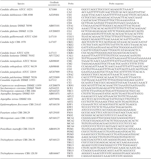
This report describes the development of an up-to-date oligo-nucleotide array for the unambiguous identification of 24 fungi, allotted into 10 diverse genera, including (i) species involved in invasive infections and frequently exhibiting a drug-resistant phenotype, such asCandida glabrata,Candida krusei, andAspergillus terreus(17, 24, 25); (ii) emerging fungal patho-gens, such asC. famata,C. kefyr, Trichosporon cutaneum, or molds such asFusarium solaniandPenicillium marneffei; and (iii) the newly defined speciesC. orthopsilosisandC. metapsi-TABLE 1. Fungal species tested and oligonucleotides used with the APEX technique
Species GenBank
accession no. Probe Probe sequence
Candida albicansATCC 10231 X71088 CA1 GGCCCAGCCTGCCGCCAGAGGTCTAAACT
CA2 ACCAATTTTTTATCAACTTGTCACACCAGATTATTAC
Candida dubliniensisCBS 8500 AJ249484 CD1 AAACTTACAACCAAATTTTTTATAAACTTGTCACGAGA
CD2 CCTGCCGCCAGAGGACATAAACTTACAACCAAAT
CD3 CAATACGACTTGGGTTTGCTTGAAAGATGA
CD4 GCTTGACAATGGCTTAGGTGTAACCAAAAACA
Candida famataDSMZ 70590 AB053101 CFA1 CTAGAAATAGTTTGGGCCAGAGGTTTACTGAAC
CFA2 AAACCTTACACACAGTGTTTTTTGTTATTACAAGAAC
Candida glabrataDSMZ 11226 AY208055 CG1 GCTCGGAGAGAGACATCTCTGGGGAGGACCAGTG
CG2 AAAGGAGGTGTTTTATCACACGACTCGACACTTTC
Candida guilliermondiiATCC 6260 L47110 CGU1 TGATACAGAACTCTTGCTTTGGTTTGGCCTAGAGA
CGU2 GGGCCAGAGGTTTAACAAAACACAATTTAATTA
Candida kefyrCBS 1970 L47107 CKE1 AGCTCGTCTCTCCAGTGGACATAAACACAAACAATAT
CKE2 CTCTGCTATCAGTTTTCTATTTCTCATCCTAAACACAA
CKE3 GATTATGAATGAATAGATTGCTGGGGGAATCGTC
CKE4 CAATTCGTGGTAAGCTTGGGTCATAGAGACTCA
Candida kruseiATCC 6258 L47113 CKR1 CGCAGAGTTGGGGGAGCGGAGCGGACGACG
Candida lusitaniaeDSMZ 70102 AF172262 CAL1 AAAATTTAATTTTTTTGTTCGCAAAAACAATGTG
CAL2 TGTTGAAAGTTTTGATATTAAGAAATTCGAATAAAAAAA
Candida metapsilosisATCC 96144 AJ698049 CM1 TAAACTCAACCAAATTTTTATTTAATTGTCAACTTGAT
CM2 TAGGAGAAGGTTGCTTAACTGCAATCCTTTTCTTTC
Candida orthopsilosisATCC 96139 AJ698048 CO1 CCAGAGATTAAACTCAACCAAATTTTATTTAAGTCAAC
CO2 CGTTGTTGAAAGTTTTGACTATTAGTTAATCAGTTGAC
Candida parapsilosisATCC 22019 AF287909 CPA1 GAATGAAAAGTGCTTAACTGCATTTTTTCTTACACA
CPA2 GGGGCCTGCCAGAGATTAAACTCAACCAAA
Candida pulcherrimaDSMZ 70336 AF235809 CPU1 CACCCTTTTAGGCACAAACTCTAAATCTTAACCG
Canadida tropicalisDSMZ 11953 L47112 CT1 TATTGAACAAATTTCTTTGGTGGCGGGAGCAATCC
CT2 GCAATCCTACCGCCAGAGGTTATAACTAAACCAAACT
Cryptococcus neoformansDSMZ 11959 AY208079 CRN1 GGCACGTTTTACACAAACTTCTAAATGTAATGAATG
Saccharomyces cerevisiaeDSMZ 70449 AJ544253 SCE1 CCAAACGGTGAGAGATTTCTGTGCTTTTGTTA
Trichosporon cutaneumCBS 2466 AF444325 TRC1 GTTTCTTAATGGATTGGATTTGGGCGCTGCCAG
Aspergillus fumigatusDSMZ 819 AB051071 AFU1 CTGTTCTGAAAGTATGCAGTCTGAGTTGATTATCG
AFU2 GAAGACCCCAACATGAACGCTGTTCTGAAAG
Aspergillus terreusDSMZ 826 AF078896 AST1 ACCTCCCACCCGTGACTATTGTACCTTGTTGCT
AST2 CCCTGTTCTGAAAGCTTGCAGTCTGAGTGTGA
Epidermophyton floccosumCBS 214.63 AF168130 EPF1 CGAAATCTCCATAGGTGGTTCAGTCTGAGCG
EPF2 TCCCCCTTCTCTCTGAATGCTGGACGGTG
EPF3 ATCCCCCGTTCCACCGGGAGAGGAGAAAGG
Fusarium solaniCBS 208.29 AF129105 FSO1 GCCGCAGCTTCCATCGCGTAGTAGCTAACACC
FSO2 GCGGGCACACGCCGTCCCCCAAATACAG
Microsporum canisCBS 113480 AF168127 MCA1 CGGGGAGGTTGCGGGCGGCGAGGGGTGCC
MCA2 CTCGCCGGAGGATTACTCTGGAAAACACACTC
MCA3 TGTGTGATGGACGACCGTCCCCCCTCCCCAG
MCA4 CGTCCCCCCTCCCCAGTAACCACCCACCGC
Penicillium marneffeiCBS 334.59 AB049129 PEM1 TGATGAAGATGGACTGTCTGAGTACCATGAAAA
PEM2 CGCCCTGTGAACCCTGATGAAGATGGACTG
PEM3 GAAGCGCCCTGTGAACCCTGATGAAGATGGAC
PEM4 AAGCACGGCTTGTGTGTTGGGTGTGGTCCC
Trichophyton rubrumCBS 286.30 AF168123 TRU1 GACCGACGTTCCATCAGGGGTGAGCAGACG
TRU2 CGCCCGCCGGAGGACAGACACCAAGAAAAAA
TRU3 AGAGCCGTCCGGCGGGCCCCTTCTGGGAGCC
TRU4 CTGTCAGTCTGAGCGTTTAGCAAGCACAATCAG
TRU5 CTAGGCGAATGGGCAGCCAATTCAGCGCCC
Trichophyton tonsuransCBS 120.30 AB166667 TRT1 AGGGCCAAACGTCCGTCAGGGGTGAGCAGATG
TRT2 CAGGATAGGGCCAAACGTCCGTCAGGGG
on May 16, 2020 by guest
http://jcm.asm.org/
[image:2.585.42.540.80.607.2]losis. The oligonucleotide probes used in this microarray are complementary to the sequence variation in the internal tran-scribed spacer 1 and 2 (ITS1 and ITS2) region of each species. Direct labeling of PCR products is not required, and signals for fungal species identification are based on the arrayed-primer extension (APEX) technique, which has been applied success-fully to discriminate natural variants of human papillomavirus type 16, to identify germ line mutations in beta-thalassemia, and for single-nucleotide polymorphism genotyping (4, 7, 8, 16). The specificity and reproducibility of the array were vali-dated with reference strains, and its application was tested with blinded clinical isolates.
MATERIALS AND METHODS
Strains.The yeast and mold reference isolates used in this study were obtained from the American Type Culture Collection (ATCC), Deutsche Sammlung von Mikroorganismen und Zellkulturen (DSMZ), and Centraalbureau voor Schim-melcultures (CBS) (Table 1). A selected panel of 20 clinical fungal isolates, previously identified with conventional laboratory tests (API32 ID and a Vitek 2 advanced colorimetry semiautomated system [bioMerieux, Marcy l’Etoile, France] for yeast identification and microscopic examination of reproductive structures for hyphomycetes), were provided by the Unita` Operativa di Micro-biologia, Ospedale Universitario, Pisa, Italy. The isolates (Table 2) were blindly submitted to the DNA microarray identification system. All of the isolates were maintained on Sabouraud agar (yeasts) or potato dextrose agar (molds) (Lio-filchem S.R.L., TE, Italy) for the duration of the study. Genomic DNAs from representative bacterial reference strains (Bacillus subtilisATCC 6633, Esche-richia coliSCS 110 [ATCC], andMycobacterium bovisbacillus Calmette-Gue´rin strain Pasteur [bioMerieux, Lyon, France]) were used as negative controls in PCR experiments.
Genomic DNA extraction. Genomic DNAs were extracted from yeasts or molds grown in Sabouraud dextrose broth (yeast) or on potato dextrose agar (molds) (Liofilchem S.R.L., TE, Italy). DNAs were extracted from yeast samples as previously described (28). Briefly, yeast cells were harvested in stationary phase, while mold suspensions were prepared by collecting conidia from 72-h
potato dextrose agar plate colonies washed with 5 ml of 0.1% Tween 20 (Sigma, St. Louis, MO)–sterile saline. Both yeast and conidia were lysed by vortexing the pellet for 3 min with 0.3 g glass beads (0.45- to 0.52-mm diameter; Sigma) in 200 l of lysis buffer (100 mM Tris-HCl, pH 8.0, 2% [vol/vol] Triton X-100, 1% [wt/vol] sodium dodecyl sulfate, and 1 mM EDTA) and 200l of 1:1 (vol/vol) phenol-chloroform. After the pellet was vortexed, 200l of TE (10 mM Tris-HCl, pH 8.0, 1 mM EDTA) was added to the lysate; the mixture was microcen-trifuged at full speed for 10 min, and the aqueous phase was transferred to a new tube. DNA was precipitated by the addition of 1 ml of ethanol. Samples were centrifuged, and the pellet was suspended in 400l of TE containing 100g RNase (Sigma). The mixture was incubated for 1 h at 37°C and subsequently treated with proteinase K (5l of a 20-mg/ml stock solution; Sigma) for 1 h at 65°C and for 30 min at 72°C. Phenol-chloroform treatment was repeated, and then DNA was precipitated with 1 ml of isopropanol and 10l of 4 M ammo-nium acetate, dried, and dissolved in 50l of TE, pH 8.0.
ITS amplification.The universal fungal primers ITS1 and ITS4 described by White et al. (29) (Sigma-Genosys, Steinheim, Germany) were used to amplify the noncoding ITS regions (ITS1 and ITS2) as well as the 5.8S rRNA gene, posi-tioned between the ITS regions. Reaction mixtures (20l) contained fungal DNA (100 ng), 0.4l primers (5M), 2l of deoxynucleoside triphosphate mix (2 mM dATP, 2 mM dCTP, 2 mM dGTP, 1.7 mM dTTP, and 0.03 mM dUTP), 2l magnesium-free buffer (10⫻; Solis Biodine, Tartu, Estonia), 2l MgCl2(25
mM; Solis Biodine), and 0.2l Hot Fire DNA polymerase (5 U/l; Solis Bio-dine). A negative control, which consisted of an equal volume of water replacing the DNA template, was included in all PCR experiments. Conditions for PCR amplification were as follows: 94°C for 15 min (hot start); 35 cycles of denatur-ation at 95°C (1 min), annealing at 56°C (30 s), and elongdenatur-ation at 72°C (75 s); and a final extension step at 72°C (10 min). Amplified DNA products were separated by electrophoresis in a 1% agarose gel containing ethidium bromide (0.5 mg/ml); the running buffer was TAE (40 mM Tris acetate [pH 8.0], 1 mM EDTA). A 100-bp DNA ladder was used as a molecular size marker (Promega). DNA bands were visualized by UV transillumination. The universal fungal primers used were tested versus representative bacterial (n⫽3) and human genomic DNA prep-arations.
Chip probe design.5⬘C-6 aminolinker-modified oligonucleotides (C-6 oligo-nucleotides) were designed by matching a region of about 35 bp within the ITS1 amplification products to the sequences of ITS deposited in GenBank (http: //www.ncbi.nlm.nih.gov/entrez/query.fcgi?db⫽Nucleotide&itool⫽toolbar) (ac-cession numbers are given in Table 1). Each C-6 oligonucleotide was designed in order to exclusively add a uracyl (fluorescein-ddUTP) at its 3⬘end during the single-base extension reaction. All of the C-6 oligonucleotides were synthesized by MWG Biotech (Ebersberg, Germany) and spotted onto silanized slides by Asperbio (Tartu, Estonia), as previously reported (1, 9). Preliminary experiments were performed by designing a large number of oligonucleotides for each species in order to test them for the ability to identify and discriminate the 24 fungal species. Details of the probes used in these experiments are provided in Table 1. For each fungal species, sets of oligonucleotides were selected among those previously designed on the basis of their discriminatory power and lack of cross-reactivity (Table 1).
Purification, hybridization of PCR products on the chip, and signal detection. PCR products were purified and concentrated using Millipore Y30 columns (7), and their sizes were reduced by fragmentation to improve hybridization with the arrayed oligonucleotides. To this end, Y30 column eluate samples (15l) were treated with 1 U uracilN-glycosylase (Epicenter Technologies, Madison, WI) and 1 U shrimp alkaline phosphatase (Amersham Biosciences, Milwaukee, WI), and the mixtures were incubated at 37°C for 1.5 h and at 95°C for 30 min. At this temperature, DNA with abasic sites is denatured and fragmented, whereas uracil
[image:3.585.44.285.87.318.2]N-glycosylase and shrimp alkaline phosphatase are inactivated. Aliquots of the treated amplicons (9l) were added to a reaction mixture containing fluores-cein-ddUTP, cyanine 3-ddATP, ROX-ddCTP, cyanine 5-ddGTP (4⫻50 pmol), 10⫻buffer, and 4 U of Thermo Sequenase (Amersham Biosciences, Uppsala, Sweden) diluted in the provided dilution buffer to give a final volume of 20l. Mixtures were quickly placed onto the spotted slide, which was previously washed with 100 mM NaOH and rinsed with distilled water at 95°C, and incu-bated at 58°C for 10 min. Slides were washed again with distilled water at 95°C to remove both traces of nonhybridized PCR products and nonincorporated labeled dideoxynucleoside triphosphates. A drop of SlowFade Light antifade reagent (Molecular Probes, Eugene, OR) was then added to prevent signal fading. Slides were imaged by a Genorama-003 four-color detector (Asper Bio-tech, Tartu, Estonia). Fluorescence intensities at each position were measured and converted to base calls according to Genorama image analysis and genotyp-ing software (Asper Biotech, Tartu, Estonia).
TABLE 2. Selected clinical fungal isolates blindly run to validate the DNA array-based identification system
Isolate Site of isolation Identification by
conventional testsa,b Array identificationb
CF1 Balanopreputial swab
Candida albicans Candida albicans
CF2 Bronchial aspirate
Candida tropicalis Candida tropicalis
CF3 Anal swab Candida
guilliermondii
Candida guilliermondii
CF4 Anal swab Candida glabrata Candida glabrata
CF5 Sputum Candida albicans Candida albicans
CF6 Sputum Candida albicans Candida albicans
CF7 Sputum Candida dubliniensis Candida dubliniensis
CF8 Urine Candida parapsilosis Candida orthopsilosis
CF9 Drainage Candida albicans Candida albicans
CF10 Feces Candida tropicalis Candida tropicalis
CF11 Scalp Microsporum canis Microsporum canis
CF12 Oral swab Candida famata Candida tropicalis
CF13 Feces Candida dubliniensis Candida dubliniensis
CF14 Nail Trichophyton rubrum Trichophyton rubrum
CF15 Bronchial aspirate
Candida tropicalis Candida tropicalis
CF16 Oral swab Candida glabrata Candida glabrata
CF17 Scalp Trichophyton
tonsurans
Trichophyton tonsurans
CF18 Vaginal swab Candida parapsilosis Candida metapsilosis
CF19 Blood Candida parapsilosis Candida parapsilosis
CF20 Balanopreputial swab
Candida lusitaniae Candida lusitaniae
a
API32 ID and Vitek 2 advanced colorimetry for yeasts and microscopic examination of reproductive structures for molds.
b
Data in bold show differences between conventional and array identification results.
on May 16, 2020 by guest
http://jcm.asm.org/
FIG. 1. (A) Representative snapshots from Genorama Basecaller software (Asperbio, Tartu, Estonia), showing the identification of fungal species by the chip. Cy3 fluorescence images (U channel) show the following identification panels: 1,Candida albicans; 2,C. dubliniensis; 3, C. famata; 4, C. glabrata; 5, C. guilliermondii; 6, C. kefyr; 7,C. krusei; 8, C. lusitaniae; 9, C. metapsilosis; 10, C. orthopsilosis; 11,C.
on May 16, 2020 by guest
http://jcm.asm.org/
Interpretation of results and identification criteria.For species identification, the following two criteria were required: (i) both APEX probes had to give a signal and (ii) the extension had to yield a signal on the U channel, as the probes were designed to extend uridine only. Although A, G, and C signals are not expected to be incorporated in the APEX reaction, ddA, ddC, and ddG were also included in the reaction mixture, thus providing a further specificity control to detect any unspecific extensions. Moreover, to ensure quality control, the fol-lowing strategies were adopted: (i) DNA samples were randomly processed by personnel who were blinded to the strain typed, (ii) each APEX oligonucleotide was spotted in duplicate, and (iii) internal positive controls were included in the microarray (Fig. 1) to verify that the intensities of the four channels (A, C, U, and G) were equilibrated.
Confirmation ofC. parapsilosisand related species identification by the array. The two-step DNA-based identification test described by Tavanti et al. (27) was used to confirm the identification ofC. parapsilosis,C. metapsilosis, andC. orthopsilosisisolates. Briefly, a 716-bp fragment of theSADH(secondary alcohol dehydrogenase) gene was amplified, purified, and digested with BanI. The dif-ferent digestion patterns were used to identify the three species, since theC. parapsilosis,C. metapsilosis, andC. orthopsilosis SADHamplicons contain 1, 3, and 0 BanI restriction sites, respectively.
RESULTS
Probe design and array setup.An up-to-date DNA
microar-ray system was developed for the unambiguous identification of 24 fungal species of clinical relevance (Table 1). Oligonu-cleotides were designed on the basis of the available ITS1 and/or ITS2 sequences (for accession numbers, see Table 1). The amplicons obtained for each species included the region carrying ITS1, the 5.8S rRNA gene, and ITS2 and varied in size from 375 bp (Candida pulcherrima) to 880 bp (Candida gla-brata) (data not shown). A check test was performed by testing the specificity of the universal fungal primers used versus bac-terial (three different strains) and human genomic DNA prep-arations. No amplification product was detected for any non-fungal genomic DNA template (data not shown). A pilot study was undertaken with a redundant number of oligonucleotide probes in order to evaluate probe reliability, discrimination power, and lack of cross-reactivity, which would potentially lead to misinterpretation of the results. The data obtained with this pilot chip led to an accurate selection of a reduced number of probes. Sets of C-6 oligonucleotides, ranging from 1 to 5 (Table 1), were proven to unambiguously identify the 24 fungal species and were used to validate their application for detect-ing/identifying fungal reference strains and blinded clinical iso-lates.
Validation of the DNA-based chip with reference strains.
The results presented in Fig. 1 were obtained from experi-ments repeated at least three times using samples containing amplicons generated from different preparations of genomic DNAs of reference species, for a total of 72 slides. As shown in Fig. 1A, the array system gave an unequivocal response for each of the 24 reference species considered. The layout of the chip allows rapid and accurate species identification (Fig. 1B): most of the species (n⫽13) were identified by two oligonu-cleotide probes, replicated twice for a total of four spots, with the exception of Epidermophyton floccosum, detected with
three replicated probes (Fig. 1A, slide 19);Candida dublinien-sis,C. kefyr,Penicillium marneffei, andMicrosporum canis, de-tected with a set of four replicated probes (Fig. 1A, slides 2, 6, 21, and 22) for a total of eight spots; andTrichophyton rubrum, detected with five replicated probes (Fig. 1A, slide 23).
Despite the fact that for five species (Candida krusei, C. pulcherrima, Cryptococcus neoformans, Saccharomyces cerevi-siae, andTrichosporon cutaneum) only one probe among the originally designed probes was found to be suitable for appli-cation in the final version of the identifiappli-cation array, the results obtained for these species provided clear and reproducible identification for each of them (Fig. 1A, slides 7, 12, 14, 15, and 16, respectively). In addition, the microarray was proven to be highly discriminative, being able to distinguish closely related species, such asC. orthopsilosis,C. metapsilosis, andC. parap-silosisas well as Aspergillus fumigatusand A. terreus.As ex-pected, the results obtained for the “psilosis” complex (Fig. 1A, slides 9 to 11) indicated that a moderate level of cross-reactivity still exists with the probes used in the final version of the chip: indeed, whenC. metapsilosis(slide 9) orC. orthopsi-losis (slide 10) was detected by the system, one of the C. parapsilosisprobes (CPA1) (Table 1) gave a simultaneous sig-nal, whereas when C. parapsilosis was identified, no other probes produced a positive signal (Fig. 1, slide 11). However, the low level of cross-reactivity observed within the “psilosis” species did not prevent their unambiguous identification. As already observed by Leinberger et al. (18), detection of As-pergillus fumigatusled to the appearance of a faint signal where one probe designed forA. terreuswas positioned (Fig. 1A, slide 17). This cross-reactivity may be explained by the close genetic relatedness of these species; nevertheless, it does not prevent the correct identification of the two Aspergillusspecies (Fig. 1A, slides 17 and 18).Penicillium marneffeiwas included in the panel of fungi identifiable by the system since it has increas-ingly been associated with opportunistic infections (31) and it shares morphological traits with the more clinically relevant organismAspergillus fumigatus. As shown in Fig. 1A, slide 22,
P. marneffeiwas clearly identified by eight characteristic spots (four probes) and did not give any cross-reaction with eitherA. fumigatusorA. terreus(Fig. 1A, slides 17 and 18, respectively). Among the three dermatophyte genera included in the array,
Microsporum caniswas identified by a clear and unique signal (four probes) (Fig. 1A, slide 21), while a weak level of cross-reactivity was observed, as expected, between the two species of theTrichophyton genus, i.e., T. rubrum(Fig. 1A, slide 23) andT. tonsurans (Fig. 1A, slide 24). When the latter species were present in an independent tested sample, one of the probes forEpidermophyton floccosum also gave a faint signal withT. rubrum(Fig. 1A, slides 23 and 24), whileE. floccosum
identification (Fig. 1A, slide 19) did not produce any cross-reactive signal. Therefore, despite the fact that faint aspecific signals were observed within related species, the identification
parapsilosis; 12, C. pulcherrima; 13, C. tropicalis; 14,Cryptococcus neoformans; 15,Saccharomyces cerevisiae; 16, Trichosporon cutaneum; 17,
Aspergillus fumigatus; 18,A. terreus; 19, Epidermophyton floccosum; 20, Fusarium solani; 21, Microsporum canis; 22,Penicillium marneffei; 23,
Trichophyton rubrum; and 24,T. tonsurans. No signals were detected in the Cy5 channel (i.e., the non-U channel, which was not displayed for brevity). (B) Layout of chip capture probes (for probe sequences, see Table 1). Control probes are positioned at the corners of each panel.
on May 16, 2020 by guest
http://jcm.asm.org/
of all dermatophytes was clear and unambiguous for any of the species tested.
Blinded test of a panel of clinical fungal isolates.In separate
experiments, a panel of clinical fungal isolates, including yeasts and molds, were blindly submitted to the array identification system. These isolates were previously identified with conven-tional laboratory tests by the Unita` Operativa di Microbiolo-gia, Ospedale Universitario, Pisa, Italy. As reported in Table 2, 17 of the isolates were unambiguously identified as the same species as those obtained by conventional methods (data not shown). Two clinical isolates identified asCandida parapsilosis
(Table 2) (CF8 and CF18) were detected asC. orthopsilosis
(CF8) andC. metapsilosis(CF18) by the array system. Molec-ular identification of these two isolates was then performed by BanI digestion of theSADHamplicon (27), and the digestion profiles confirmed the results obtained with the array (data not shown). Isolate CF12, identified asCandida famata(Table 2), turned out to beC. tropicalison the basis of the spot position on the array; the conventional identification was therefore repeated, confirming that the array identification was indeed correct and that mislabeling of the isolate plate may have occurred.
DISCUSSION
This paper describes the setup of an up-to-date version of a DNA microarray-based system for the identification of the most common fungal pathogens in humans, encompassing 24 different species belonging to 10 genera. Despite the fact that a panmicrobial oligonucleotide array was recently proposed for the diagnosis of infectious diseases (23), it appears to be better suited for molecular surveillance of infectious agents in high-throughput/reference laboratories than in small mycology units. Indeed, the use of the panmicrobial chip may supersede the effective needs of the local diagnostic routine and may have high running costs. Moreover, with this array, the use of fungal probes designed based on the 18S rRNA sequence does not provide enough discriminatory power to identify each single species of the 73 fungal genera included in the chip.
The microarray we propose utilizes oligonucleotide probes complementary to the ITS region (ITS1 or ITS2) of each species and is based on the APEX technique. The present work describes the first application of APEX to the field of medical mycology. The technique proposed here relies both on specific hybridization between probes and their targets, like conven-tional microarray-based methods (11, 18, 23), and on the spe-cific extension of a single base at the 3⬘side of the anchored probe. In other words, APEX provides a further level of anal-ysis and thus, at least theoretically, should give more clear-cut results and reduced misclassification of fungal species than do hybridization-only array-based methods.
Optimization of the detection system was a crucial point for ensuring the reproducibility, specificity, and sensitivity of the results obtained; in many cases, it was necessary to reduce the number of probes originally designed and spotted on the pilot chip (such as those forCandida krusei) due to cross-reaction among species, which could possibly cause misinterpretation of results. The optimized microarray detection system was proven to be highly discriminative, and in its final layout, it allowed the unequivocal identification of each of the 24 species, including
yeasts and hyphomycetes. Despite a very low level of cross-reactivity still detectable in the identification of genetically related species, the signals obtained provided effective discrim-ination of the “psilosis” group,Aspergillus, and dermatophyte species tested.
Furthermore, the system was validated by using a selected panel of clinical fungal isolates previously identified with con-ventional laboratory tests, which were blindly submitted to the array identification system. The majority of the isolates were detected unambiguously in perfect concordance with the re-sults obtained with the laboratory tests. In two cases, the mi-croarray resulted in more discriminative results than did con-ventional biochemical tests, suggesting that this system could efficaciously flank the diagnostic routine, providing a rapid and sensitive tool for the identification of closely related species such asC. orthopsilosis,C. metapsilosis, andC. parapsilosisor aspergilli.
The chip we have developed is currently used in our clinical laboratory in parallel with conventional identification proce-dures. Our aims are both to widen the collection of clinically significant fungi run with the array system and to confirm its performance traits, such as reproducibility, specificity, and un-equivocal interpretation of the results. Indeed, high-through-put processing of clinical samples will be necessary to validate the system by testing fungal isolates undergoing selective pres-sure during the complex interactions occurring within the in-fected host.
ACKNOWLEDGMENTS
This study was supported by research grant 2005068754 from the Italian Ministero dell’Istruzione, dell’Universita` e della Ricerca (MIUR) and by an Associazione Italiana Ricerca sul Cancro (AIRC) regional grant.
REFERENCES
1.Auffray, C., C. Mundy, and A. Metspalu.2000. DNA arrays: methods and applications: report on HUGO Meeting, Tartu, Estonia, 23–26 May, 1999. Eur. J. Hum. Genet.8:236–238.
2.Bedini, A., C. Venturelli, C. Mussini, G. Guaraldi, M. Codeluppi, V. Borghi, F. Rumpianesi, F. Barchiesi, and R. Esposito.2006. Epidemiology of can-didaemia and antifungal susceptibility patterns in an Italian tertiary-care hospital. Clin. Microbiol. Infect.12:75–80.
3.Bolehovska, R., L. Pliskova, V. Buchta, J. Cerman, and P. Hamal.2006. Detection of Aspergillus spp. in biological samples by real-time PCR. Biomed. Pap. Med. Fac. Univ. Palacky Olomouc Czech Repub.150:245–248. 4.Campa, D., S. Zienolddiny, H. Lind, D. Ryberg, V. Skaug, F. Canzian, and A. Haugen.2007. Polymorphisms of dopamine receptor/transporter genes and risk of non-small cell lung cancer. Lung Cancer56:17–23.
5.Chang, S. E., K. J. Kim, W. S. Lee, J. H. Choi, K. J. Sung, K. C. Moon, and J. K. Koh.2003. A case ofTrichosporon cutaneumfolliculitis and septicae-mia. Clin. Exp. Dermatol.28:37–38.
6.Colombo, A. L., T. Guimaraes, L. R. Silva, L. P. de Almeida Monfardini, A. K. Cunha, P. Rady, T. Alves, and R. C. Rosas.2007. Prospective obser-vational study of candidemia in Sao Paulo, Brazil: incidence rate, epidemi-ology, and predictors of mortality. Infect. Control Hosp. Epidemiol.28:570– 576.
7.Gemignani, F., S. Landi, A. Chabrier, A. Smet, I. Zehbe, F. Canzian, and M. Tommasino.2004. Generation of a DNA microarray for determination of E6 natural variants of human papillomavirus type 16. J. Virol. Methods119:95– 102.
8.Gemignani, F., C. Perra, S. Landi, F. Canzian, A. Kurg, N. Tonisson, R. Galanello, A. Cao, A. Metspalu, and G. Romeo.2002. Reliable detection of beta-thalassemia and G6PD mutations by a DNA microarray. Clin. Chem. 48:2051–2054.
9.Guo, Z., R. A. Guilfoyle, A. J. Thiel, R. Wang, and L. M. Smith.1994. Direct fluorescence analysis of genetic polymorphisms by hybridization with oligo-nucleotide arrays on glass supports. Nucleic Acids Res.22:5456–5465. 10.Ha, Y. S., S. F. Covert, and M. Momany.2006. FsFKS1, the 1,3-beta-glucan
synthase from the caspofungin-resistant fungusFusarium solani. Eukaryot. Cell5:1036–1042.
on May 16, 2020 by guest
http://jcm.asm.org/
11.Huang, A., J. W. Li, Z. Q. Shen, X. W. Wang, and M. Jin.2006. High-throughput identification of clinical pathogenic fungi by hybridization to an oligonucleotide microarray. J. Clin. Microbiol.44:3299–3305.
12.Iwen, P. C., A. G. Freifeld, T. A. Bruening, and S. H. Hinrichs.2004. Use of a panfungal PCR assay for detection of fungal pathogens in a commercial blood culture system. J. Clin. Microbiol.42:2292–2293.
13.Kardjeva, V., R. Summerbell, T. Kantardjiev, D. Devliotou-Panagiotidou, E. Sotiriou, and Y. Graser.2006. Forty-eight-hour diagnosis of onychomycosis with subtyping ofTrichophyton rubrumstrains. J. Clin. Microbiol.44:1419– 1427.
14.Kocsube, S., M. Toth, C. Vagvolgyi, I. Doczi, M. Pesti, I. Pocsi, J. Szabo, and J. Varga.2007. Occurrence and genetic variability ofCandida parapsilosis
sensu lato in Hungary. J. Med. Microbiol.56:190–195.
15.Krcmery, V., and A. J. Barnes.2002. Non-albicansCandidaspp. causing fungaemia: pathogenicity and antifungal resistance. J. Hosp. Infect.50:243– 260.
16.Landi, S., F. Gemignani, L. Gioia-Patricola, A. Chabrier, and F. Canzian. 2003. Evaluation of a microarray for genotyping polymorphisms related to xenobiotic metabolism and DNA repair. BioTechniques35:816–827. 17.Lass-Florl, C., K. Griff, A. Mayr, A. Petzer, G. Gastl, H. Bonatti, M. Freund,
G. Kropshofer, M. P. Dierich, and D. Nachbaur. 2005. Epidemiology and outcome of infections due toAspergillus terreus: 10-year single centre experience. Br. J. Haematol.131:201–207.
18.Leinberger, D. M., U. Schumacher, I. B. Autenrieth, and T. T. Bachmann. 2005. Development of a DNA microarray for detection and identification of fungal pathogens involved in invasive mycoses. J. Clin. Microbiol.43:4943– 4953.
19.Luo, G., and T. G. Mitchell.2002. Rapid identification of pathogenic fungi directly from cultures by using multiplex PCR. J. Clin. Microbiol.40:2860– 2865.
20.Marr, K. A.2004. InvasiveCandidainfections: the changing epidemiology. Oncology (Williston Park)18:9–14.
21.Martin, C., D. Roberts, M. van Der Weide, R. Rossau, G. Jannes, T. Smith, and M. Maher.2000. Development of a PCR-based line probe assay for identification of fungal pathogens. J. Clin. Microbiol.38:3735–3742. 22.McNeil, M. M., S. L. Nash, R. A. Hajjeh, M. A. Phelan, L. A. Conn, B. D.
Plikaytis, and D. W. Warnock.2001. Trends in mortality due to invasive mycotic diseases in the United States, 1980–1997. Clin. Infect. Dis.33:641– 647.
23.Palacios, G., P. L. Quan, O. J. Jabado, S. Conlan, D. L. Hirschberg, Y. Liu, J. Zhai, N. Renwick, J. Hui, H. Hegyi, A. Grolla, J. E. Strong, J. S. Towner, T. W. Geisbert, P. B. Jahrling, C. Buchen-Osmond, H. Ellerbrok, M. P. Sanchez-Seco, Y. Lussier, P. Formenty, M. S. Nichol, H. Feldmann, T. Briese, and W. I. Lipkin.2007. Panmicrobial oligonucleotide array for diag-nosis of infectious diseases. Emerg. Infect. Dis.13:73–81.
24.Pfaller, M. A., D. J. Diekema, D. L. Gibbs, V. A. Newell, J. F. Meis, I. M. Gould, W. Fu, A. L. Colombo, and E. Rodriguez-Noriega.2007. Results from the ARTEMIS Disk Global Antifungal Surveillance study, 1997 to 2005: an 8.5-year analysis of susceptibilities ofCandidaspecies and other yeast species to fluconazole and voriconazole determined by CLSI standardized disk dif-fusion testing. J. Clin. Microbiol.45:1735–1745.
25.Pfaller, M. A., S. A. Messer, L. Boyken, S. Tendolkar, R. J. Hollis, and D. J. Diekema.2004. Geographic variation in the susceptibilities of invasive iso-lates ofCandida glabratato seven systemically active antifungal agents: a global assessment from the ARTEMIS Antifungal Surveillance Program conducted in 2001 and 2002. J. Clin. Microbiol.42:3142–3146.
26.Pryce, T. M., S. Palladino, D. M. Price, D. J. Gardam, P. B. Campbell, K. J. Christiansen, and R. J. Murray.2006. Rapid identification of fungal patho-gens in BacT/ALERT, BACTEC, and BBL MGIT media using polymerase chain reaction and DNA sequencing of the internal transcribed spacer re-gions. Diagn. Microbiol. Infect. Dis.54:289–297.
27.Tavanti, A., A. D. Davidson, N. A. Gow, M. C. Maiden, and F. C. Odds.2005.
Candida orthopsilosisandCandida metapsilosisspp. nov. to replaceCandida parapsilosisgroups II and III. J. Clin. Microbiol.43:284–292.
28.Tavanti, A., L. A. Hensgens, E. Ghelardi, M. Campa, and S. Senesi.2007. Genotyping ofCandida orthopsilosisclinical isolates by amplification frag-ment length polymorphism reveals genetic diversity among independent isolates and strain maintenance within patients. J. Clin. Microbiol.45:1455– 1462.
29.White, T., T. Bruns, S. Lee, and J. Taylor.1990. Amplification and direct sequencing of fungal ribosomal RNA genes for phylogenetics, p. 315–322.In
M. Innis, D. Gelfand, J. Sninsky, and T. White (ed.), PCR protocols: a guide to methods and application. Academic Press, San Diego, CA.
30.Wingard, J. R.2005. The changing face of invasive fungal infections in hematopoietic cell transplant recipients. Curr. Opin. Oncol.17:89–92. 31.Xi, L., X. Xu, W. Liu, X. Li, Y. Liu, M. Li, J. Zhang, and M. Li.2007.
Differentially expressed proteins of pathogenicPenicillium marneffeiin yeast and mycelial phases. J. Med. Microbiol.56:298–304.