CD1 presentation of microbial nonpeptide
antigens to T cells.
D Jullien, … , W A Ernst, R L Modlin
J Clin Invest.
1997;99(9):2071-2074. https://doi.org/10.1172/JCI119378.
Perspective
Find the latest version:
http://jci.me/119378/pdf
Perspectives Series: Host/Pathogen Interactions
J. Clin. Invest.
© The American Society for Clinical Investigation, Inc. 0021-9738/97/05/2071/04 $2.00
Volume 99, Number 9, May 1997, 2071–2074
CD1 Presentation of Microbial Nonpeptide Antigens to T Cells
Denis Jullien,* Steffen Stenger,* William A. Ernst,§ and Robert L. Modlin*‡
*Division of Dermatology, ‡Department of Microbiology and Immunology, UCLA School of Medicine, Los Angeles, California 90095;
and §Department of Microbiology and Molecular Genetics, University of California, Los Angeles, California 90095
Introduction
It has long been recognized that T cells are an essential compo-nent of efficient cell-mediated immunity against intracellular pathogens. The central paradigm has been that T cells recog-nize foreign peptides in the context of MHC class I and II mol-ecules. Upon activation, these T cells lyse infected macro-phages, depleting the reservoir of cells harboring the pathogen. Furthermore, T cell recognition of antigen leads to the release of cytokines which activate macrophages to kill microbial or-ganisms. For many such pathogens, considerable effort has been invested towards identifying peptide antigens which can engender cell-mediated immunity with the ultimate goal of de-veloping vaccines against infectious disease. The drawback of this approach lies in the polymorphic nature of MHC mole-cules. Therefore, peptides which bind to one individual’s MHC and activate that person’s T cells, may not bind to another per-son’s MHC and therefore cannot be recognized by their T cells. Thus, an effective vaccine requires the delineation of peptide antigens which can bind to and be presented by a vari-ety of MHC molecules.
A new advance in our understanding of T cell biology has been the demonstration of T cell recognition of nonpeptide antigens. In one system, gd T cells were shown to recognize isopentenyl pyrophosphate and related structures in the iso-prenoid family (1). The ability of gd T cells to recognize these antigens may occur in a manner independent of antigen-pre-senting molecules. In a second system, ab T cells have been shown to recognize lipid and lipoglycan antigen in the context of CD1 molecules (2–4).
CD1 family of MHC-related proteins
The human cluster of differentiation I (CD1)1 gene family
con-sists of five nonpolymorphic genes, CD1A, -B, -C, -D, and -E, mapped to a cluster on chromosome 1 (for review see refer-ence 5). Human CD1 has a unique tissue distribution, with
CD1a, -b, and -c present on thymocytes but not peripheral blood T cells. Human CD1 are also present on professional an-tigen-presenting cells including dendritic cells, Langerhans cells, and mantle zone B cells. The structural homology of CD1 with class I and class II molecules suggests an antigen-presenting function. However, the amino acids encoded in the predicted antigen binding site, the a1 and a2 domains, are ex-tremely hydrophobic.
Recognition of CD1 by human T cells
A unique function of human CD1 is the ability to present non-peptide antigen to T cells. Specifically, human CD1b and CD1c have been shown to present lipid and lipoglycan anti-gens of Mycobacterium tuberculosis and Mycobacterium leprae
to T cells (2–4, 6). Two major antigens have been elucidated, both derived from the cell wall of mycobacteria: mycolic acids and lipoarabinomannan. Mycolic acids are branched long chain fatty acids specifically found in mycobacteria. Lipoarabi-nomannan (LAM) contains an arabinose head, branched man-nose core, and phosphatidyl inositol which contains two fatty acids: tuberculostearic acid and palmitic acid. For lipoarabi-nomannan, the data suggested that T cell recognition was at the level of the branched mannose core. These data do not preclude the possibility that human CD1 can present peptide antigen, as convincingly shown for murine CD1 (7).
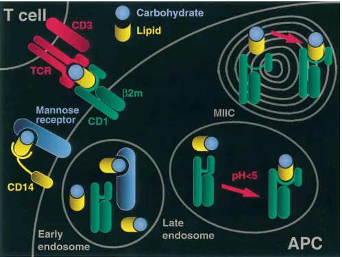
CD1 antigen presentation pathway
CD1-restricted T cell recognition of nonpeptide antigens in-volves an intracellular presentation pathway (Fig. 1). The CD1 antigen presentation pathway has novel aspects, although it shares some features in common with the classical MHC class II pathway. It has been demonstrated that the macrophage mannose receptor is involved in the uptake of LAM, through binding of its mannose core. The mannose receptor delivers LAM to late endosomal and lysosomal vesicles, as well as to the MHC class II antigen loading compartment (MIIC). The uptake of LAM into endosomes and its presentation to LAM-reactive T cells are blocked by reagents which interfere with the mannose receptor (8).
In the MIIC compartment, the mannose receptor, LAM, and CD1b colocalize, suggesting that this is the place where LAM is loaded onto CD1b molecules. CD1b is endocytosed at the plasma membrane in coated pits and coated vesicle struc-tures, transits to early endosomes, and is then delivered to the MIIC. In striking contrast to other MHC molecules, CD1b en-docytosis and trafficking to the MIIC compartment were shown to be directed by a YXXZ (where Y is a tyrosine, X is any amino acid, and Z is a hydrophobic residue) motif con-tained in the cytoplasmic tail of the molecule (9). The endo-somal localization motif is essential for antigen presentation by CD1b, suggesting that trafficking of CD1b through lysosomal compartments is required for loading of antigen into CD1b.
Address correspondence to Dr. Robert L. Modlin, UCLA School of Medicine, 52-121 CHS, 10822 Le Conte Ave., Los Angeles, CA 90095. Phone: 310-825-6214; FAX: 310-206-9878; E-mail: rmod [email protected]
Received for publication 26 March 1997 and accepted in revised form 1 April 1997.
The presentation of both LAM and mycolic acids is also dependent on the acidification of endosomes, since lysosomo-tropic drugs which prevent endosomal acidification block anti-gen presentation. At pH , 5, the biophysical properties of CD1b and CD1c are dramatically altered to facilitate the di-rect interaction of CD1 with nonpeptide ligands. Principally, at acid pH there is unfolding of the a-helical part of CD1. This can influence antigen binding, since the two a-helixes of anti-gen-presenting molecules form the walls of the antigen-bind-ing groove. This groove is believed to be deeper and narrower than the class I molecule groove as shown for the mouse CD1 (10). Indeed, for human CD1b and CD1c, the unfolding of the
a-helixes exposes a hydrophobic binding site which accommo-dates the lipid portion of LAM. At acid pH, the direct physical binding of LAM to CD1b is of high affinity, 3.2 3 1028 M,
comparable to high affinity peptide–MHC interactions (Ernst, W.A., M. Kronenberg, J. Maher, S. Cho, D. Chatterjee, and R.L. Modlin, manuscript submitted for publication).
This novel antigen presentation pathway bridges two arms of the immune system. Innate immunity pertains to those cells
which are preprogrammed to respond in certain ways, includ-ing macrophages, natural killer cells, and mast cells. The man-nose receptor, a pattern recognition marker, is part of the in-nate immune response. In this manner, the mannose receptor is able to traffic antigen first recognized by the innate immune system to T cells, which are part of the acquired immune re-sponse since their selection and expansion involves the devel-opment of memory. In that manner, the CD1 antigen presen-tation pathway links together the innate immune system and the acquired immune response.
CD1-restricted T cells in microbial immunity
[image:3.612.57.556.58.433.2]Investigation of human leprosy has provided direct evidence for the involvement of the CD1-restricted T cells in host re-sponse to infection. CD1a, -b, and -c were expressed on den-dritic cells in the granulomas within the skin lesions of leprosy patients. Furthermore, the frequency of CD11 cells correlated with the level of cell-mediated immunity to M. leprae, being 10-fold more abundant in the granulomas of patients with the immunologically responsive tuberculoid form of the disease, as
compared with the unresponsive lepromatous form (Sieling, P.A., M. Mehrali, T.H. Rea, M.B. Brenner, R.L. Modlin, and S. Porcelli, manuscript in preparation). It is likely that these differences are related to the level of GM-CSF in the lesions, being greater in the tuberculoid lesions. In addition, IL-10, which is specifically found at the site of infection in the lepro-matous form, has been shown to downregulate CD1 expres-sion (11). Further evidence for a role of CD1 presentation of antigens in immunity to mycobacterial infection in vivo in-cludes the isolation of CD1-restricted T cells from patients with mycobacterial infection. Initially, a CD1b-restricted, M. leprae– specific T cell line was derived from a cutaneous leprosy lesion (4) and more recently, M. tuberculosis–reactive CD1-restricted T cell lines from the peripheral blood of patients with tubercu-losis (12).
The study of CD1-restricted T cells from patients with my-cobacterial infection as well as normal healthy donors has re-vealed certain shared functional features. First, all the CD1-restricted T cell lines isolated produce high levels of IFN-g, but little or no IL-4 upon stimulation with mycobacterial antigen. This Th1 cytokine pattern can directly contribute to cell-medi-ated immunity against intracellular infection by enhancing T cell proliferation and macrophage activation. Secondly, CD1-restricted T cells also show a high degree of cytolytic activity against antigen-pulsed macrophages. Furthermore, these T cells can lyse macrophages infected with virulent M. tuberculo-sis. The induction of cytotoxicity is dependent on antigen pre-sentation and T cell recognition since lysis is blocked by anti-bodies to CD1b. Lysis of highly infected macrophages can contribute to host defense either by directly killing the bacteria or indirectly by disbursing the pathogen and thereby allowing freshly recruited macrophages to take up and more effectively dispose of the bacteria (13).
Two mechanisms of cell-mediated cytotoxicity differen-tially contribute to the outcome of infection with intracellular pathogens. CD4-CD82 (double negative) CD1-restricted T cells lyse targets through the Fas/Fas-ligand pathway, while the lysis of targets by CD81 cells depends on the release of gran-ules containing perforin and granzymes. However, only the CD81 subset of CD1-restricted T cells kills the intracellular
M. tuberculosis during the lysis of the target cell. While the double negative T cells may have an immunoregulatory role by reducing the local cellular infiltration and thereby limiting tissue damage, the CD81 T cells may have the greatest impact in combating intracellular pathogens (12). The mechanisms by which CD81 T cells kill the bacteria remain to be determined and are of potentially important therapeutic interest.
Nonpeptide antigens of other pathogens
Description of the CD1 presentation pathway, its unique abil-ity to present nonpeptide antigens, and evidence for its role in host immune responses were pioneered in the study of myco-bacterial infections. However, nonpeptide antigens sharing very high structural similarity with LAM or mycolic acids are present in many other pathogens. Lipoteichoic acid in Staphy-lococcus and Streptococcus are analogous in structure to LAM. Similarly, the lipopolysaccharides of gram-negative organisms share structural homology with LAM. The capsular polysac-charides of some gram-negative organisms are linked to pal-mitic acid, making them logical candidates for presentation by CD1 to T cells. Homology can be found in the structural com-ponents of some parasitic organisms, for instance the
lipophos-phoglycan of Leishmania. We anticipate studies delineating involvement of CD1-restricted T cells in these and other infec-tious diseases.
Advantage of nonpeptide antigens as vaccine candidates
The discovery of nonpeptide lipid and glycolipid antigen rec-ognition by CD1-restricted T cells defines a new paradigm for immune recognition and provides a distinct mechanism for host responses to infection. It may be possible to exploit this aspect of the immune response in the area of vaccine develop-ment. Current immunoprophylactic strategies use protein sub-unit vaccines, which may vary in effectiveness according to the MHC haplotype of the individual.
The advantage of antigens presented by CD1 resides in the nonpolymorphic nature of these antigen-presenting molecules. That is, everyone has the same CD1 molecules. Therefore, these nonpeptide antigens will bind to everyone’s CD1 and can be recognized by everyone’s T cells. The next critical step will be to determine whether CD1-restricted T cell responses to nonpeptide antigens are sufficient for protective immune re-sponses to microbial pathogens.
Acknowledgments
D. Jullien was supported by the French research ministry (grant Lavoisier) and the Cilag Corporation (grant Marc Chaptal). S. Stenger was supported by the AIDS Stipendium, Deutsches Krebs-forschungszentrum, Heidelberg. This work was supported by grants from the National Institutes of Health (AI-22553, AI-36069, AR-40312) and the UNDP/World Bank/World Health Organization Spe-cial Program for Research and Training in Tropical Diseases (IMMLEP) (R.L. Modlin).
References
1. Tanaka, Y., C.T. Morita, E. Nieves, M.B. Brenner, and B.R. Bloom. 1995. Natural and synthetic nonpeptide antigens recognized by human gamma/
delta T cells. Nature (Lond.). 375:155–158.
2. Porcelli, S., C.T. Morita, and M.B. Brenner. 1992. CD1b restricts the
re-sponse of human CD4-82 T lymphocytes to a microbial antigen. Nature
(Lond.). 360:593–597.
3. Beckman, E.M., S.A. Porcelli, C.T. Morita, S.M. Behar, S.T. Furlong, and
M.B. Brenner. 1994. Recognition of a lipid antigen by CD1-restricted ab1 T
cells. Nature (Lond.). 372:691–694.
4. Sieling, P.A., D. Chatterjee, S.A. Porcelli, T.I. Prigozy, T. Soriano, M.B. Brenner, M. Kronenberg, P.J. Brennan, and R.L. Modlin. 1995. CD1-restricted
T cell recognition of microbial lipoglycans. Science (Wash. DC). 269:227–230.
5. Porcelli, S. 1995. The CD1 family: a third lineage of antigen presenting
molecules. Adv. Immunol. 59:1–98.
6. Beckman, E.M., A. Melian, S.M. Behar, P.A. Sieling, D. Chatterjee, S.T. Furlong, R. Matsumoto, J.P. Rosat, R.L. Modlin, and S.A. Porcelli. 1996. CD1c restricts responses of mycobacteria-specific T cells. Evidence for antigen
pre-sentation by a second member of the human CD1 family. J. Immunol. 157:
2795–2803.
7. Castano, A.R., S. Tangri, J.E.W. Miller, H.R. Holcombe, M.R. Jackson, W.D. Huse, M. Kronenberg, and P.A. Peterson. 1995. Peptide binding and
pre-sentation by mouse CD1. Science (Wash. DC). 269:223–226.
8. Prigozy, T.I., P.A. Sieling, D. Clemens, P.L. Stewart, S.M. Behar, S.A. Porcelli, M.B. Brenner, R.L. Modlin, and M. Kronenberg. 1997. The mannose receptor delivers lipoglycan antigens to endosomes for presentation to T cells
by CD1b molecules. Immunity. 6:187–197.
9. Sugita, M., R.M. Jackman, E. van Donselaar, S.M. Behar, R.A. Rogers, P.J. Peters, M.B. Brenner, and S.A. Porcelli. 1996. Cytoplasmic tail-dependent
localization of CD1b antigen-presenting molecules to MIICs. Science (Wash.
DC). 273:349–352.
10. Zheng, Z.H., A.R. Castano, B. Segelke, E.A. Stura, P.A. Peterson, and I.A. Wilson. 1997. The crystal structure of murine CD1: an MHC-like fold but
with a large hydrophobic binding groove. Science (Wash. DC). In press.
interleukin-10 on the expression of HLA class II and CD1 molecules induced
by granulocyte/macrophage colony-stimulating factor/interleukin-4. Eur. J.
Im-munol. 25:2465–2470.
12. Stenger, S., R.J. Mazzaccaro, K. Uyemura, S. Cho, P.F. Barnes, J.P. Ro-sat, M.B. Brenner, S.A. Porcelli, B.R. Bloom, and R.L. Modlin. 1997.
Differen-tial effects of cytolytic T cell subsets on intracellular infection. Science (Wash.
DC). In press.
13. Kaufmann, S.H.E. 1988. CD81 T lymphocytes in intracellular microbial
infections. Immunol. Today. 9:168–174.
“Host/Pathogen Interactions: Understanding the Strategies of Microbial Virulence and Host Defense”
Series Editors, Donald G. Guiney and Martin F. Kagnoff
February 1, 1997 Arthropod- and host-specific gene expression by Borrelia burgdorferi ... Aravinda M. de Silva and Erol Fikrig
February 15, 1997 Regulation of bacterial virulence gene expression by the host environment ... Donald G. Guiney
March 1, 1997 Bacterial toxins that target Rho proteins... Klaus Aktories March 15, 1997 Yersinia proteins that target host cell signaling pathways ... Maria Fällman,
Cathrine Persson, and Hans Wolf-Watz April 1, 1997 Cytotoxic T cells and viral hepatitis... Francis V. Chisari April 15, 1997 Membrane-protein traffic in pathogen-infected cells ... Keith A. Joiner
May 1, 1997 CD1 presentation of microbial nonpeptide antigens to T cells... Denis Jullien,
Steffen Stenger, William A. Ernst, and Robert L. Modlin May 15, 1997 Conscription of the cytoskeleton by invasive bacteria ... Pascale Cossart June 1, 1997 Dynamics of HIV replication in vivo... David D. Ho June 15, 1997 Mechanisms of nitric oxide–related antimicrobial activity ... Ferric C. Fang July 1, 1997 Epithelial cells as sensors for microbial infection... Martin F. Kagnoff July 15, 1997 Invasion and intracellular sorting of bacteria... Stanley Falkow August 1, 1997 Pathogen-induced apoptosis ... Philippe Sansonetti
August 15, 1997 Mechanisms of the long-term interaction between Helicobacter pylori