PERFORMANCE EVALUATION OF HMSK AND SQFD
ALGORITHMS FOR COMPUTER TOMOGRAPHY (CT)
IMAGE SEGMENTATION OF EFFECTIVE RADIOTHERAPY
1V.V.GOMATHI, 2S.KARTHIKEYAN
1
Ph.D Research Scholar, Research and Development Centre, Bharathiar University, Coimbatore 2
Assistant Professor, Department of Information Technology, College of Applied Sciences, Sohar, Oman
E-mail: [email protected], [email protected]
ABSTRACT
Medical Image segmentation plays a significant role in many medical image processing for effective diagnosis. Manual segmentation of medical image by the radiologist is not only a tiresome and time consuming process, also not a very accurate with the increasing medical imaging modalities and unmanageable quantity of medical images. Therefore it is essential to examine current methodologies of image segmentation. Enormous research has been done in medical image segmentation, but it is still difficult to evaluate all the medical images. However the problem remains challenging, with no general and unique solution. In this paper, we present a HMSK (Hybrid Medoid shift and K-Means) algorithm and Signature Quadratic form distance (SQFD) algorithm for Computer tomography image segmentation. The performance of the two algorithms is investigated. Experimental results with real patient images indicate the SQFD algorithm is effective and efficient and reduce the number of fragments. Their pros and cons were analyzed and proposed SQFD algorithm for slices of CT images to give effective radiation therapy.
Keywords: HMSK, SQFD, K-Means, Medical image segmentation, Computer tomography, Radiotherapy
1. INTRODUCTION
Image segmentation is the process of partitioning a digital image into segments. Segmentation refers to simplifying and/or change the representation of an image into more meaningful and easier to analyze [1]. Image Segmentation is the most interesting and challenging problems in computer vision generally and especially in medical imaging applications. Accurate, fast and reproducible image segmentation techniques are required for various applications. Segmentation algorithms available vary widely depending on the specific application, image modality and other factors.
Medical image segmentation is the process of outlining relevant anatomical structures in an image dataset. It is a problem that is central to a variety of medical applications including image enhancement and reconstruction, surgical planning, disease classification, data storage and compression, and 3D visualization [2].
The segmentation of the medical image faces the challenges of existence in a large number of diverse structures of human anatomy and inevitable
artifacts induced from the imaging procedure. Most medical image segmentation algorithms are either semi-automatic or require some form of human intervention to perform satisfactorily.
One fundamental problem in medical image analysis is image segmentation which identifies the edges of objects such as organs or abnormal regions (e.g. tumors) in images. Good segmentations will help clinicians and patients as they provide vital information for 3-D visualization, surgical planning and early disease recognition. The most common images used in medicine, particularly in radiation therapy are the Computer Tomography Images (CT), because of its electron density. It is a diagnostic tool used to detect cancer and find out the cancer’s stage. It has the gold standard for radiation therapy planning. CT has the most well defined source detector geometry and also has the highest contrast rate than other modalities [3].
2030. The most useful way to reduce deaths due to cancer is to treat the disease in the early stages. Early treatment needs early diagnosis, and early diagnosis requires an accurate and reliable diagnostic procedure that allows physicians to differentiate tumors from normal organs. Manual segmentation process requires at least three hours completing by the radiologist or radiation oncologist. It is the time-consuming process and also inter and intra-expert variability residing between persons to persons. Hence, manual segmentation becomes less efficient in clinical operations and human interpretation may not be produce correct information. So intellectual techniques are needed to segment automatically [3].
The main objective of this research work is to develop an efficient segmentation algorithm for segmenting the different organs in computer tomography images. If the number of fragments (connected components) increases, then the complexity is also more. A very good segmentation algorithm should give only less number of exact fragments. In this paper, we have compared Hybrid medoid shift with K-Means algorithm (HMSK) and Signature Quadratic Form distance (SQFD) algorithm. The Experimental result proves that the SQFD algorithm gives most promising results with less number of fragments.
The rest of this paper is organized as follows. Section 2 depicts the related works. Section 3 describes Materials and Methods presents Dataset, Hybrid medoid shift with K-Means algorithm, Signature Quadratic form distance segmentation algorithm. Computational results and discussion are given in Section 4. Finally, some conclusions are drawn in Section 5.
2. RELATED WORKS
Mohammad Talebi et al proposed a segmental method combines genetic algorithm and active contour with an energy minimization procedure based on genetic algorithms for ultrasound images[4]. Kaihua Zhang et al proposed a novel region-based active contour model (ACM) is. It was implemented with a special processing named Selective Binary and Gaussian Filtering Regularized Level Set (SBGFRLS) method, which first selectively penalizes the level set function to be binary, and then uses a Gaussian smoothing kernel to regularize it. This method can segment not only the desired object but also the other objects [5]. Djamal Boukerroui proposed an adaptive region segmentation algorithm based on global and local statistics in a Bayesian framework for 2D
breast ultrasound data and on echocardiographic sequences( 2D+ T). This algorithm gives good segmentation results [6].
Miin-Shen Yang et al proposed an alternative fuzzy C-mean (AFCM) clustering algorithm for MRI segmentation in Ophthalmology [7]. Beloit Caldairou et al proposed to integrate into the FCM segmentation methodology concepts with the non-local (NL) framework for MRI Image segmentation. This non-local framework is used to handle intensity inhomogeneities and noise in the data [8]. Keh-Shih Chuang et al presented a fuzzy c-means (FCM) algorithm that incorporates spatial information into the membership function for clustering. This method is efficient for noisy image segmentation and works for both single and multiple-feature data with spatial information for MRI Images [9]. Jie Wu1 et al proposed a new texture feature based seeded region growing algorithm for automated segmentation of organs in abdominal MR images [10].
Segmentation of the lungs in chest computed tomography [16]. Jianhua Liu et al proposed watershed algorithm and region merging approach, to solve over-segmentation for identifying Liver Cancer CT image [17]. Shojaii et al presented a novel lung segmentation technique based on watershed transform which eliminates the tasks of finding an optimal threshold and separating the attached left and right lungs, which are two common practices in most lung segmentation methods and require a significant amount of time [18].
3. MATERIALS AND METHODS
3.1 Real CT Image Data set
Different type of Tumor patient dataset was collected by a SIEMENS SOMATOM EMOTION SPIRAL CT scanner located at Multi Speciality Hospital, Coimbatore. Besides a normal scan performed at a routine clinical dosage (130 mA), an additional scan from the same patient was acquired at a much lower tube current, i.e. 20 mA. The dataset is very difficult in that the volumes come from various sources. The volumes have various dimensionalities due to the different scanning protocols.
3.2 Methodology
An efficient SQFD algorithm is proposed to avoid the drawbacks of HMSK algorithm. One of the major drawbacks was over fragmentation produced by the HMSK algorithm. This will produce different connected components. So classifying many connected components is not possible. This will cause wrong segmentation and also lead to wrong decision making by the radiologist. Here SQFD is designed to avoid the above said limitation and segmenting the required organs efficiently.
3.2.1HMSK algorithm
The HMSK Algorithm is as follows:
Step I: Consider the Single Dicom image or slices
of Dicom images
Step II: Apply the ECFT (Enhanced Curvelet Filter
Technique) algorithm to get a noiseless image
Step III: Find the Histogram of the input image
Step IV: Initialize the control parameter
Step V: Find the gray level cluster values based on
an initialized control parameter
Step V: Find no of pixel values present between
each range of all gray level cluster values.
Step VI: Cluster the pixels which lies between the
range to the respective gray level cluster value
Step VII: Each cluster are considered as data points
Step VIII: Find the distance between each cluster to
all the data points
Step IX: Make the data point allocation by using
(
)
(
)
( )
( )
(
∑
ci=1∗
∑
cj=1x
i−
v
j)
2
Step X: Find the new cluster center by using
(
1
/
C
i)
∗
(
∑
Cj=i1( )
x
i)
Step XI: Obtain the Clustered Image
In the HMSK algorithm, initially the ECFT algorithm (Enhanced Curvelet Filtering Technique) has been applied for removal of noise in the CT images. This noiseless image has taken as the input image. First the histogram has taken for the input image and initializes the control parameter. Based on the control parameter the random cluster value or points has taken. The number of occurrences of cluster value was found and then found the closest cluster based on the occurrences. Here each cluster is considered as data points. The distance between data points and cluster points (Closest cluster) has been calculated and found the new cluster. Finally the similar cluster image has been obtained.
Limitation of the HMSK Algorithm
The HMSK segmentation algorithm gives more number of fragments and also the number of fragments are not consistent when executed for a certain number of times i.e. when the same image executed for different number of times the result were not holding the same number of fragments and also position of fragment and size of fragment were also dynamic. For diminishing these drawbacks, the SQFD algorithm has been proposed for effective segmentation.
3.2.2SQFD Algorithm
between the well known concept of Quadratic Form Distances and feature signatures. The SQFD is a way to measure the similarity between two objects. The SQFD algorithm described detail in the following
Step 1: Initialize the cluster step value
Step 2: Generate the Intensity Vector (cluster centers) based on the cluster step value.
Step 3: Extract the feature signatures from intensity vector and input image pixel i.e. P= Xk , Q= Yij
Where
X - Intensity vector
Xk – kth Value of Intensity vector Yij – Input Image Pixel
P, Q - Feature Signatures
Step 4: Calculate the Similarity Matrix by using the following formula=
(
)
(
)
(
1
2 2)
/
1
n n m
m
P
Q
P
Q
A
=
+
−
+
−
Where
A-Similarity Matrix
Qm and Qn – X and Y position of Q elements respectively
Pm and Pn – X and Y position of P elements respectively
Step 5: Compute Signature Quadratic Form Distance value (SQFD) between the each pixel from the input image and all cluster values in the intensity vector by using the following formula
SQFD Mk =
(
(
)
(
)
)
T k ij k
ij
X
A
Y
X
Y
|
−
*
*
|
−
WhereA-Similarity Matrix X - Intensity vector
Xk – kth Value of Intensity vector Yij – Input Image Pixel
M - Output vector
Mk - kth Value of output vector
Step 6: Find the minimum distance value i.e.
Zij
=
min(
M
)
Where
M – Output Vector Zij - Output image pixel
Step 7: Find the cluster center value with respect to minimum difference value
Step 8: Assign that cluster center value to the respective pixel position in the image
Step 9: Repeat the step 3,4,5,6,7 and 8 until convergence is attained (i.e. no pixels change clusters).
In this SQFD algorithm, the initial cluster step value has chosen manually. Then find the cluster
centers based on the initialized cluster step value. The number of clusters in the image is equal to the number of cluster centers. Then the distance measure has been calculated between every pixel and the cluster centers.
Figure.1. Flow diagram detailing the SQFD algorithm used for segmentation
Signature Quadratic form distance is used to find the distance. For this process, the two vectors P and
Input Image
Cluster Step
Intensity vector (having values incremented by cluster step)
I, J< (RxC)
K<=Length of Intensity
vector
Y=Input Image (I, J) X=Intensity vector (K)
Similarity Matrix (A, P, Q)
SQFD (A, P, Q)
M (K) =SQFD
Z= Min (M)
Segmented Image
No
No
Q is formed. By using P and Q the similarity matrix is generated. Then the SQFD similarity between the P and Q is identified using the SQFD formula. Then identify the position with minimum SQFD value. Then the intensity vector having the cluster value in that minimum value position is replaced with the original pixel value in the same position. Then the respective cluster center is assign to the respective pixel in the image. Repeat the process till the convergence attained (ie there is no change in the pixel value of an image). Finally the image will have the cluster number equal to the cluster centers.
4 COMPUTATIONAL RESULTS AND
DISCUSSION
Experimentation was carried out on 100 numbers of different tumor patients contains 100 to 1000 slices of Computer Tomography images using different Segmentation algorithms. The image format is DICOM (Digital Imaging Communications in Medicine). The algorithm has been implemented in Matlab environment. Manual Segmentation done by the experts. Experimental results of the images are illustrated here.
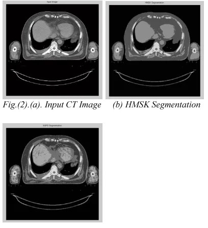
Fig.(2).(a). Input CT Image (b) HMSK Segmentation
(c) SQFD Segmentation
Fig 2(a) shows input CT image (b) shows segmentation result generated with HMSK (c) shows SQFD algorithm on the CT images. Results generated with HMSK and SQFD algorithm are shown for comparison.
Fig.(3).(a) HMSK
(
b) SQFD Segmentation Segmentation(c)Manual Segmentation done by the experts
Fig 3 (a) shows segmentation result generated with HMSK for Liver region (b) shows results generated with SQFD algorithm for Liver Region (c) depicts the manual segmentation results contoured by the medical experts.
A real set of CT images are used to evaluate the accuracy of the proposed algorithm in segmenting the medical images. This paper mainly focuses on segmentation algorithm in an efficient manner by using two different algorithms namely HMSK and SQFD. In our previous research, we have compared many segmentation algorithms. The segmentation results of HMSK and SQFD are considered the best algorithm that gives better segmentation results for real CT images. The HMSK algorithm is based on clustering based segmentation algorithm and proposed SQFD algorithm is a distance based algorithm.
[image:5.595.295.507.71.375.2] [image:5.595.85.289.423.647.2]Manhattan Distance, Minkowski distance, Chebyshev distance and Signature Quadratic form Distance(SQFD) measures[20]. The SQFD algorithm finds the similarity between all cluster elements and every pixel values such that the feature space have the highest possible similarity values of cluster vector. So it is suitable for computer tomography image segmentation.
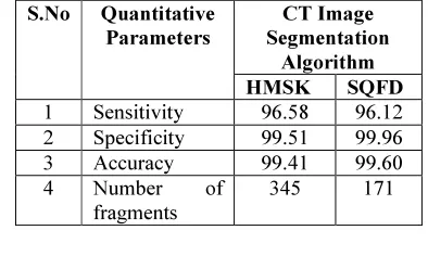
[image:6.595.92.289.444.562.2]The four important parameters are used to determine the precision of the HMSK and SQFD algorithms are Sensitivity, specificity, accuracy and number of fragments. The important parameters mentioned above i.e. Sensitivity, specificity, accuracy and number of fragments are specified in Table 1, for the HMSK and SQFD algorithms. In this paper, the liver organ has taken for demonstrating the segmentation performance. In fig(3) describes the liver segmentation done by HMSK and SQFD algorithm. SQFD having less number of fragments compared to HMSK algorithm based on the visible part and analysis part. From the results obtained it can be concluded that our proposed SQFD algorithm performed well in segmenting the real CT images with less number of fragments (connected components).
Table 1. Performance analysis of HMSK and SQFD Segmentation algorithm
S.No Quantitative Parameters
CT Image Segmentation
Algorithm
HMSK SQFD
1 Sensitivity 96.58 96.12
2 Specificity 99.51 99.96
3 Accuracy 99.41 99.60
4 Number of
fragments
345 171
5 CONCLUSION AND FUTURE WORK
In this study, two medical image segmentation algorithms have been considered for Computer tomography images. Large amount medical image segmentation techniques are exist in the current scenario and still emerging the new techniques need to overcome the limitations. Here HMSK and SQFD segmentation algorithm have implemented and analyzed the performance. An experimental result shows that the SQFD algorithm is effective, more robust and outperforms the HMSK algorithm. The SQFD gives good segmentation results with less number of fragments.
In this research work, the Cluster step value is fixed and also suitable for this computer tomography image contrast levels. Future work will be aimed to determine the cluster step value automatically for any type of computer tomography image contrast levels.
ACKNOWLEDGEMENT
The authors like to thank Dr.M.Hemalatha, Professor, Department of Computer Science, Karpagam University for her valuable suggestions, comments and words of encouragement. It helped us to make this research work successful.
REFERENCES
[1] Shapiro, L.G., Stockman, G.C,” Computer Vision”, Prentice-Hall, New Jersey (2001) ISBN 0-13-030796-3, pp. 279–325.
[2] Tsai, A., Wells, W., Tempany, C., Grimson, E., Willsky, A,”Mutual information in coupled multi-shape model for medical image segmentation”, Medical Image Analysis, 2004, pp.429–445.
[3] Gomathi V.V, Karthikeyan.S, “A Proposed Hybrid Medoid Shift with K-Means (HMSK) Segmentation Algorithm to Detect Tumor and Organs for Effective Radiotherapy”, International Conference on Mining Intelligence and Knowledge Exploration,
Lecture Notes in Computer
Science(Springer), Volume 8284, 2013,
pp.139-147.
[4] Mohammad Talebi, Ahamd Ayatollahi, Ali Kermani,”Medical ultrasound image segmentation using genetic active contour”,
Journal of Biomedical Science and
Engineering, 2011, pp.105-109.
[5] Kaihua Zhanga, Lei Zhanga, Huihui Song , Wengang Zhou, “Active contours with selective local or global segmentation: A new formulation and level set method”, Image and Vision Computing (Elsevier), Vol. 28 ,2010,pp. 668–676.
[6] Djamal Boukerroui , Atilla Baskurt , J. Alison Noble , Olivier Basset ,” Segmentation of ultrasound images––multiresolution 2D and 3D algorithm based on global and local statistics, Pattern Recognition Letters(Elsevier), 2003, pp. 779-790.
algorithms”, Magnetic Resonance Imaging(Elsevier), 2002, pp.173-179.
[8] Beloit Caldairou, Nicolas Passata, Piotr A. Habasb, Colin Studholme, François Rousseaua, ”A non-local fuzzy segmentation method: Application to brain MRI”, Pattern Recognition, Vol. 44, No. 9, 2011, pp.1916-1927.
[9] Keh-Shih Chuang , Hong-Long Tzeng , Sharon Chen, Jay Wu , Tzong-Jer Chen, “Fuzzy c-means clustering with spatial information for image segmentation”, Computerized Medical Imaging and Graphics (Elsevier), Vol. 30, No. 1, 2006, pp. 9-15.
[10]Jie Wu1, Skip Poehlman, Michael D. Noseworthy, Markad V. Kamath,” Texture feature based automated seeded region growing in abdominal MRI segmentation”, Journal of Biomedical Science and Engineering, 2009, pp.1-8.
[11]Seong-Jae Lim , Yong-Yeon Jeong , Yo-Sung Ho, “Automatic liver segmentation for volume measurement in CT Images”, Journal of Visual
communication and image
representation(Elsevier), Vol. 17, No.4, 2006, pp.860-875.
[12]Oussema Zayane, Besma Jouini, Mohamed Ali Mahjoub,” Automatic liver segmentation method in CT images” , Canadian Journal on Image Processing & Computer Vision ,Vol. 2, No. 8, 2011.
[13]Chih-Chin Lai, Chuan-Yu Chang, “A hierarchical evolutionary algorithm for automatic medical image segmentation”, Expert Systems with Applications (Elsevier), Vol . 36, No. 1, 2009, pp. 248–259.
[14]Daw-Tung Lin, Member, Chung-Chih Lei, and Siu-Wan Hung,” Computer-Aided Kidney Segmentation on Abdominal CT Images”, IEEE Transactions On Information Technology In Biomedicine, Vol. 10, No. 1, 2006.
[15]Ali Hassan Al-Fayadh, Hind Rostom Mohamed, Raghad Saaheb Al-Shimsah, “CT Angiography Image Segmentation by Mean Shift Algorithm and Contour with Connected Components Image”, International Journal of Scientific & Engineering Research ,Vol.3, No.8, 2012, ISSN 2229-5518.
[16]Jiantao Pu, Justus Roos, Chin A Yi, Sandy Napel, Geoffrey D. Rubin and David S. Paik, “Adaptive Border Marching Algorithm: Automatic Lung Segmentation on Chest CT Images”, Computerized Medical Imaging Graphics,Vol.32, No.6, 2008, pp.452–462.
[17]Liu, J., Wang, Z., Zhang, R,”Liver Cancer CT Image Segmentation Methods Based on Watershed Algorithm”, International Conference on Computational Intelligence and Software Engineering, 2009.
[18]Shojaii, R., Alirezaie, J., Babyn, P,”Automatic lung segmentation in CT images using watershed transform”, International Conference on Image Processing, 2005. [19]Beecks.C, Uysal M.S, Seidl.T, “Signature
Quadratic Form Distances for Content-based Similarity”, ACM CVIR, 2010.
[20]Gomathi V.V, Karthikeyan.S, “Performance Analysis of Distance Measures for Computer tomography Image Segmentation”, International Journal of Computer Technology and Applications, Vol 5, No.2, pp. 400-405. [21]Beecks C, Uysal M.S, Seidl.T, “Signature
Quadratic Form Distances for Content-Based Similarity,” in Proceeding of ACM
International Conference on multimedia,
2009, pp. 697–700.
[22]Beecks.C, Uysal M.S, Seidl.T, “A Comparative Study of Similarity Measures for Content-Based Multimedia Retrieval”, International Proceeding. IEEE International Conference on Multimedia & Expo, pages 1552–1557, 2010.
[23]Christian Beecks, Anca Maria Ivanescu, Steffen Kirchhoff and Thomas Seidl ,” Modeling Image Similarity by Gaussian Mixture Models and the Signature Quadratic Form Distance”, IEEE International
Conference on Computer Vision, 2011.