Tales from the crypt
Eric A. Schon
J Clin Invest.
2003;
112(9)
:1312-1315.
https://doi.org/10.1172/JCI20249
.
Intestinal colonic crypts are derived from a stem cell population located at the base of each
crypt. A new analysis of mitochondrial function and of the rates of mitochondrial DNA
(mtDNA) mutation in individual crypts shows that mtDNA mutations arise in stem cells —
and at a surprisingly high frequency. Because crypts turn over extremely rapidly (about once
per week), somatic mtDNA mutations can “take over the system” and even become
homoplasmic, in a manner similar to what has been shown to occur in tumors.
Commentary
Find the latest version:
1. Sakaguchi, S. 2000. Regulatory T cells: key con-trollers of immunologic self-tolerance. Cell.
101:455–458.
2. Shevach, E.M. 2000. Regulatory T cells in autoim-munity. Annu. Rev. Immunol. 18:423–449. 3. Maloy, K.J., and Powrie, F. 2001. Regulatory T
cells in the control of immune pathology. Nat. Immunol.2:816–822.
4. Sakaguchi, S., Sakaguchi, N., Asano, M., Itoh, M., and Toda, M. 1995. Immunologic tolerance main-tained by activated T cells expressing IL-2 recep-tor α-chains (CD25): breakdown of a single mech-anism of self-tolerance causes various autoimmune diseases. J. Immunol.155:1151–1164. 5. Shevach, E.M. 2001. Certified professionals: CD4+CD25+ suppressor T cells. J. Exp. Med.
193:F41–F46.
6. Gambineri, E., Torgerson, T.R., and Ochs, H.D. 2003. Immune dysregulation, polyendocrinopa-thy, enteropapolyendocrinopa-thy, and X-linked inheritance (IPEX), a syndrome of systemic autoimmunity caused by mutations of FOXP3, a critical regula-tor of T-cell homeostasis. Curr. Opin. Rheumatol.
15:430–435.
7. Brunkow, M.E., et al. 2001. Disruption of a new forkhead/winged-helix protein, scurfin, results in the fatal lymphoproliferative disorder of the scurfy mouse. Nat. Genet.27:68–73.
8. Chatila, T.A., et al. 2000. JM2, encoding a fork
head-related protein, is mutated in X-linked autoimmunity-allergic disregulation syndrome. J. Clin. Invest.106:R75–R81.
9. Wildin, R.S., et al. 2001. X-linked neonatal dia-betes mellitus, enteropathy and endocrinopathy syndrome is the human equivalent of mouse scurfy. Nat. Genet.27:18–20.
10. Bennett, C.L., et al. 2001. The immune dysregu-lation, polyendocrinopathy, enteropathy, X-linked syndrome (IPEX) is caused by mutations of FOXP3. Nat. Genet.27:20–21.
11. Hori, S., Nomura, T., and Sakaguchi, S. 2003. Control of regulatory T cell development by the transcription factor Foxp3. Science.
299:1057–1061.
12. Fontenot, J.D., Gavin, M.A., and Rudensky, A.Y. 2003. Foxp3 programs the development and function of CD4+CD25+regulatory T cells. Nat.
Immunol.4:330–336.
13. Khattri, R., Cox, T., Yasayko, S.A., and Ramsdell, F. 2003. An essential role for Scurfin in CD4+CD25+T regulatory cells. Nat. Immunol.
4:337–342.
14. Walker, M.R., et al. 2003. Induction of FoxP3 and acquisition of T regulatory activity by stimulated human CD4+CD25– T cells. J. Clin. Invest.
112:1437–1443. doi:10.1172/JCI200319441. 15. Thorstenson, K.M., and Khoruts, A. 2001.
Gener-ation of anergic and potentially
immunoregula-tory CD25+CD4 T cells in vivo after induction of
peripheral tolerance with intravenous or oral antigen. J. Immunol.167:188–195.
16. Apostolou, I., Sarukhan, A., Klein, L., and von Boehmer, H. 2002. Origin of regulatory T cells with known specificity for antigen. Nat. Immunol.
3:756–763.
17. Annacker, O., Burlen-Defranoux, O., Pimenta-Araujo, R., Cumano, A., and Bandeira, A. 2000. Regulatory CD4 T cells control the size of the peripheral activated/memory CD4 T cell com-partment. J. Immunol. 164:3573–3580. 18. Gavin, M.A., Clarke, S.R., Negrou, E., Gallegos, A.,
and Rudensky, A. 2002. Homeostasis and anergy of CD4+CD25+suppressor T cells in vivo. Nat.
Immunol.3:33–41.
19. Stephens, L.A., and Mason, D. 2000. CD25 is a marker for CD4+ thymocytes that prevent
autoimmune diabetes in rats, but peripheral T cells with this function are found in both CD25+and CD25–subpopulations. J. Immunol.
165:3105–3110.
20. Levings, M.K., et al. 2002. Human CD25+CD4+T
suppressor cell clones produce transforming growth factor beta, but not interleukin 10, and are distinct from type 1 T regulatory cells. J. Exp. Med. 196:1335–1346.
21. O’Garra, A., and Vieira, P., et al. 2003. Twenty-first century Foxp3. Nat. Immunol. 4:304–306.
Tales from the crypt
Eric A. Schon
Department of Neurology and Department of Genetics and Development, Columbia University, New York, New York, USA
Intestinal colonic crypts are derived from a stem cell population located at the base of each crypt. A new analysis of mitochondrial function and of the rates of mitochondrial DNA (mtDNA) mutation in individual crypts shows that mtDNA mutations arise in stem cells — and at a surprisingly high frequency (see the related article beginning on page 1351). Because crypts turn over extremely rapidly (about once per week), somatic mtDNA mutations can “take over the system” and even become homoplasmic, in a manner similar to what has been shown to occur in tumors.
J. Clin. Invest.112:1312–1316 (2003). doi:10.1172/JCI200320249.
Stem cells are the progenitors of specif-ic cell lineages that become the body’s organs and tissues during embryonic development. After birth, however, stem cells continue to play an equally impor-tant role in tissue maintenance, as they are called upon to repopulate cells that
turn over constantly. Hematopoietic stem cells were among the earliest iden-tified exemplars of this role, but stem cells exist even in long-lived tissues — for example, muscle “satellite” cells — and, with the discovery in the last few years of stem cell lineages in brain and heart, our whole view of the idea of a “terminally differentiated” tissue has undergone a complete overhaul.
Mitochondrial dysfunction in stem cells
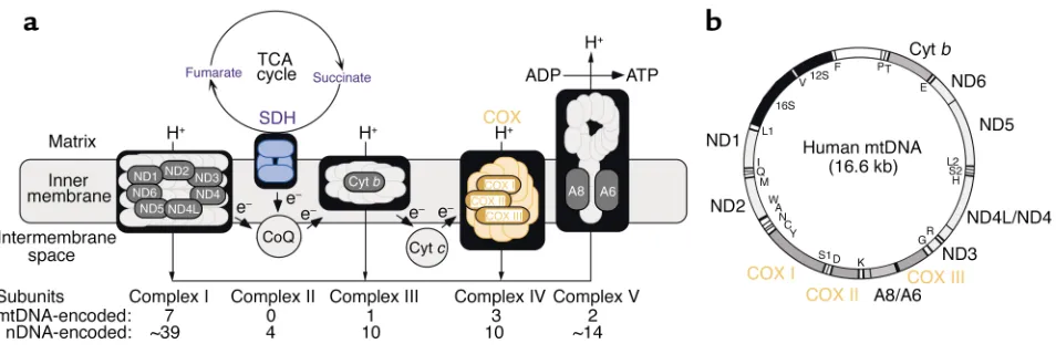
Mitochondria are semiautonomous organelles that are present in
essen-housekeeping functions. Foremost among these is the production of energy via the respiratory chain and oxidative phosphorylation, an intri-cate system composed of five com-plexes and two electron carriers (Fig-ure 1a). The mtDNA (Fig(Fig-ure 1b), a tiny 16.6 kb maternally inherited cir-cular genome present in multiple copies in each organelle (there are about 10,000 mtDNAs in a typical cell), encodes 2 rRNAs, 22 tRNAs, and only 13 polypeptides, all of which are subunits of the respiratory complexes. In the last 15 years, mutations in mtDNA, all of which impair oxidative energy metabolism, have been found to cause a wide spectrum of disorders (1). In these patients, the mutations are typically heteroplasmic; that is, mutated mtDNAs coexist with wild-type mtDNAs in varying proportions, resulting in a mosaic pattern of respi-ratorily competent and incompetent cells. Respiratorily deficient cells must typically contain at least 80% mutated mtDNA to initiate dysfunction.
Heteroplasmic populations of mtDNA mutations can also arise ran-domly in somatic cells and can accu-mulate at low levels in individual cells during the course of normal aging (2). Even more intriguingly, somatic
Address correspondence to: Eric A. Schon, Department of Neurology, Room 4-431, Columbia University, 630 West 168th Street, New York, New York 10032, USA.
Phone: (212) 305-1665; Fax: (212) 305-3986; E-mail: [email protected].
Conflict of interest: The author has declared that no conflict of interest exists.
relationship between mtDNA muta-tions and tumorigenesis has not yet been established.
Mitochondria in every cell, even those that do not divide, turn over, because they replicate their DNA and divide independently of the cell cycle. In a sense, then, as befitting an organelle that is derived evolutionari-ly from bacteria, mitochondria are each cell’s own “stem” population, continually dividing and replacing themselves within their “hosts.”
If mtDNA is always replicating within cells, it stands to reason that somatic mtDNA mutations could arise in stem cells as well, but the con-firmation of this supposition, both qualitatively (does it happen?) and quantitatively (at what rate?), has been lacking. This question is not an academic one, as it goes to the heart of issues relating to the accumulation of mutated mtDNAs in disease (espe-cially in the brain, a particularly sus-ceptible tissue), in aging, and in tumorigenesis. It is also a hard ques-tion to answer for the simple reason that it has been extremely difficult to identify a stem cell population amenable to be studied easily and in sufficient quantity — until now. In this issue of the JCI, Taylor et al. (4) provide convincing evidence that at least one population of stem cells — those giving rise to intestinal colonic crypts — do indeed harbor somatic mtDNA mutations, which, even more
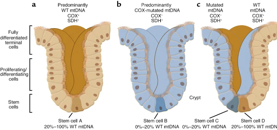
surprisingly, arise at a relatively high frequency. The decision to examine colonic crypts may have been inspired by earlier work on the segregation of mtDNA haplotypes in “transmito-chondrial mice” by Shoubridge’s group (5). The advantage of studying a colonic crypt is that it is a macro-scopically observable clonal popula-tion of cells derived from one or two single cells — the stem cells — located at the base of each crypt (Figure 2). Thus, any mtDNA mutation found in the crypt in toto must perforce have been amplified from that very muta-tion in the stem cell itself, as the crypt is the surrogate of the stem cell.
The assumptions underlying the approach of Taylor et al. (4) were based on a simple syllogism: if (i) a stem cell harbors an mtDNA muta-tion that disrupts respiratory chain function and if (ii) that mutation expands (in the stem cell) to a level exceeding the threshold for dys-function (typically >80% mutated mtDNA), then (iii) one ought to be able to observe both the dysfunction (by histochemical and/or biochemical means) and the mtDNA mutation (by genetic means) in the daughter popu-lation comprising the entire crypt. In order to assess function, Taylor et al. (4) applied a powerful method of mitochondrial analysis on serial transverse sections from individual human crypts: two-color histochem-istry (6) to detect simultaneously the
enzymatic activities of cytochrome c
oxidase (COX; complex IV of the res-piratory chain), which contains both mtDNA- and nuclear DNA (nDNA)-encoded subunits; and succinate dehydrogenase (SDH; complex II of the respiratory chain), which contains only nDNA-encoded subunits. Im-portantly, when stained for both COX (brown) and SDH (blue) simul-taneously, crypts with normal COX activity stained brown (the blue was hidden by the brown stain), whereas those with impaired COX activity stained blue (the brown was absent, revealing the blue). The authors then performed PCR coupled with mtDNA sequencing on the same transverse sections to search for somatic muta-tions that might correlate with the histochemistry. The two-color ap-proach also allowed them to make a three-dimensional reconstruction of the COX activity in the entire crypt (Figure 2).
[image:3.576.59.536.51.211.2]What Taylor et al. (4) found was, literally most illuminating. They observed three types of crypts: a major-ity of all-brown crypts, plus a minormajor-ity consisting of both all-blue crypts and “mosaic” crypts containing ribbons of brown and blue cells. The first two pat-terns are consistent with the idea that the crypt has been repopulated from a single stem cell, either COX-normal (i.e., brown) or COX-deficient (blue). The most likely explanation for the third pattern (brown/blue mosaicism)
Figure 1
is that at least two stem cells — one COX-normal and the other COX-defi-cient — were involved in crypt forma-tion. Thus, all three patterns are con-sistent with the notion that a colonic crypt is indeed a clonal population derived from one or more stem cells. Upon sequencing the sectioned crypts, they found many crypts with a normal mtDNA genotype (no surprise here), but in many crypts they found numerous mtDNA mutations, both in COX-negative crypts (many, but not all, mutations were in COX or protein synthesis genes) and in some of the ostensibly normal COX-positive crypts (mainly neutral mutations or muta-tions in non-COX genes). Interesting-ly, there were some COX-negative crypts in which no mtDNA mutations were found at all — presumably there were nuclear mutations in these stem cells that affected COX function. Over-all, the amount of mutated mtDNA was extremely high (on average, one mtDNA point mutation per crypt), and, as has been known for a dozen years now (2), the mutational “load” increased with age.
Using mitochondrial function
stem cell biologists? First, we now know that mtDNA mutations can indeed arise in stem cells. Second, by mathematical modeling, Taylor et al. (4) were able to estimate the rate of somatic mtDNA mutation: it is approximately 5 ×10⁻⁵per genome per day, a rate far greater than that of nuclear DNA. Third, the accumula-tion of mtDNA mutaaccumula-tions, often to homoplasmy, in crypt stem cells is highly reminiscent of the dramatic shifts in mtDNA genotypes in solid tumors (3), which, of course, are clonal expansions of a tumor “stem cell.” However, unlike stem cells, tumors are aneuploid. Moreover, they also amplify segments of their chromosomes as “double minutes,” up to 5 Mb in size, that likely contain nuclear-embedded mtDNA pseudo-genes. If these are amplified by PCR, pseudogene-derived polymorphisms may be attributed erroneously to mutations in authentic mtDNA.
Finally, the use of mitochondrial markers may allow researchers to track the progeny of multiple stem cells simultaneously. In fact, one can envision the use of COX-negative cells, especially in tissues from aged
random distribution of COX-negative neurons in the hippocampus of brains of the elderly is likely due to random mtDNA somatic mutations arising in ependyma-derived single
neuronal stem cells that repopulate
groups of COX-negative neurons in
the hippocampus (7). Similarly, the accumulation of COX-negative neu-rons in aging parvocellular, but not magnocellular, neurons of the lateral geniculate nucleus (8) may reflect the fact that a stem cell population exists for the former but not the latter.
Clearly, the use of colonic crypts as a stem cell “model system” allows investigators to address new types of questions in stem cell biology (9). From a mitochondrial standpoint, crypts may be of particular value, especially with regard to studying the mitochondrial population “bot-tleneck” that occurs during early oogenesis, and in understanding the dynamics of shifts from heteroplas-my to homoplasheteroplas-my in mitochondri-al disease.
1. DiMauro, S., and Schon, E.A. 2003. Mitochondr-ial respiratory-chain diseases. N. Engl. J. Med.
348:2656–2668.
[image:4.576.55.526.55.277.2]2. Khrapko, K., Nekhaeva, E., Kraytsberg, Y., and
Figure 2
mitochondrial genome in human colorectal tumours. Nat. Genet.20:291–293.
4. Taylor, R.W., et al. 2003. Mitochondrial DNA muta-tions in human colonic crypt stem cells. J. Clin. Invest.112:1351–1360. doi:10.1172/JCI200319435. 5. Jenuth, J.P., Peterson, A.C., Fu, K., and Shoubridge, E.A. 1996. Random genetic drift in the female germline explains the rapid segrega-tion of mammalian mitochondrial DNA. Nat.
Genet.14:146–151.
6. Bonilla, E., et al. 1992. New morphological approaches to the study of mitochondrial encephalomyopathies. Brain Pathol.2:113–119. 7. Bonilla, E., et al. 1999. Mitochondrial involve-ment in Alzheimer’s disease. Biochim. Biophys. Acta.
1410:171–182.
8. DiMauro, S., Tanji, K., Bonilla, E., Pallotti, F., and Schon, E.A. 2002. Mitochondrial abnormalities
in muscle and other aging cells: classification, causes, and effects. Muscle Nerve.26:597–607. 9. Kim, K.M., and Shibata, D. 2002. Methylation
reveals a niche: stem cell succession in human colon crypts. Oncogene.21:5441–5449. 10. Schon, E.A., and Manfredi, G. 2003. Neuronal
degeneration and mitochondrial dysfunction. J. Clin. Invest. 111:303–312. doi:10.1172/ JCI200317741.
The TRAIL to arthritis
George C. Tsokos
1and Maria Tsokos
21Department of Cellular Injury, Walter Reed Army Institute of Research, Silver Spring,
Maryland, USA
2Laboratory of Pathology, National Cancer Institute, Bethesda, Maryland, USA
Antigen-specific lymphocytes are involved in synovial proliferation with-in with-inflamed jowith-ints. Activated lymphocytes and synoviocytes from patients with rheumatoid arthritis express receptors that can bind TNF-related apoptosis-inducing ligand (TRAIL). A new study demonstrates that DCs pulsed with collagen and transduced with an adenovirus-based vector able to express TRAIL limit the incidence of arthritis in a model of colla-gen-induced arthritis and joint inflammation (see the related article beginning on page 1332). These results suggest that gene-modified cell therapy represents a therapeutic option for systemic rheumatic diseases.
J. Clin. Invest.112:1315–1317 (2003). doi:10.1172/JCI200320297.
TNF-related apoptosis-inducing ligand (TRAIL) is a type II transmembrane protein that belongs to the TNF super-family. It binds to death receptors (DRs) 4 and 5, two decoy receptors, and a soluble receptor called osteoprote-gerin. The TRAIL signaling pathway was identified recently, and it has gen-erated a great deal of interest since TRAIL induces apoptosis preferential-ly in tumor but not in normal cells, thus providing exciting opportunities for development of novel therapeutic strategies in cancer. TRAIL, like FasL, induces apoptosis by cross-linking and oligomerizing its receptors and form-ing a death-inducform-ing signalform-ing
com-plex through recruitment of an adapter molecule and the initiator caspase-8 and subsequent mitochondria-depend-ent or -independmitochondria-depend-ent activation of the downstream effector caspase-3. Resis-tance of tumor cells to TRAIL has been associated either with low expression of its receptors or with defects in the downstream signaling (1).
Rheumatoid arthritis is a chronic inflammatory disorder that affects up to 1% of the population. The exact ori-gin and pathogenesis of the disease are still unknown, and numerous disease-modifying drugs and biologics have been tested. There is a significant need for increased efficacy and safety of these agents (2).
TRAIL controls negative selection of T cells in the thymus
Recent reports have claimed a central role for TRAIL in thymocyte selection. TRAIL–/–mice have larger thymi, and immature CD4+CD8+cells expressing high levels of heat-stable antigen are resistant to anti-CD3 antibody–medi-ated cell death. Similarly, TRAIL–/– mice fail to reduce ovalbumin-specif-ic cells following exposure to
ovalbu-min. Both experiments clearly show that TRAIL is essential for negative selection of T cells in the thymus (3). In vitro, TRAIL blockade enhances the accumulation of concanavalin-stimu-lated spleen T cells into the S-G2/M cell cycle phase, supporting that TRAIL is important in the control of the lymphocyte cell cycle (4).
TRAIL suppresses the development of arthritis
TRAIL–/–mice are sensitive to the devel-opment of collagen-induced arthritis, probably because they fail to delete rel-evant T cell specificities and because they fail to properly silence activated T cells (3). As predicted, blockade of TRAIL with soluble DR5 administered systemically exacerbates arthritis, whereas direct transfer of a nonreplica-tive adenovirus expressing TRAIL into the joints of arthritic mice reduces arthritis (4). Injection of a TRAIL-expressing adenovirus into IL-1β– induced arthritic joints also signifi-cantly limits synovial proliferation (5). An anti–TRAIL receptor antibody has been shown to be quite effective in treating bone-erosive disease in a model that involves transfer of fibrosarcoma cells into mice (6). How-ever, antibodies to DRs, including those against CD95, may be associated with hepatotoxicity (7, 8), precluding their use in the treatment of tumors and autoimmune diseases.
Collagen-pulsed TRAIL-expressing DCs suppress arthritis
In this issue of the JCI, Liu et al. (9) report suppression of collagen-induced arthritis using DCs pulsed with colla-gen and transfected with an aden-ovirus-based vector expressing the TRAIL gene under the control of the doxycycline-inducible (DOX-inducible) tetracycline response element. The sys-tem offered two novel features: DCs were primed to recognize
collagen-spe-Address correspondence to: George C. Tsokos, Walter Reed Army Institute of Research, Building 503, Room 1A32, Robert Grand Road, Silver Spring, Maryland 20910, USA. Phone: (301) 319-9911;
Fax: (301) 319-9133; E-mail: [email protected].
Conflict of interest: The authors have declared that no conflict of interest exists. The opinions expressed herein are the private views of the authors and do not represent those of the Department of Defense.