Copyright © 1999, American Society for Microbiology. All Rights Reserved.
Multicenter Laboratory Validation of Susceptibility Testing of
Mycobacterium tuberculosis
against Classical Second-Line and
Newer Antimicrobial Drugs by Using the Radiometric
BACTEC 460 Technique and the Proportion
Method with Solid Media
GABY E. PFYFFER,1* DONALD A. BONATO,2ADELEH EBRAHIMZADEH,3WENDY GROSS,2 JACQUELINE HOTALING,4JOHN KORNBLUM,3ADALBERT LASZLO,5GLENN ROBERTS,6
MAX SALFINGER,4FRANZISKA WITTWER,1ANDSALMAN SIDDIQI7
Swiss National Center for Mycobacteria, Department of Medical Microbiology, University of Zurich, Zurich, Switzerland1;
Veterans Affairs Medical Center, West Haven, Connecticut2; New York City Department of Health, New York,3and
Wadsworth Center, New York State Department of Health, Albany,4New York; Laboratory Center
for Disease Control, Ottawa, Canada5; Mayo Clinic, Rochester, Minnesota6; and
Becton Dickinson Microbiology Systems, Sparks, Maryland7
Received 10 May 1999/Returned for modification 11 July 1999/Accepted 14 July 1999
In a large multicenter study involving six major study sites in the United States, Canada, and Europe, the
susceptibilities of 272Mycobacterium tuberculosis strains to classical second-line antituberculosis (anti-TB)
drugs (capreomycin, cycloserine, ethionamide, and kanamycin) and newer compounds (amikacin, clofazimine, ofloxacin, and rifabutin) were determined by the radiometric BACTEC 460 procedure and the conventional proportion method on Middlebrook 7H10 agar. Previously established critical concentrations for classical second-line anti-TB drugs were compared with several concentrations in liquid medium to establish equiva-lence. MICs of newer compounds determined in liquid medium were either the same or up to four times lower than those determined in agar medium. After establishing critical concentrations (breakpoints) in the extended testing of clinical isolates, we obtained an excellent overall correlation between the two systems, with no errors with amikacin, kanamycin, and ofloxacin and very few major or very major errors with the other drugs; however, for cycloserine, no breakpoint concentration could be recommended due to repeatedly inconsistent results by both methods. Based on these data we conclude that the BACTEC 460 procedure is a simple and rapid method requiring 4 to 8 days on average to generate accurate antimicrobial susceptibility testing (AST) results for eight anti-TB drugs other than those considered primary ones. These data not only fill a major gap of knowledge regarding the critical test concentrations of secondary anti-TB drugs but also provide a baseline
for future evaluations ofM. tuberculosisAST with the more recently developed, nonradiometric broth-based
culture systems.
With the recent global resurgence of tuberculosis (TB) and concomitant rise in multidrug-resistant strains of Mycobacte-rium tuberculosis(37, 38), there is an increasing demand for determining the in vitro susceptibilities of clinical isolates to antimicrobial agents other than those considered primary drugs (isoniazid, rifampin, ethambutol, pyrazinamide, and streptomycin). Conventional antimicrobial susceptibility test-ing (AST) with solid media such as Lo¨wenstein-Jensen (LJ) or Middlebrook 7H10 agar requires 3 or more weeks to be com-pleted, and for some classical second-line drugs as well as newer antimicrobial agents, appropriate critical drug concen-trations have not fully been established.
AST by the radiometric BACTEC method (Becton Dickin-son Diagnostic Instrument Systems, Sparks, Md.), introduced in 1980, is an efficient way to test frontline drugs (27, 30–33) and results in a significantly shorter turnaround time than that of the traditional proportion method. Susceptibility testing by this procedure has become the current method of choice (34),
even when the hazards arising from the use of radioisotopes are taken into account. However, data on critical concentra-tions for classical second-line and other newer drugs are largely fragmentary or lacking altogether. Some data from AST of second-line drugs with BACTEC 12B medium were presented quite early (11) but have never been published. Allen et al. (1) defined the MICs on Middlebrook agar of aminoglycosides while Heifets’ group undertook several investigations into AST with the BACTEC 12B medium, e.g., with aminoglycosides (13), ethionamide (16, 22), and quinolones (15), the last group of compounds having also been studied by others (4, 5, 8, 9). The MICs of rifabutin, generated either with the radiometric BACTEC 12B medium (17) or with Middlebrook 7H9 broth (7), are also available. However, as a whole, most of these studies have either included only a narrow spectrum of drugs and/or tested only a rather limited number ofM. tuberculosis strains.
In light of this situation, the primary aim of our study was to develop a basic protocol which defines appropriate critical concentrations for secondary drugs to allow reliable testing with both BACTEC 12B and agar medium. With more than 270 strains ofM. tuberculosis, the reproducibility of results gener-ated by different laboratories was also assessed. Once vali-* Corresponding author. Mailing address: Swiss National Center for
Mycobacteria, Department of Medical Microbiology, University of Zurich, Gloriastrasse 30, 8028 Zurich, Switzerland. Phone: 41 1 634 27 86. Fax: 41 1 634 49 18. E-mail: [email protected].
3179
on May 15, 2020 by guest
http://jcm.asm.org/
dated, the radiometric procedure should allow clinical labora-tories to provide physicians with accurate and timely drug susceptibility information. In addition, since quite a few non-radiometric mycobacterial culture systems based on liquid me-dium, such as the Mycobacteria Growth Indicator Tube (man-ual version [23]; BACTEC 960 automated version [12]), the MB/BacT (2), and the ESP Culture System II (35), have re-cently been developed, this study may also provide a guideline for future development of AST procedures with these novel devices.
MATERIALS AND METHODS
Study sites.This study was conducted at six mycobacteriology laboratories, including the Mycobacteriology Laboratory, Veterans Affairs Medical Center, West Haven, Conn.; the Bureau of Laboratories, New York City Department of Health, New York, N.Y.; the Laboratory Center for Disease Control, Ottawa, Ontario, Canada; the Mycobacteriology Laboratory, Mayo Clinic, Rochester, Minn.; the Mycobacteriology Laboratory, Wadsworth Center, New York State Department of Health, Albany, N.Y.; and the Swiss National Center for Myco-bacteria, Department of Medical Microbiology, University of Zurich, Zurich, Switzerland. A seventh site, the Research and Development Division, Becton Dickinson Microbiology Systems, Sparks, Md., was the coordination site, from which cultures and antimicrobial agents were supplied and at which data were collected.
Antimicrobial agents.All drugs were obtained from the manufacturers in a chemically pure form. The drugs used were capreomycin sulfate,D-cycloserine, ethionamide, and kanamycin monosulfate from Sigma, St. Louis, Mo.; amikacin from Bristol-Myers Squibb, Syracuse, N.Y.; clofazimine from Ciba-Geigy, Suf-fern, N.Y.; ciprofloxacin from Miles, West Haven, Conn.; ofloxacin from Ortho/ R. W. Johnson Pharmaceutical, Raritan, N.J.; and rifabutin from Adria Labo-ratories, Dublin, Ohio. The compounds were supplied by the coordinator of the study. Amikacin, capreomycin, ciprofloxacin, and kanamycin were dissolved in sterile distilled water (DW). Ofloxacin was dissolved in a 0.1 N NaOH solution. Ethionamide was dissolved in ethylene glycol and incubated overnight for ster-ilization. Subsequent dilutions of these two drugs were made in sterile DW. Clofazimine was dissolved in dimethyl sulfoxide and stored at room temperature in the dark. Subsequent dilutions were made in the same solvent. For D-cy-closerine a 0.1% solution of Na2CO3was prepared and added to 100 ml of DW
until a pH of 10 was achieved. Dilutions were made in sterile DW. Rifabutin was dissolved in methanol and diluted in DW. Stock solutions of each drug were made at least 40 times higher than the highest test concentration used. Except for clofazimine and ethionamide, which are considered self-sterilizing, all stock solutions were filtered in a sterile manner with a 0.22-m-pore-size polycarbon-ate filter; approximpolycarbon-ately 20% of the initial filtrpolycarbon-ate was discarded. All stock solutions except clofazimine were stored at ⫺70°C in small aliquots. Once thawed, the remaining solutions were discarded.
Culture media and quality control.Middlebrook 7H12 broth (BACTEC 12B; Becton Dickinson Microbiology Systems) was used for radiometric testing, and Middlebrook 7H10 agar plates (BBL, Cockeysville, Md.) were used for AST by the proportion method. All media were supplied by the coordinator of the study. M. tuberculosisH37Rv (ATCC 27294; susceptible to all drugs tested) was used as a strain for internal quality control, which was done on a weekly basis, along with the other strains included in the study.
AST. (i) BACTEC 460.For radiometric AST, the standard protocol was fol-lowed (31). Each drug was tested at several concentrations. Antimicrobials were always added in 0.1-ml quantities to a BACTEC 12B vial to achieve the desired concentrations. Susceptibility and resistance were judged by comparison of the change in the growth index of the control with that of the test drug as recom-mended. This interpretation gave susceptibility results at the 1% proportion basis.
(ii) Traditional proportion method.AST was carried out with Middlebrook 7H10 agar according to the standard procedure (18, 20). For some classical second-line antituberculotics (capreomycin, cycloserine, ethionamide, and kana-mycin), the recommended critical concentrations (20) were used. For all other drugs, several concentrations were tested to establish the MICs and critical concentrations. The MIC was defined as the lowest concentration of drug that inhibited more than 99% of the bacterial population. As a consequence, the critical concentration was defined as the concentration of drug required to prevent growth above the 1% threshold (critical proportion) of the test popula-tion of TB bacilli (18). Results were read 21 days after inoculapopula-tion of the medium.
Study design and strains.The study was carried out concomitantly in three phases at all six centers.
Phase I.Phase I was designed to establish a basic test procedure and to determine a working range for the antimicrobial drug concentrations to be used in liquid medium (BACTEC 12B) and on conventional solid media. In addition, the reproducibility of the results of the test systems was evaluated. For this purpose, a total of 10 clinical isolates ofM. tuberculosiswhich were susceptible to all primary drugs in earlier tests and stored in a strain collection were
subcul-tured onto LJ slants. As soon as growth appeared on the slants (10 to 15 days after inoculation) these isolates were shipped by overnight delivery to the six clinical testing sites. In an effort to minimize variations of results, the cultures were used directly for AST without subculturing. For those antibiotics whose critical concentrations had been established previously on solid medium, i.e., capreomycin, cycloserine, ethionamide, and kanamycin, only one drug concen-tration was tested on Middlebrook 7H10 agar but at least three concenconcen-trations were tested in BACTEC 460. For all other drugs at least three different concen-trations were tested by the proportion method as well as with BACTEC 460.
Phase II.The aims of phase II were to establish equivalent breakpoint drug concentrations for the BACTEC 460 and conventional solid medium methodol-ogies and to evaluate interlaboratory reproducibility of test results for susceptible and resistant isolates. Drug concentrations used in phase II were adjusted and finalized based on the information obtained from phase I. Strains ofM. tuber-culosiswith known drug resistance, especially to the test drugs, were selected from several clinical sources. Twenty isolates were selected and subcultured onto LJ medium. Once growth appeared, sets of 20 isolates each were shipped to the six test sites and were used for AST as in phase I.
Phase III.Phase III was extended testing with both BACTEC 460 and Middle-brook 7H10 agar with the drug concentrations established in phase II. Staff at each site independently tested at least 24 strains ofM. tuberculosiswhich had recently been isolated from clinical specimens at their own laboratory.
RESULTS
In phases I and II of this multicenter AST study sets of TB strains containing 10 and 20 isolates, all from stock cultures, were analyzed. Strains tested in phase I were confirmed to be susceptible to all drugs. Among the 20 strains tested in phase II, 6 were resistant to capreomycin, 0 were resistant to cy-closerine, 10 were resistant to ethionamide, 9 were resistant to kanamycin, 8 were resistant to amikacin, 1 was resistant to clofazimine, 1 was resistant to ofloxacin, and 13 were resistant to rifabutin. In phase III, AST was performed with a total of 242 M. tuberculosisstrains which had recently been isolated from clinical specimens by staff at the six participating labora-tories. Nineteen strains were resistant to capreomycin, 19 were resistant to cycloserine, 56 were resistant to ethionamide, 34 were resistant to kanamycin, 32 were resistant to amikacin, 0 were resistant to clofazimine, 20 were resistant to ofloxacin, and 83 were resistant to rifabutin.
Phase I.The 10 fully drug-susceptible isolates supplied to all
sites were tested against various concentrations of eight drugs. Most BACTEC 460 MICs determined at the test sites agreed within⫾1 serial dilution (Table 1). For example, the MIC of amikacin wasⱕ1.0g/ml for 100% of the strains and that of capreomycin wasⱕ1.25g/ml for 98.3% of the strains at all test sites combined. With a few drugs, MICs extended over a wider range than that noted above. This held true for the quinolones, whose MICs varied by more than 1 dilution from site to site, although in the majority of cases the values were betweenⱕ0.5 and 1.0g/ml. Cycloserine gave the least-repro-ducible results for both BACTEC 460 and conventional testing (proportion method). Based on phase I results, drug concen-trations were adjusted for subsequent testing in phase II. For amikacin, clofazimine, and ethionamide, drug concentrations to be used with BACTEC 460 were lowered by 1 dilution, whereas for cycloserine and ofloxacin, concentrations were in-creased for both solid and liquid medium systems. Ciprofloxa-cin testing was discontinued; on the other hand, rifabutin test-ing was included in phases II and III.
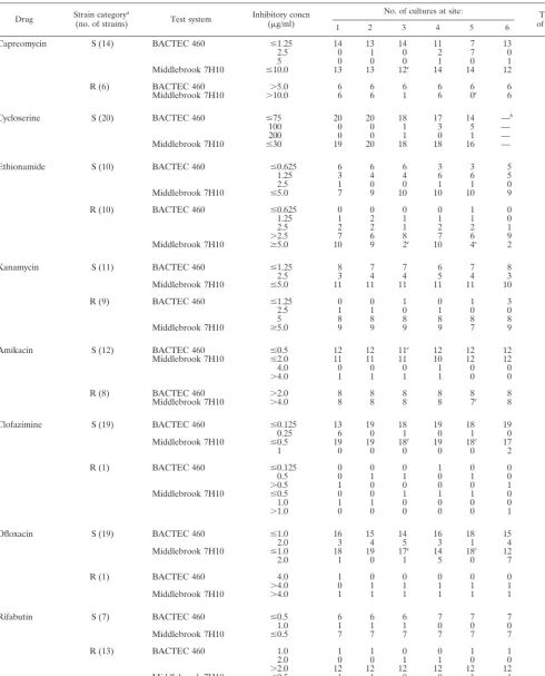
Phase II.Results of phase II testing of the 20 susceptible and
resistant strains ofM. tuberculosissent to all six study sites are reported in Table 2. Interlaboratory reproducibility of results by the BACTEC 460 and conventional AST methods was good, with MICs being within⫾1 dilution of each other in most cases. It appeared that agar AST results varied more from site to site than those obtained by the BACTEC 460 method. In general, interlaboratory variation in results was observed more frequently with cycloserine and to some extent
on May 15, 2020 by guest
http://jcm.asm.org/
with ethionamide and capreomycin than with the other drugs, regardless of the AST method applied.
For some drugs BACTEC 460 medium-based MICs were lower than agar-based MICs (up to four times), while for others they were slightly lower than or equal to agar-based MICs. MICs for susceptible strains in 12B medium were mainly betweenⱕ1.25 and 2.5g/ml for capreomycin,ⱕ0.625 and 1.25g/ml for ethionamide, andⱕ1.25 and 2.5g/ml for kanamycin. Amikacin susceptibility test results with MICs of ⱕ0.5g/ml in 12B medium andⱕ2.0 and 4.0g/ml in Middle-brook 7H10 medium were equivalent. Clofazimine was more active in liquid medium, with MICs betweenⱕ0.125 and 0.25
g/ml in the BACTEC 460 and ⱕ0.5 and 1.0 g/ml in the Middlebrook 7H10 medium. Ofloxacin and rifabutin were found to be equally active in BACTEC 12B medium and on Middlebrook 7H10 medium, with MICs betweenⱕ1.0 and 2.0
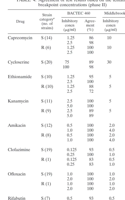
g/ml andⱕ0.5 and 1.0 g/ml, respectively. Cycloserine sus-ceptibility testing continuously yielded inconsistent results. Therefore, no breakpoint could be established. Results for cycloserine on Middlebrook 7H10 agar were even less reliable. Overall, the results allowed distinct MICs to be determined for susceptible and resistant strains by both techniques. This information was used to establish tentative breakpoint concen-trations for further data analysis (Table 3). When results were analyzed with these critical concentrations, there was good agreement of results with both susceptible and resistant TB strains (Table 4). In some instances (clofazimine and ofloxa-cin), the total number of resistant strains was, however, too small to draw any definite conclusions.
Phase III.Phase III extended testing with 242 clinical
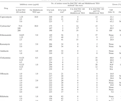
iso-lates of M. tuberculosis among the six study sites, with 27 isolates at site 1, 42 isolates at site 2, 48 isolates at site 3, 24 isolates at site 4, 68 isolates at site 5, and 33 isolates at site 6. Tentative breakpoint concentrations elaborated in phase II (Tables 3 and 4) were acceptable with some minor changes (Table 5). Generally, we looked at the concentrations which gave the fewest total errors (ethionamide at 1.25g/ml in the BACTEC 460 and 5.0g/ml in conventional AST, kanamycin at 5.0g/ml in the BACTEC 460 and 5.0g/ml in conventional AST, amikacin at 1.0g/ml in the BACTEC 460 and 4.0g/ml in conventional AST, clofazimine at 0.5g/ml in the BACTEC 460 and 1.0g/ml in conventional AST, ofloxacin at 2.0g/ml in the BACTEC 460 and 2.0g/ml in conventional AST, and rifabutin at 0.5g/ml in the BACTEC 460 and 1.0g/ml in conventional AST). For kanamycin, amikacin, clofazimine, cip-rofloxacin, and rifabutin these concentrations also gave the fewest very major errors. The only exception was capreomycin. Although overall agreement for this drug at 2.5g/ml (8 very major errors, 2 major errors) was better than at 1.25g/ml (4 very major errors, 13 major errors), we have chosen 1.25g/ml as the breakpoint (only 4 very major errors instead of 8). Cycloserine testing was, again, very inconsistent by both the BACTEC 460 and agar methodologies. At one site, cycloserine testing by the method of proportion did not yield reportable results at all, with false resistance being shown even with theM. tuberculosisH37Rv control. These data were excluded from our analysis (Table 5).
Results obtained from the BACTEC 460 and the conven-tional agar methods were compared by constructing 2-by-2 tables for each drug concentration. Critical concentrations were eventually established in such a way that they yielded the least number of very major errors (false susceptibility) and major errors (false resistance). With the critical test concen-trations established in this phase, neither false resistance be-tween BACTEC 460 and conventional AST (resistance by BACTEC 460, susceptibility by conventional AST) nor false susceptibility (susceptibility by BACTEC 460, resistance by conventional AST) was observed for amikacin, kanamycin, or ofloxacin. Results with other antimicrobial agents like clofazi-mine and rifabutin showed very minor discordance. Ethio-namide results produced 7 (12.5%) very major disagreements, while capreomycin results produced 13 major but only 4 (21.1%) very major disagreements. Cycloserine testing re-sulted in 17 (89.5%) very major disagreements (Table 5).
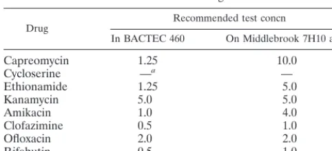
Eventually, this information led to the definition of the crit-ical concentrations (breakpoints) for testing in BACTEC 12B medium and on Middlebrook 7H10 agar (Table 6).
DISCUSSION
The data presented here constitute the first comprehensive, multicenter AST study of second-line anti-TB drugs and newer compounds currently being used for the treatment of TB, val-idating both broth-based and solid-medium-based systems. Given the fact that MICs, as a whole, are highly dependent on a large array of different factors such as medium composition, pH, inoculum size, and incubation time, the primary aim of this study was to establish critical concentrations with which to perform AST on a wide spectrum of anti-TB drugs under defined conditions.
Our data, encompassing more than 17,000 individual sus-ceptibility test results generated with 272 strains ofM. tuber-culosisin six centers, corroborate the fact that the radiometric BACTEC 460 method is feasible for AST of most of these drugs as it has previously been shown to be for frontline an-TABLE 1. Results of phase I generated by the six study sites in
AST of 10 susceptible strains ofM. tuberculosis
Antimicrobial agent
BACTEC 460 medium Middlebrook 7H10 agar Inhibitory concn
(g/ml) Count(%)a Inhibitory concn(g/ml) Count(%) Capreomycin ⱕ1.25 59 (98.3) 10.0 59 (98.3)
2.5 1 (1.7)
Cycloserine ⱕ50 27 (45) 25.0 36 (60)
60.0 23 (38.3) 70.0 9 (15) ⬎70.0 1 (1)
Ethionamide ⱕ1.25 59 (98.3) 5.0 59 (98.3) 2.5 1 (1.7)
Kanamycin ⱕ1.25 48 (80) 5.0 60 (100)
2.5 12 (20)
Amikacin ⱕ1.0 60 (100) ⱕ2.0 54 (90)
⬎4.0 6 (10) Clofazimine ⱕ0.25 59 (98.3) ⱕ0.25 47 (78.3)
0.5 1 (1.7) 0.5 13 (21.7)
Ciprofloxacin ⱕ0.5 46 (76.7) ⱕ0.5 41 (68.3)
1.0 13 (21.7) 1.0 18 (30)
2.0 1 (1.7) 2.0 1 (1.7)
Ofloxacin ⱕ0.5 21 (35) ⱕ0.5 19 (31.7) 1.0 34 (56.7) 1.0 38 (63.3)
2.0 4 (6.7) 2.0 3 (5)
⬎4.0 1 (1.7) aThe count is the total number of susceptible strains.
on May 15, 2020 by guest
http://jcm.asm.org/
TABLE 2. Reproducibility data generated at the six study sites (phase II) during AST of 20 strains ofM. tuberculosis
Drug Strain category(no. of strains)a Test system Inhibitory concn(g/ml) No. of cultures at site: of culturesTotal no.
1 2 3 4 5 6
Capreomycin S (14) BACTEC 460 ⱕ1.25 14 13 14 11 7 13 72
2.5 0 1 0 2 7 0 10
5 0 0 0 1 0 1 2
Middlebrook 7H10 ⱕ10.0 13 13 12c 14 14 12 78
R (6) BACTEC 460 ⬎5.0 6 6 6 6 6 6 36
Middlebrook 7H10 ⬎10.0 6 6 1 6 0c 6 25
Cycloserine S (20) BACTEC 460 ⱕ75 20 20 18 17 14 —b 89
100 0 0 1 3 5 — 9
200 0 0 1 0 1 — 2
Middlebrook 7H10 ⱕ30 19 20 18 18 16 — 91
Ethionamide S (10) BACTEC 460 ⱕ0.625 6 6 6 3 3 5 29
1.25 3 4 4 6 6 5 28
2.5 1 0 0 1 1 0 3
Middlebrook 7H10 ⱕ5.0 7 9 10 10 10 9 55
R (10) BACTEC 460 ⱕ0.625 0 0 0 0 1 0 1
1.25 1 2 1 1 1 0 6
2.5 2 2 1 2 2 1 10
⬎2.5 7 6 8 7 6 9 43
Middlebrook 7H10 ⱖ5.0 10 9 2c 10 4c 2 37
Kanamycin S (11) BACTEC 460 ⱕ1.25 8 7 7 6 7 8 43
2.5 3 4 4 5 4 3 23
Middlebrook 7H10 ⱕ5.0 11 11 11 11 11 10 65
R (9) BACTEC 460 ⱕ1.25 0 0 1 0 1 3 1
2.5 1 1 0 1 0 0 3
5 8 8 8 8 8 8 48
Middlebrook 7H10 ⱖ5.0 9 9 9 9 7 9 52
Amikacin S (12) BACTEC 460 ⱕ0.5 12 12 11c 12 12 12 71
Middlebrook 7H10 ⱕ2.0 11 11 11 10 12 12 67
4.0 0 0 0 1 0 0 1
⬎4.0 1 1 1 1 0 0 4
R (8) BACTEC 460 ⬎2.0 8 8 8 8 8 8 48
Middlebrook 7H10 ⬎4.0 8 8 8 8 7c 8 47
Clofazimine S (19) BACTEC 460 ⱕ0.125 13 19 18 19 18 19 106
0.25 6 0 1 0 1 0 8
Middlebrook 7H10 ⱕ0.5 19 19 18c 19 18c 17 110
1 0 0 0 0 0 2 2
R (1) BACTEC 460 ⱕ0.125 0 0 0 1 0 0 1
0.5 0 1 1 0 1 0 3
⬎0.5 1 0 0 0 0 1 2
Middlebrook 7H10 ⱕ0.5 0 0 1 1 1 0 3
1.0 1 1 0 0 0 0 2
⬎1.0 0 0 0 0 0 1 1
Ofloxacin S (19) BACTEC 460 ⱕ1.0 16 15 14 16 18 15 94
2.0 3 4 5 3 1 4 20
Middlebrook 7H10 ⱕ1.0 18 19 17c 14 18c 12 98
2.0 1 0 1 5 0 7 14
R (1) BACTEC 460 4.0 1 0 0 0 0 0 1
⬎4.0 0 1 1 1 1 1 5
Middlebrook 7H10 ⬎4.0 1 1 1 1 1 1 6
Rifabutin S (7) BACTEC 460 ⱕ0.5 6 6 6 7 7 7 39
1.0 1 1 1 0 0 0 3
Middlebrook 7H10 ⱕ0.5 7 7 7 7 7 7 42
R (13) BACTEC 460 1.0 1 1 0 0 1 1 4
2.0 0 0 1 1 0 0 2
⬎2.0 12 12 12 12 12 12 72
Middlebrook 7H10 ⱕ0.5 1 1 0 0 1 1 4
1.0 0 0 0 1 2 0 3
2.0 0 0 3 0 2 0 5
⬎2.0 12 12 9c 12 5c 12 62 aS, susceptible; R, resistant.
bSite 6 was excluded (see Results).
cResults of some susceptibility tests are missing.
on May 15, 2020 by guest
http://jcm.asm.org/
ti-TB drugs (30–33). Complying with the recommendation of the Centers for Disease Control and Prevention of using broth-based methods for AST (34), more and more laboratories in the United States have now adopted the BACTEC 460 tech-nique so that test results can be reported to the clinician with a minimal turnaround time, i.e., within 4 to 8 days, as was the average in our study. Besides the longer turnaround time, there are several other drawbacks of AST on solid media, such as drug inactivation during agar preparation and degradation of the antimicrobial agent during the extended period of incu-bation (10), which can, at least to some extent, be circum-vented easily by using broth-based media. Another important goal of the study was to achieve a high degree of interlabora-tory reproducibility of AST results. As indicated by our data, the agreement in results among the six laboratories was quite high throughout the three phases.
The drug panel remained the same throughout the study, except that (i) ciprofloxacin was dropped after phase I because of its known poor in vivo response against TB, and (ii) rifabu-tin, a promising anti-TB drug, was added for phases II and III. For the first two phases, each of the six sites was provided with identical sets of M. tuberculosis strains. In testing of these strains by all participants, susceptibility data generated in these two initial phases were found to be highly reliable and repro-ducible. Further testing eventually led to the establishment of the final critical test concentrations for seven of the eight second-line drugs. Basically, establishment of the final concen-trations was achieved in three major steps: (i) by establishing MICs by testing fully susceptible TB strains (phase I), (ii) by adjusting drug concentrations and testing susceptible and re-sistant strains (phase II), and (iii) by testing a large number of clinicalM. tuberculosisisolates (n ⫽242) with particular em-phasis on isolates that are resistant to primary drugs. This process helped in establishing the final breakpoints by selecting those drug concentrations which gave minimum false suscep-tible and false resistant results with both test systems.
Allen et al. (1) have defined critical concentrations in Middlebrook 7H11 agar as 9.5 g/ml for capreomycin, 2.5
g/ml for kanamycin, and 1.0g/ml for amikacin. These con-centrations are different from ours on Middlebrook 7H10, as we tested 10.0g of capreomycin per ml and 5.0g of kana-mycin per ml, which have been established previously as critical concentrations, and 4.0g of amikacin per ml.
Important work in radiometric AST, though with a very limited number of strains ofM. tuberculosis, has been carried out by Heifets et al. (13–17). This group has suggested the following critical drug concentrations for BACTEC 12B me-dium: 5.0g/ml (MIC range, 1.5 to 3.0) for capreomycin, 4.0
g/ml (indicating an MIC range of 0.5 to 1.0) for amikacin, 2.0
g/ml (MIC range, 0.25 to 2.0) for ofloxacin, and 0.12g/ml (MIC range, 0.015 to 0.06) for rifabutin. In general, these MICs differ by 1 to 2 serial dilutions from ours. This divergence is not surprising when one considers those authors’ notion that, at these suggested critical concentrations, the test strain, if susceptible, should be considered only moderately susceptible. In their hands, fully susceptible strains were those which were susceptible to a concentration 1 dilution (twofold) lower than the critical concentration while resistant strains were those which were resistant to a concentration 1 serial dilution higher than the critical concentration. Even a fourth category, desig-nated very resistant, was suggested. Though the concept of four categories of susceptibility, i.e., susceptible, moderately susceptible, resistant, and very resistant, might be helpful to some physicians, in particular when dealing with multidrug-resistant TB, this concept has, however, not been widely ac-cepted.
[image:5.612.53.293.93.200.2]Additional information is available for the quinolones (4, 5, 19, 21). Chen et al. (5) established MIC ranges in BACTEC 12B medium for ciprofloxacin between 0.25 and 2.0g/ml and for ofloxacin between 0.5 and 2.0g/ml and set the breakpoint concentration at 2.0g/ml. These values agree perfectly with TABLE 3. Tentative breakpoints and ranges of presumptive critical
concentrations for AST in liquid and on solid media (phase II)
Drug Range of critical concn or critical concn a
In BACTEC 460 On Middlebrook 7H10 agar
Capreomycin 1.25–2.5 10
Cycloserine 75–100 30
Ethionamide 1.25–2.5 5
Kanamycin 2.5–5.0 5
Amikacin 0.5–1.0 2.0–4.0
Clofazimine 0.125–0.25 0.5–1.0
Ofloxacin 1.0–2.0 1.0–2.0
Rifabutin 0.5–1.0 0.5–1.0
aSingle values indicate previously established critical concentrations (20).
TABLE 4. Agreement of test results based on the tentative breakpoint concentrations (phase II)
Drug
Strain categorya
(no. of strains)
BACTEC 460 Middlebrook 7H10 Inhibitory
concn (g/ml)
Agree-ment
(%)
Inhibitory concn (g/ml)
Agree-ment
(%)
Capreomycin S (14) 1.25 86 10 94
2.5 98
R (6) 1.25 100 10 71
2.5 100
Cycloserine S (20) 75 89 30 91
100 98
Ethionamide S (10) 1.25 95 5 92
2.5 100
R (10) 1.25 88 5 65
2.5 72
Kanamycin S (11) 2.5 100 5 98
5.0 100
R (9) 2.5 89 5 96
5.0 89
Amikacin S (12) 0.5 100 2.0 93
1.0 100 4.0 94
R (8) 0.5 100 2.0 100
1.0 100 4.0 100
Clofazimine S (19) 0.125 93 0.5 98
0.25 100 1.0 100
R (1) 0.125 83 0.5 50
0.25 83 1.0 17
Ofloxacin S (19) 1.0 100 1.0 88
2.0 100 2.0 100
R (1) 1.0 100 1.0 100
2.0 100 2.0 100
Rifabutin S (7) 0.5 93 0.5 100
1.0 100 1.0 100
R (13) 0.5 100 0.5 95
1.0 95 1.0 91
aS, susceptible; R, resistant.
on May 15, 2020 by guest
http://jcm.asm.org/
[image:5.612.310.552.331.717.2]our ofloxacin data (MICs, 1.0 to 2.0g/ml; breakpoint, 2.0
g/ml). Others, however, have reported MICs of ofloxacin between 0.5 and 1.0g/ml and a critical concentration of 1.0
g/ml (4). The range of MICs for our test strains on solid medium appeared, however, to be narrow (1.0 to 4.0g/ml, with a critical concentration of 2.0g/ml), compared to MICs of 0.3 to 4.0g/ml and 0.125 to 4.0g/ml for ciprofloxacin and ofloxacin, respectively, in Middlebrook 7H10 medium (5).
The reported MICs of rifabutin in BACTEC 12B medium extend over a broad range. Our values (0.5 to 1.0g/ml) are perfectly concordant with those of Della Bruna and Olliaro (6). However, other groups have found both lower ranges (0.015 to 0.125g/ml [5] and 0.015 to 0.6g/ml [17]) and a higher range (3.6g/ml [7]), the last having been generated in Middlebrook
7H9 broth. One of the most recent studies of AST of secondary anti-TB drugs (25) confirms largely our values for capreomycin (1.0 to 2.0 versus 1.25 to 5.0g/ml [this study]), kanamycin (5.0 versus 2.0 to 4.0g/ml), amikacin (0.5 to 1.0 versus 0.5g/ml), and clofazimine (0.5 versus 0.1 to 0.4g/ml). Such low MICs of the latter drug were also found by others (26). Rastogi et al. (25) used a human macrophage system and demonstrated that, of a large variety of drugs tested, clofazimine was the only one which yielded discrepant results between its extracellular and intracellular activities. This may be due to the fact that this drug concentrates within phagosomes and phagolysosomes of infected macrophages (24), which might, at least to some ex-tent, explain its ineffectiveness as an anti-TB agent.
[image:6.612.55.557.90.508.2]Even though for some of the compounds tested it was not TABLE 5. Analysis of false susceptibility (very major error) and false resistance (major error)
results at various drug concentrations (phase III)
Drug
Inhibitory concn (g/ml) No. of isolates tested by BACTEC 460 and Middlebrook 7H10methodsathat were: Errors (%)
In BACTEC
460 medium On Middlebrook7H10 agar S by bothtests R by bothtestsb
S by BACTEC 460 and R by Middlebrook 7H10
R by BACTEC 460 and S by Middlebrook 7H10
VMc
(false S) M d (false R)
Capreomycin 1.25 10.0 210 15 4 13 21.1 6.2h
2.5 221 11 8 2 42.1 0.9
5.0 223 7 12 0 63.2 None
Cycloserinee 75.0 30.0 175 5 14 15 73.7 8.6
100 187 2 17f 3 89.5 1.6
200 190 0 19 0 100 None
Ethionamide 0.625 5.0 149 56 0 37 None 24.8
1.25 177 49 7 9 12.5 5.1h
2.5 182 41 15 4 26.8 2.2
Kanamycin 2.5 5.0 206 34 0 2 None 1.0
5.0 208 34 0 0 None Noneh
Amikacin 0.5 4.0 209 32 0 1 None 0.5
1.0 210 32 0 0 None Noneh
Clofazimine 0.125 0.5 202 3 2 35 40.0 17.3
0.25 227 2 3 10 60.0 4.4
0.5 234 0 5 3 98.7 1.3
0.125 1.0 204 0 0 38 NAg 18.6
0.25 230 0 0 12 NA 5.2
0.5 239 0 0 3 NA 1.3h
Ofloxacin 1.0 1.0 211 21 4 6 16.0 2.8
2.0 217 20 5 0 20.0 None
4.0 217 18 7 0 28.0 None
1.0 2.0 215 20 0 7 None 3.3
2.0 222 20 0 0 None Noneh
4.0 222 18 2 0 10.0 None
1.0 4.0 215 18 0 9 None 4.2
2.0 222 18 0 2 None 0.9
4.0 223 17 1 1 5.6 0.5
Rifabutin 0.5 1.0 158 83 0 1 None 0.6h
1.0 158 82 1 1 1.2 0.6
0.5 2.0 158 81 0 3 None 1.9
1.0 159 81 0 2 None 1.3
aS, susceptible; R, resistant.
bReliability of the analyses was questionable due to the very low number of resistant isolates. cVM, very major disagreement (susceptible by BACTEC 460 and resistant by the proportion method). dM, major disagreement (resistant by BACTEC 460 and susceptible by the proportion method).
eSite 6 was excluded. For cycloserine no recommendation concerning the critical concentration is being made.
fFourteen of 17 isolates that were resistant by the Middlebrook 7H10 method but susceptible by the BACTEC 460 method were from a single laboratory (site 3). gNA, not applicable since none of the tested organisms were resistant at the conventional concentration.
hCritical concentration.
on May 15, 2020 by guest
http://jcm.asm.org/
possible to include a fair amount of resistant strains in the test panels, which would call for additional studies with genetically well-characterized, resistant strains, several important conclu-sions can be drawn from our study. (i) Except with cycloserine, the radiometric BACTEC 460 procedure is a simple and rapid method requiring 4 to 8 days on average to generate reliable AST results for second-line anti-TB drugs. (ii) Cycloserine testing should be discontinued, due to difficulties with the drug at all six study sites, despite repeated testing. (iii) With these comprehensive AST data it is anticipated that our evaluation may also provide a guideline for future studies, especially when AST procedures are established for the newer, nonradiomet-ric, broth-based systems such as the MB/BacT (28), the ESP Culture System II (3), the manual Mycobacteria Growth Indi-cator Tube (29, 36) or its automated version (BACTEC 960). The drug concentrations defined here for BACTEC 12B me-dium and Middlebrook 7H10 agar will undoubtedly serve as a reliable baseline for establishing critical test concentrations for AST by these recently developed growth-based technologies.
ACKNOWLEDGMENTS
The expert technical collaboration of T. Dolgaia (New York City Department of Health, New York, N.Y.), V. Handze and N. Pearson (Laboratory Center for Disease Control, Ottawa, Canada), A. Loder (Wadsworth Center, Albany, N.Y.), and K. Doerr (Mayo Clinic, Roch-ester, Minn.) is gratefully acknowledged.
REFERENCES
1.Allen, B. W., D. A. Michison, Y. C. Chan, W. W. Yew, and W. G. L. Allan. 1983. Amikacin in the treatment of pulmonary tuberculosis. Tubercle64: 111–118.
2.Benjamin, W. H., Jr., K. B. Waites, A. Beverly, L. Gibbs, M. Waller, S. Nix, S. A. Moser, and M. Willert.1998. Comparison of the MB/BacT system with a revised antibiotic supplement kit to the BACTEC 460 system for detection of mycobacteria in clinical specimens. J. Clin. Microbiol.36:3234–3238. 3.Bergman, J. S., and G. L. Woods.1998. Evaluation of the ESP Culture
System II for testing susceptibilities ofMycobacterium tuberculosisisolates to four primary antituberculous drugs. J. Clin. Microbiol.36:2940–2943. 4.Casal, M., F. Rodriguez, J. Gutierrez, P. Ruiz, and R. Villalba.1988. In vitro
activity of antimicrobial agents against mycobacteria. Drugs Exp. Clin. Res. 14:741–745.
5.Chen, C. C., J. Shin, P. Lindholm-Levy, and L. B. Heifets.1989. Minimal inhibitory concentrations of rifabutin, ciprofloxacin, ofloxacin against Myco-bacterium tuberculosisisolated before treatment of patients in Taiwan. Am. Rev. Respir. Dis.140:987–989.
6.Della Bruna, C., and P. Olliaro.1994. Setting breakpoints for assessing the sensitivity of mycobacteria to rifabutin in vitro. J. Antimicrob. Chemother. 34:184–186.
7.Dhillon, J., and D. A. Mitchison.1992. Activity in vitro of rifabutin, FCE 22807, rifapentine, and rifampin againstMycobacterium microtiandM. tu-berculosisand their penetration into mouse peritoneal macrophages. Am. Rev. Respir. Dis.145:212–214.
8.Fenlon, C. H., and M. H. Cynamon.1986. Comparative in vitro activities of ciprofloxacin and other 4-quinolones againstMycobacterium tuberculosisand Mycobacterium intracellulare. Antimicrob. Agents Chemother.29:386–388.
9.Gay, J. D., D. R. DeYoung, and G. D. Roberts.1984. In vitro activities of norfloxacin and ciprofloxacin againstM. tuberculosis,M. aviumcomplex,M. chelonei,M. fortuitum, andM. kansasii. Antimicrob. Agents Chemother. 26:94–96.
10. Griffith, M. E., and H. L. Bodily.1992. Stability of antimicrobial drugs in susceptibility testing. Antimicrob. Agents Chemother.36:2398–2402. 11. Gross, W. M., and J. E. Hawkins.1986. Radiometric susceptibility testing of
Mycobacterium tuberculosiswith secondary drugs, abstr. C-378, p. 391.In Abstracts of the 86th General Meeting of the American Society for Micro-biology. American Society for Microbiology, Washington, D.C.
12. Hanna, B. A., A. Ebrahimzadeh, L. B. Elliott, M. A. Morgan, S. M. Novak, S. Ru¨sch-Gerdes, M. Acio, D. F. Dunbar, T. M. Holmes, C. H. Rexer, C. Savthyakumar, and A. M. Vannier.1999. Multicenter evaluation of the BACTEC 960 System for recovery of mycobacteria. J. Clin. Microbiol.37: 782–784.
13. Heifets, L., and P. Lindholm-Levy.1989. Comparison of bactericidal activi-ties of streptomycin, amikacin, kanamycin, and capreomycin against Myco-bacterium aviumandM. tuberculosis. Antimicrob. Agents Chemother.33: 1298–1301.
14. Heifets, L. B.1991. Drug susceptibility in the chemotherapy of mycobacterial infections. CRC Press, Boca Raton, Fla.
15. Heifets, L. B., and P. J. Lindholm-Levy.1987. Bacteriostatic and bactericidal activity of ciprofloxacin and ofloxacin againstMycobacterium tuberculosisand Mycobacterium aviumcomplex. Tubercle68:267–276.
16. Heifets, L. B., P. Lindholm-Levy, and M. Flory.1991. Comparison of bac-teriostatic and bactericidal activity of isoniazid and ethionamide against Mycobacterium aviumandMycobacterium tuberculosis. Am. Rev. Respir. Dis. 143:268–278.
17. Heifets, L. B., P. Lindholm-Levy, and M. A. Flory.1996. Bactericidal activity in vitro of various rifamycins againstMycobacterium aviumand Mycobacte-rium tuberculosis. Am. Rev. Respir. Dis.141:626–630.
18. Inderlied, C. B., and K. A. Nash.1996. Antimycobacterial agents: in vitro susceptibility testing, spectra of activity, mechanisms of action and resistance, and assays for activity in biologic fluids, p. 127–175.InV. Lorian (ed.), Antibiotics in laboratory medicine, 4th ed. Williams & Wilkins, Baltimore, Md.
19. Kennedy, N., R. Fox, G. M. Kisyombe, A. O. S. Satrium, L. O. Uiso, A. R. C. Ramsy, F. I. Nigowi, and S. H. Gillespie.1993. Early bactericidal and ster-ilizing activities of ciprofloxacin in pulmonary tuberculosis. Am. Rev. Respir. Dis.148:1547–1551.
20. Kent, P. T., and G. P. Kubica.1985. Public health mycobacteriology. A guide for the level III laboratory. U.S. Department of Health and Human Services, Centers for Disease Control and Prevention, Atlanta, Ga.
21. LaBombardi, V. J., and L. Cataldo-Caputzal.1993. Ciprofloxacin suscepti-bility testing by MIC and disc elution of drug-resistantMycobacterium tuber-culosisandMycobacterium aviumcomplex. Antimicrob. Agents Chemother. 37:1556–1557.
22. Lee, C., and L. B. Heifets.1987. Determination of minimal inhibitory con-centrations of antituberculosis drugs by radiometric and conventional meth-ods. Am. Rev. Respir. Dis.136:349–352.
23. Pfyffer, G. E., H.-M. Welscher, P. Kissling, C. Cieslak, M. J. Casal, J. Gutierrez, and S. Ru¨sch-Gerdes.1997. Comparison of the Mycobacteria Growth Indicator Tube (MGIT) with radiometric and solid culture for re-covery of acid-fast bacilli. J. Clin. Microbiol.35:364–368.
24. Rastogi, N.1993. Mycobacteria as intracellular pathogens: current notions of pathogenicity, virulence, and drug resistance and their relation to effective therapy, p. 245–300.InD. Raoult (ed.), Antimicrobial agents and intracel-lular pathogens. CRC Press, Boca Raton, Fla.
25. Rastogi, N., V. Labrousse, and K. S. Goh.1996. In vitro activities of fourteen antimicrobial agents against drug susceptible and resistant clinical isolates of Mycobacterium tuberculosisand comparative intracellular activities against the virulent H37Rv strain in human macrophages. Curr. Microbiol.33:167– 175.
26. Reddy, V. M., G. Nadadhur, D. Daneluzzi, J. F. O’Sullivan, and P. R. Gangadharam.1996. Antituberculosis activities of clofazimine and its new analogs B4154 and B4157. Antimicrob. Agents Chemother.40:633–636. 27. Roberts, G. D., N. L. Goodman, L. Heifets, H. W. Larsch, T. H. Lindner,
J. K. McClatchy, M. R. McGinnis, S. H. Siddiqi, and P. Wright.1983. Evaluation of the BACTEC radiometric method for recovery of mycobac-teria and drug susceptibility testing ofMycobacterium tuberculosisfrom acid-fast smear-positive specimens. J. Clin. Microbiol.18:689–696.
28. Ruiz-Serrano, M. J., M. S. Diaz, L. Martinez-Sanchez, J. Albadalejo, A. Ortega, and E. Bouza.1998. Evaluation of the MB/BacT for susceptibility testing ofMycobacterium tuberculosis, abstr. D-101, p. 157.InAbstracts of the 38th Interscience Conference on Antimicrobial Agents and Chemother-apy. American Society for Microbiology, Washington, D.C.
29. Ru¨sch-Gerdes, S., C. Domehl, G. Nardi, M. R. Gismondo, H.-M. Welscher, and G. E. Pfyffer.1999. Multicenter evaluation of the Mycobacteria Growth Indicator Tube for testing susceptibility ofMycobacterium tuberculosisto first-line drugs. J. Clin. Microbiol.37:45–48.
[image:7.612.53.294.91.200.2]30. Salfinger, M., L. B. Reller, B. Demchuk, and Z. T. Johnson.1989. Rapid radiometric method for pyrazinamide susceptibility testing ofMycobacterium TABLE 6. Final recommendations for critical test concentrations
for second-line drugs
Drug Recommended test concn
In BACTEC 460 On Middlebrook 7H10 agar
Capreomycin 1.25 10.0
Cycloserine —a —
Ethionamide 1.25 5.0
Kanamycin 5.0 5.0
Amikacin 1.0 4.0
Clofazimine 0.5 1.0
Ofloxacin 2.0 2.0
Rifabutin 0.5 1.0
aNo recommendation due to inconsistent results.
on May 15, 2020 by guest
http://jcm.asm.org/
tuberculosis. Res. Microbiol.140:301–309.
31.Siddiqi, S. H.1995. BACTEC 460 TB System. Product and procedure man-ual, revision D. Becton Dickinson Microbiology Systems, Sparks, Md. 32.Siddiqi, S. H., J. P. Libonati, and G. Middlebrook.1981. Evaluation of a
rapid radiometric method for drug susceptibility testing ofMycobacterium tuberculosis. J. Clin. Microbiol.13:908–912.
33.Siddiqi, S. H., J. E. Hawkins, and A. Laszlo.1985. Interlaboratory drug susceptibility testing ofMycobacterium tuberculosisby radiometric and two conventional methods. J. Clin. Microbiol.22:919–923.
34. Tenover, F. C., J. K. Crawford, R. E. Huebner, L. H. Geiter, R. Horsburgh, Jr., and R. Good.1993. The resurgence of tuberculosis: is your laboratory ready? J. Clin. Microbiol.31:767–770.
35. Tortoli, E., P. Cichero, M. G. Chirillo, M. R. Gismondo, L. Bono, G. Gesu,
M. T. Simonetti, G. Volpe, G. Nardi, and P. Marone.1998. Multicenter comparison of ESP Culture System II with BACTEC 460TB and with Lo-wenstein-Jensen medium for recovery of mycobacteria from different clinical specimens, including blood. J. Clin. Microbiol.36:1378–1381.
36. Walters, S. B., and B. A. Hanna.1996. Testing of susceptibility of Mycobac-terium tuberculosisto isoniazid and rifampin by the Mycobacteria Growth Indicator Tube method. J. Clin. Microbiol.34:1565–1567.
37. World Health Organization.1998. Report on the global tuberculosis epi-demic. World Health Organization, Geneva, Switzerland.
38. World Health Organization.1997. Anti-tuberculosis drug resistance in the world. The WHO/IUATLD global project on anti-tuberculosis drug resis-tance surveillance 1994–1997. World Health Organization, Geneva, Switzer-land.