RESEARCH ARTICLE
The
C. elegans
heterochronic gene
lin-28
coordinates the timing of
hypodermal and somatic gonadal programs for hermaphrodite
reproductive system morphogenesis
Sungwook Choi and Victor Ambros*
ABSTRACT
C. elegansheterochronic genes determine the timing of expression of specific cell fates in particular stages of developing larvae. However, their broader roles in coordinating developmental events across diverse tissues have been less well investigated. Here, we show that loss of
lin-28, a central heterochronic regulator of hypodermal development, causes reduced fertility associated with abnormal somatic gonadal morphology. In particular, the abnormal spermatheca-uterine valve morphology of lin-28(lf)hermaphrodites traps embryos in the spermatheca, which disrupts ovulation and causes embryonic lethality. The same genes that act downstream oflin-28in the regulation of hypodermal developmental timing also act downstream of lin-28in somatic gonadal morphogenesis and fertility. Importantly, we find that hypodermal expression, but not somatic gonadal expression, oflin-28
is sufficient for restoring normal somatic gonadal morphology in
lin-28(lf) mutants. We propose that the abnormal somatic gonadal morphogenesis of lin-28(lf) hermaphrodites results from temporal discoordination between the accelerated hypodermal development and normally timed somatic gonadal development. Thus, our findings exemplify how a cell-intrinsic developmental timing program can also control proper development of other interacting tissues, presumably by cell non-autonomous signal(s).
This article has an associated‘The people behind the papers’interview. KEY WORDS: Heterochronic pathway, Lin-28, Morphogenesis, Reproductive system, Somatic gonad
INTRODUCTION
Heterochrony refers to modes of developmental alterations of an organism in which genetic changes lead to either accelerated or delayed development of certain body parts relative to others, in the context of evolution (Keyte and Smith, 2011; Klingenberg, 1998) or in experimental organisms (Ambros and Horvitz, 1984, 1987). Genetic mutations causing heterochrony in the nematode Caenorhabditis elegans identified a gene regulatory network, the ‘heterochronic pathway’, that governs the relative timing of developmental events during the four larval stages (L1-L4). Loss-of-function (lf ) mutations of lin-14 or lin-28 result in precocious development via the skipping of hypodermal cell fates specific to one or more larval stages (Ambros and Horvitz, 1984, 1987). In contrast,
lin-4(lf )and let-7(lf )mutations prevent the normal progression of certain stage-specific cell fates, leading to abnormal repetition of larval stages (Ambros and Horvitz, 1984; Reinhart et al., 2000).
lin-28encodes a conserved RNA-binding protein containing one cold shock domain and two zinc-finger domains. The mammalian homolog of LIN-28 is implicated in diverse biological processes, including tumorigenesis, pluripotency and metabolism (Piskounova et al., 2011; Yu et al., 2007; Zhang et al., 2016; Zhu et al., 2011). In C. elegans, expression of LIN-28 is highest in the late embryo and L1 stages, and decreases from the L2 stage onwards. LIN-28 is primarily expressed in the hypodermis, neurons and muscle. lin-28(lf )mutants skip L2-specific hypodermal cell fates, resulting in precocious expression of L3, L4 and adult hypodermal fates (Moss et al., 1997; Seggerson et al., 2002). Thus, in the wild type,lin-28 functions in early larval stages to specify the proper timing of the L2-to-L3 cell fate transitions. lin-28 does this by promoting expression of the HBL-1 transcription factor, and by preventing premature expression of mature let-7 microRNA (Abbott et al., 2005; Vadla et al., 2012; Van Wynsberghe et al., 2011).
Here, we have investigated the role oflin-28in maintaining the fertility ofC. elegans hermaphrodites. The hermaphrodite gonad consists of germ cells, which originate from proliferation of two germline precursor cells (Z2 and Z3), and somatic gonadal tissues, which are derived from two somatic precursor cells (Z1 and Z4) located within the gonadal primordium of L1 larvae (Kimble and Hirsh, 1979). Somatic gonadal development occurs during the L1-L4 larval stages and is characterized by stage-specific patterns of cell division, morphogenesis and differentiation into tissues with specific functions, including the gonadal sheath cells, spermatheca, spermathecal-uterine valve (Sp-Ut valve), uterus and uterine seam (utse) cells. During ovulation, mature oocytes are released to the spermatheca for fertilization. Post-fertilization, the embryo exits from the spermatheca into the uterus. Efficient ovulation, fertilization and spermathecal exit are crucial for optimal reproductive capacity ofC. eleganshermaphrodites, and mutants defective in these processes exhibit reduced fertility (Iwasaki et al., 1996; Kariya et al., 2004; Kovacevic and Cram, 2010).
Our results show that certain aspects of somatic gonadal development are abnormal inlin-28(lf ) hermaphrodites, reflected by abnormal morphology of the uterus, uterine seam and Sp-Ut valve. These morphological defects, particularly the abnormal Sp-Ut valve, dramatically limit lin-28(lf ) fertility. Our results further indicate that the normal development of the somatic gonad relies on temporal coordination of hypodermal developmental events with somatic gonadal events, and that lin-28 acts in the hypodermis to specify a schedule of hypodermal events that is properly coordinated with a corresponding schedule of somatic gonadal developmental events. We demonstrate that the hypodermal function of lin-28 is sufficient to regulate somatic Received 14 February 2018; Accepted 4 February 2019
Program in Molecular Medicine, University of Massachusetts Medical School, Worcester, MA 01605, USA.
*Author for correspondence ([email protected])
S.C., 0000-0003-0328-2421; V.A., 0000-0002-5562-5345
DEVEL
O
gonadal development non-autonomously, consistent with a role for lin-28 and downstream heterochronic genes, in controlling the hypodermal components of crucial developmental signaling between the gonad and hypodermis.
RESULTS
lin-28(lf)mutants exhibit defects in embryo production and embryonic viability
lin-28(n719) hermaphrodites produce fewer larval progeny than wild-type hermaphrodites (Fig. 1A). lin-28(n719) mutants are unable to lay eggs, owing to precocious vulva development, which results in abnormal vulva morphogenesis (Euling and Ambros, 1996). Like other egg-laying defective mutants, lin-28(lf ) hermaphrodites contain their entire brood of embryos trapped inside the limited space of the somatic gonad. To test whether the reduced number of progeny of lin-28(lf ) hermaphrodites could be simply the result of their egg-laying defect, we compared the number of progeny of lin-28(n719) hermaphrodites with that of
lin-2(e1309). lin-2(e1309) mutants exhibit defects in egg laying due to their vulvaless phenotype, which results from cell lineage defects not related to developmental timing (Hoskins et al., 1996). lin-28(n719) mutants produce substantially fewer progeny than lin-2(e1309)(Fig. 1A), suggesting the reduced brood oflin-28(n719) animals is not merely the result of an egg-laying defect. To quantify embryo production inlin-28(n719) hermaphrodites, we dissected mature gravid hermaphrodites and counted the embryos retained inside.lin-28(n719)gravid adults contained fewer embryos than lin-2(e1039)animals (Fig. 1B), indicating thatlin-28(n719)mutants are defective in embryo production. To test embryonic viability, we harvested embryos from dissected gravid adults, and counted the number that hatched and developed into larvae. Approximately 70% oflin-28(n719)embryos failed to develop, whereas essentially all of thelin-2(e1309)embryos were viable (Fig. 1C). These results suggest that reduced embryo production and embryonic lethality contribute to the reduced progeny number oflin-28(n719)mutants. lin-28(n719) mutants also displayed these same defects at 20°C, although at a somewhat reduced penetrance compared with 25°C (Fig. S1). We conducted all our subsequent experiments at 25°C, where those defects are most prominent.
We also performed tissue-specific RNAi experiments using rrf-1(lf )and ppw-1(lf ), and observed that somatic knockdown of lin-28caused greater reduction of total number of progeny than did germline knockdown oflin-28(Fig. S2). This suggests the fertility defects oflin-28(lf ) mutants are mainly due to somatic absence oflin-28.
Defects in ovulation and spermathecal exit cause reduced embryo production inlin-28(lf)mutants
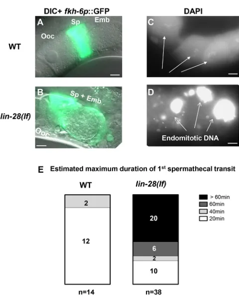
To investigate the cause of reduced embryo production in lin-28(n719)mutants, we checked whether ovulation and spermathecal exit proceed normally. We examined the spermatheca of lin-28(n719)mutants using the expression of a spermatheca reporter, fkh-6p::GFP (Chang et al., 2004). Approximately 70% of lin-28(n719)adults contained embryos in their spermathecae, whereas fewer than 10% of wild-type spermathecae contained embryos (Fig. 2A,B). Unlike in the wild type, many embryos inlin-28(n719) hermaphrodites had undergone multiple rounds of cell division inside the spermathecae. This suggests thatlin-28(n719) mutants have defects in the process of spermathecal exit. We used time-lapse video microscopy to monitor the first ovulation and spermathecal exit (Movies 1, 2). In wild-type hermaphrodites, ovulation, fertilization and spermathecal exit of an individual embryo happens within 20 min (McCarter et al., 1999). By contrast, we observed that for∼50% oflin-28(n719)hermaphrodites, the first ovulated embryo remained in the spermatheca for more than 60 min (Fig. 2E; Movie 2).
[image:2.612.106.241.266.606.2]Next, we addressed whether ovulation is also defective in lin-28(n719)mutants. The presence of endomitotically replicating (Emo) oocytes is a characteristic of ovulation mutants (Iwasaki et al., 1996), where oocytes undergo several rounds of DNA replication without ovulation and fertilization. Gonadal DAPI staining revealed that some lin-28(n719) hermaphrodites contain endomitotic oocytes (Fig. 2D) in the oviduct. We speculate that the defective ovulation in lin-28(n719) mutants may result from impairment of the spermathecal exit process, wherein the presence of fertilized embryos trapped within the limited spermathecal space would prevent subsequent entry of mature oocytes. We conclude that the poor fertility of lin-28(n719) mutants is the consequence of embryos becoming trapped in the spermathecae, stalling subsequent ovulation.
Fig. 1.lin-28(lf)hermaphrodites have reduced brood size and exhibit defects both in embryo production and embryonic viability.(A) Number of live progeny per hermaphrodite for wild type (strain N2),lin-2(e1309)and
lin-28(n719)mutants at 25°C.lin-2(e1309)mutants are used as egg-laying defective controls; bothlin-2(e1309)andlin-28(n719)hermaphrodites are unable to lay eggs. (B) Number of embryos per hermaphrodite forlin-28(n719)
andlin-2(e1309)mutants at 25°C (dissected from gravid adults 60 h after initiation of synchronized larval development by feeding starved L1s). (C) Viability of mutant embryos dissected fromlin-28(n719)and
lin-2(e1309)hermaphrodites at 25°C. % viability=100×(viable hatched larvae/total embryos). Number of animals≥15 per assay; number of independent replicate assays=11 forlin-28(n719), 4 forlin-2(e1309). Data are mean±s.d. analyzed using an unpairedt-test, ****P<0.0001.
RESEARCH ARTICLE Development (2019) 146, dev164293. doi:10.1242/dev.164293
DEVEL
O
Sp-Ut valve morphogenesis, uterine lumen formation and utse cell migration are abnormal inlin-28(lf)mutants
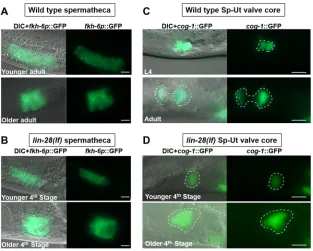
Spermathecae inC. eleganshermaphrodites consist of two rows of 12 cells forming a long tube structure at the young adult stage (Gissendanner et al., 2008; Kimble and Hirsh, 1979). Long spermathecal tube structures, labeled using fkh-6p::GFP, are observed in the lin-28(n719) mutants at the 4th stage (Fig. 3B), which corresponds to L4 and adult stage in wild type (see Fig. 6C). Wild-type spermathecae become constricted horizontally as somatic gonadal tissues continue morphogenesis before the first ovulation. lin-28(n719) spermathecae exhibited a similar constricted and extended morphology as in the wild type (Fig. 3A,B). Overall, we did not detect appreciable differences in spermathecal morphology between wild-type animals andlin-28(n719)mutants.
The Sp-Ut valve connects the spermatheca to the uterus in wild-type hermaphrodites, and serves as a portal through which fertilized embryos exit the spermatheca into the uterus (McCarter et al., 1999). The mature Sp-Ut valve consists of the toroidal syncytium and the core cell syncytium. We characterized Sp-Ut valve structure in lin-28(n719) mutants using a cog-1::GFP reporter, which is
expressed in the Sp-Ut valve core cell syncytium from the late L3 or early L4 stage (Palmer et al., 2002). In wild-type animals, the core cell syncytium stretches during the L4 stage, and in young adults, the Sp-Ut valve core forms a dumbbell-like structure with one end in the spermatheca and the other end in the uterus (Fig. 3C). Although we observed apparently normal expression of cog-1:: GFPin the Sp-Ut valve region oflin-28(n719)mutants in late 3rd larval stage, the core cell stretching did not occur and the Sp-Ut valve core remained as a single lobe structure in 4th stage animals (Fig. 3D). This observation indicates that Sp-Ut valve morphology is abnormal in lin-28(n719) mutants, suggesting the connection between the spermatheca and the uterus is also disrupted. We conclude that the aberrant connection between the uterus and the spermatheca associated with the abnormal morphology of the Sp-Ut valve causes the spermathecal exit defect in lin-28(lf ) mutants. To assess whether the abnormal Sp-Ut valve morphology of lin-28(n719) could reflect somatic gonadal cell lineage defects analogous to the precocious hypodermal cell lineages exhibited by lin-28(n719)(Ambros and Horvitz, 1984), we investigated whether the Sp-Ut valve syncytium in the mutant contains a normal number of nuclei as in the wild type. The Sp-Ut valve is derived from daughter cells of the dorsal uterine lineage, and the valve core syncytium comprises a fusion of two cells (Kimble and Hirsh, 1979). We used confocal microscopy to count the number of nuclei in the Sp-Ut valve core region based on labeling bycog-1::GFP. Two nuclei were present in the Sp-Ut valve core cell of both wild-type andlin-28(n719)mutants, suggesting the morphological defect is not caused by abnormal cell division in the lineage generating the Sp-Ut valve core (Fig. S3A,B).
In addition to the abnormal Sp-Ut valve morphology, other somatic gonadal defects were evident in lin-28(n719)mutants. In wild-type animals, the uterine lumen forms between the dorsal and ventral uterus during the L4 stage, when uterine toroidal cells fuse to generate a syncytium (Newman et al., 1996). Inlin-28(n719)mutants at the 4th stage, we observed abnormally small, incompletely connected and/or less elongated uterine luminal structures, compared with the wild type (Fig. S4B,C).
The uterine seam (utse) is a component of the hermaphrodite somatic gonad that mediates structural attachment between the hypodermis and uterus (Newman and Sternberg, 1996). The utse syncytium forms in the early L4 stage and extends laterally during progression to mid L4. The utse connects the uterus to the seam cells laterally and also to uv1 and the vulva.egl-13p::GFPis expressed in the nuclei ofπcell lineage, the products of which include the utse cells, from late L3 to L4 stages (Ghosh and Sternberg, 2014). Utse cells labeled by egl-13p::GFP migrated laterally in wild-type hermaphrodites (Fig. S5A,B). However, the utse nuclei did not migrate during the 4th stage inlin-28(n719)mutants, although egl-13p::GFP(+)cells were detected in the utse region (Fig. S5C,D).
lin-28(n719);lin-2(e1309) double mutants, which lack vulva formation, have the same somatic gonadal defects aslin-28(n719) mutants, implying these defects are not indirect consequences of the abnormal vulval morphology inlin-28(n719)mutants (Fig. S6). The defects in uterine lumen formation and utse cell migration in lin-28(n719) mutants, together with their Sp-Ut valve morphological defects, suggest thatlin-28activity is required for multiple aspects of proper hermaphrodite somatic gonadal development.
Eggshell integrity is compromised inlin-28(lf)mutant embryos
[image:3.612.54.294.52.357.2]To investigate the embryonic lethality inlin-28(n719)mutants, we dissected embryos from adult hermaphrodites and imaged the Fig. 2.lin-28(lf)hermaphrodites have defects in exit of embryos from
the spermatheca, and defects in ovulation.(A,B) Spermatheca labeled byfkh-6p::GFPof representative wild-type andlin-28(n719)adult
hermaphrodites. (A) In the wild type, oocytes (Ooc) pass into the spermatheca (Sp), where they are fertilized, and rapidly exit as a one-cell embryo (Emb). Therefore, most spermathecae are not observed to contain an embryo. (B) In a
lin-28(n719)hermaphrodite, an embryo (around∼150 cells) was trapped in the spermatheca. (C,D) DAPI staining of oocytes in wild-type (C) andlin-28(n719)
hermaphrodites. (C) In the wild type, individual oocytes (Ooc) contained a haploid complement of condensed chromosomes (arrows). (D) In the
lin-28(n719) mutant, endomitotic DNA was evidenced by an excessively bright DAPI signal in the oviduct before spermathecae, a characteristic of ovulation-defective mutants. Scale bars: 10 µm. (E) Distribution of estimated maximum duration of 1st spermathecal transit to uterus in wild type and
lin-28(lf )mutants (see Materials and Methods).
DEVEL
O
isolated embryos. Thelin-28(n719) embryos displayed abnormal irregular shapes (Fig. 4A), reminiscent of the misshapen phenotypes exhibited by egg-shell mutants (Johnston et al., 2006; Maruyama et al., 2007; Zhang et al., 2005). TheC. eleganseggshell, formed rapidly after fertilization in the spermatheca, consists of chitin, lipid and structural proteins, and functions as a protective barrier around the embryo. Eggshell abnormalities often result in embryo lethality (Johnston et al., 2010).
Chitin-binding domain-protein 1 (CBD-1) is a component of the eggshell cortex, andcbd-1::mcherryexpression marks the periphery of wild-type embryos (Allen et al., 2014; Johnston et al., 2010). cbd-1::mcherryexpression was evident surrounding embryos from lin-28(n719)hermaphrodites, indicating that an eggshell does form (Fig. 4B). The wild-type eggshell is impermeable to the lipophilic dye FM4-64 (Johnston et al., 2006), but, in our study, about 50% of lin-28(n719) embryos were permeable to FM4-64, while around 10% oflin-2(e1309)embryos were permeable (Fig. 4C).
This finding indicates that eggshell integrity is compromised in embryos produced by lin-28(n719) mutants. In support of this conclusion, lin-28(n719) embryos also exhibited osmotic stress sensitivity. Embryos from wild-type hermaphrodites maintained their oval shape upon exposure to 0, 150 and 300 mM KCl,whereas embryos fromlin-28(n719)animals swelled up in 0 or 150 mM KCl (Fig. 4D). Interestingly, embryos offln-1(tm545) mutants, which have defective spermathecal exit (Kovacevic and Cram, 2010), also exhibited permeability to FM4-64 (Fig. 4C), suggesting that abnormally prolonged residence in the spermatheca may result in compromised eggshell integrity (see Discussion).
In summary, lin-28 is required for normal somatic gonadal development, including uterine lumen formation, utse cell migration and proper Sp-Ut valve formation. We suggest that the abnormal Sp-Ut valve structure in lin-28(n719) mutants causes defects in spermathecal exit and ovulation, resulting in reduced embryo production. In addition, the embryos stalled in the spermatheca seem to suffer eggshell damage, resulting in embryonic lethality (Fig. 4E).
Embryonic lethality oflin-28(lf)is rescued by maternal expression of wild-typelin-28
[image:4.612.150.463.56.307.2]We speculated that the lethality of embryos produced bylin-28(n719) hermaphrodites results from defects in maternal somatic gonadal morphology (which results in trapping of embryos in the spermatheca), rather than from an absence oflin-28function in the embryos per se. If so, then maternal expression of lin-28 should rescue the embryonic lethality oflin-28(n719)homozygous embryos. To test this, we crossedlin-28(n719)hermaphrodites with wild-type males to obtain heterozygous mutants [lin-28(n719)/+], and then assessed the viability of lin-28(n719) homozygous self-progeny from these lin-28(n719)/+ hermaphrodites. Viable lin-28(n719) homozygotes were identified by their characteristic egg-laying defective phenotype as adults. Among the self-progeny of heterozygous [lin-28(n719)/+] hermaphrodites, we observed a ratio of wild-type progeny [+/+orlin-28(n719)/+] to egg-laying defective progeny [lin-28(n719)/lin-28(n719)] of 2.89(±0.2):1 on average, which is very close to the expected 3:1 ratio for complete maternal rescue of embryonic lethality (Table 1). This finding Fig. 3.lin-28(lf)mutants show essentially normal spermathecal primordium structure, but display defects in spermathecal-uterine (Sp-Ut) valve morphology.Spermathecal primordium visualized byfkh-6p::GFPexpression (A,B) and Sp-Ut valve core structure visualized bycog-1::GFPexpression (C,D) in wild type andlin-28(n719)hermaphrodites at successive times in the advancement towards the first ovulation. In each panel, the upper images are of hermaphrodites at the L4 or early young adult stage; the lower images are of hermaphrodites somewhat later in development, just before the time of first ovulation. [lin-28(n719)hermaphrodites skipped one larval stage; therefore, the‘4th stage’corresponds to L4 and adult stage in wild type (see Fig. 6).] (A,B) In both wild type andlin-28(n719)mutants, similar tube-shaped spermathecal primordia were detected, which contracted horizontally to form similar sac-like structures in older adults (A,B, lower panels). (C,D) Sp-Ut valve core structure (outlined) labeled bycog-1::GFPat successive stages in L4-adult developmental progression of a wild type andlin-28(n719)hermaphrodite. (C) In wild type, the Sp-Ut valve core stretches to form a fully developed ‘dumbbell’structure, with one lobe residing in the spermatheca and the other lobe in the uterus. The distance between each lobe shown here was∼10 µm. (D) Inlin-28(n719)mutants, the Sp-Ut valve core does not stretch and remains as a‘single lobe’structure, indicating an abnormal connection between the spermatheca and uterus in the mutants. Scale bars: 10 µm.
RESEARCH ARTICLE Development (2019) 146, dev164293. doi:10.1242/dev.164293
DEVEL
O
suggests that lin-28(n719) embryonic lethality reflects a requirement for lin-28 activity in the mother to enable proper development of her somatic gonad, which is required for the production of viable embryos.
Genes downstream oflin-28in developmental timing regulation also function downstream oflin-28in somatic gonadal morphogenesis and fertility
Functional interactions oflin-28with other heterochronic genes for the control ofC. elegans hypodermal cell lineage developmental timing have been described previously (Ambros, 2011; Resnick et al., 2010) (see Fig. 5E). lin-28 functions upstream of lin-46 and hbl-1 to control the timing of L2 to L3 fate transitions in hypodermal cell lineages, and lin-28 also acts via a pathway consisting oflet-7,hbl-1,lin-41andlin-29to regulate the transition from L4 fates to adult fates in the hypodermis.
[image:5.612.50.421.54.510.2]To determine whether these same heterochronic genes that act downstream of lin-28 for hypodermal cell fate timing also function in fertility and embryonic lethality, we examined those phenotypes in double mutants of the heterochronic genes (Fig. 5A-C). The number of live progeny per animal produced by the lin-28(n719);let-7(mn112)andlin-28(n719);lin-29(n836) double mutants was significantly higher than that produced by thelin-28(n719) mutants (Fig. 5A). In addition, the number of
Fig. 4.lin-28(lf)embryos are misshapen and are defective in eggshell integrity.(A) DIC images of wild-type,lin-2(e1309)andlin-28(n719)
embryos from dissected adult animals. Wild type andlin-2(e1309)mutants produce ovoid embryos, butlin-28(n719)
embryos exhibit irregular shapes. (B)cbd-1::mCherryexpression indicates that eggshells are present inlin-28(n719)
embryos despite their misshapen morphology (lower panels). (C) Eggshell permeability of embryos produced by
lin-2(e1309),lin-28(n719),egg-1(tm1071)
andfln-1(tm545)hermaphrodites. Eggshell mutantegg-1(tm1071)served as a control. Embryos fromlin-28(n719),
fln-1(tm545)andegg-1(tm1071)were more permeable to the lipophilic dye FM 4-64 thanlin-2(e1309)embryos. Likelin-28(n719),fln-1(tm545)
hermaphrodites exhibit defects in spermathecal exit. Permeability was calculated as the percentage of permeable embryos/total embryos from dissected adult animals. Number of animals≥15 per each assay; number of independent replicate assays=3 for each strain. Data are mean±s.d. analyzed using an unpairedt-test compared with
lin-2(e1309), *P<0.05. (D) Morphology of embryos under different osmotic conditions.lin-28(n719)embryos were more sensitive than wild type to low-salt conditions, indicating a lack of protection from osmotic stress. (E) Model for physiological causes of fertility defects in
lin-28(lf )mutants.lin-28(lf )animals have abnormal Sp-Ut valve structure, which leads to defects in spermathecal exit and ovulation, and hence a reduced embryo production. In addition, retention of embryos in the spermatheca compromises eggshell integrity, which causes embryonic lethality.
Table 1. Ratio of egg laying and egg laying defective progeny fromlin-28 heterozygous mutants
lin-28(+)/lin-28(−)
heterozygote number
Number of egg laying progeny
Number of egg laying defective progeny
Ratio (egg laying/egg laying defective)*
1 133 43 3.09
2 108 40 2.70
3 89 31 2.87
4 71 27 2.63
5 142 46 3.09
6 111 41 2.71
7 102 35 2.91
8 131 42 3.12
*Average ratio±s.d.=2.89±0.20.
DEVEL
O
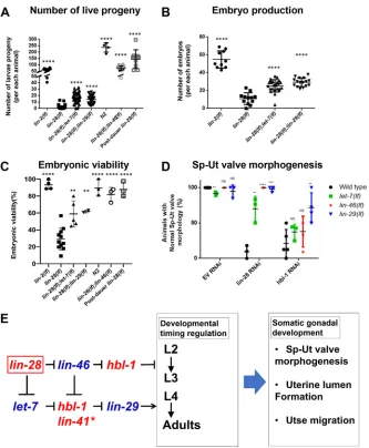
[image:5.612.50.301.614.728.2]live progeny of lin-28(n719);lin-46(ma164) was even greater than that of lin-2(e1309), because the loss of lin-46 rescued the vulva and egg-laying defects of lin-28(n719) mutants. lin-28(n719);let-7(mn112) and lin-28(n719);lin-29(n836) mutants produced more embryos per animal than lin-28(n719) mutants (Fig. 5B) and the embryonic lethality was suppressed in these double mutants, compared with lin-28(n719) (Fig. 5C). The
embryonic viability oflin-28(n719);lin-46(ma164)was similar to wild-type animals (Fig. 5C).
[image:6.612.140.473.55.459.2]Consistent with the genetic epistasis observed above for fertility and embryonic viability,lin-46(lf ),let-7(lf )andlin-29(lf)were also epistatic tolin-28(lf )for somatic gonadal morphogenesis. Our RNAi knockdown experiments showed that genetic absence oflet-7,lin-46 orlin-29suppresses the Sp-Ut valve defects caused bylin-28(RNAi). Fig. 5. Genetic epistasis analysis oflin-28and other heterochronic genes for effects on fertility, embryonic lethality and Sp-Ut valve morphogenesis. (A) Total number of viable larva progeny oflin-2(e1309),lin-28(n719),lin-28(n719);let-7(mn112),lin-28(n719);lin-29(n836),lin-46(ma164);lin-28(n719)mutants, post-dauerlin-28(n719)and wild type. (B) Embryo production oflin-2(e1309),lin-28(n719),lin-28(n719);let-7(mn112)andlin-28(n719);lin-29(n836)mutants. (C) Embryonic viability oflin-2(e1309),lin-28(n719),lin-28(n719);let-7(mn112),lin-28(n719);lin-29(n836)andlin-46(ma164);lin-28(n719)mutants, post-dauer
lin-28(n719)and wild type. Number of animals≥15 per each assay; each dot represents an independent replicate assay. The number of progeny oflin-28(n719)
mutants is increased by loss oflet-7,lin-29orlin-46.(A). Both embryo production (B) and embryo viability (C) are improved by loss oflet-7,lin-29orlin-46. Total number of progeny and embryonic viability oflin-28(lf )mutants are also improved by post-dauer development (A,C). Data are mean±s.d. analyzed using an unpairedt-test compared withlin-28(lf ); NS; not significant, **P<0.01, ****P<0.0001. (D) The percentage of animals with normal Sp-Ut valve core morphology, visualized bycog-1::GFPexpression in wild type,let-7(mn112),lin-46(ma164)andlin-29(n836)mutants, treated with control RNAi (EV, empty vector strain L4440),lin-28(RNAi) orhbl-1(RNAi).Wild type,lin-46(ma164)andlin-29(n836)mutants treated with L4440 empty vector RNAi rarely showed Sp-Ut valve defects.
let-7(mn112)mutants with control RNAi showed less than 10% of Sp-Ut valve defects.lin-28(RNAi) treatment of wild type led to∼95% Sp-Ut valve defects, an effect that was partially or fully suppressed bylet-7(mn112),lin-46(ma164)andlin-29(n836). hbl-1(RNAi) treatment of wild type also led to Sp-Ut valve defects that were rarely suppressed bylet-7(mn112)or bylin-46(ma164), or by moderately suppressed (∼70%) bylin-29(n836). Number of animals≥12 per assay; each dot represents an independent replicate assay. Data are mean±s.d. analyzed using an unpairedt-test. Significance was calculated by comparing each mutant with wild type in each RNAi set. NS, not significant; **P<0.01; ***P<0.001; ****P<0.0001. (E) The genetic regulatory pathway model for somatic gonadal morphogenesis, derived from the results of epistasis experiments presented in Fig. 5A-D, is highly similar to the model for temporal regulation of hypodermal cell fates derived from previous studies (Ambros, 2011; Resnik et al., 2010). (See Discussion regarding the possible involvement oflin-41in somatic gonadal morphogenesis.) We suggest that the specification of proper hypodermal developmental timing by these genes is essential for somatic gonadal morphogenesis.
RESEARCH ARTICLE Development (2019) 146, dev164293. doi:10.1242/dev.164293
DEVEL
O
hbl-1(RNAi) phenocopied the fertility phenotypes of lin-28(n719) mutants, including abnormal Sp-Ut valve core morphology (Fig. S7A,B). Loss of lin-46 or let-7 function rarely affected the Sp-Ut valve morphological defect inhbl-1(RNAi)animals, indicating hbl-1is epistatic to these genes. Finally,∼70% oflin-29(lf );cog-1:: GFP animals showed the wild-type Sp-Ut valve core cell morphology when hbl-1 function was compromised (Fig. 5D). In addition, a normal uterine lumen was observed in ∼50% of lin-28(n719);let-7(mn112)mutants (Fig. S4D).Mostlin-28(n719); lin-29(n836) and lin-28(n719);lin-46(ma164) mutants showed complete uterine lumen formation (Fig. S4E,F). Utse cell migration defects oflin-28(lf )mutants were also partially suppressed by loss of eitherlet-7orlin-46(Fig. S5G-J). Overall, our findings indicate that let-7,lin-46,hbl-1andlin-29act downstream oflin-28for somatic gonadal development in a network configuration that is essentially identical to that previously described for the control of hypodermal cell fate timing by these same genes (Fig. 5E).
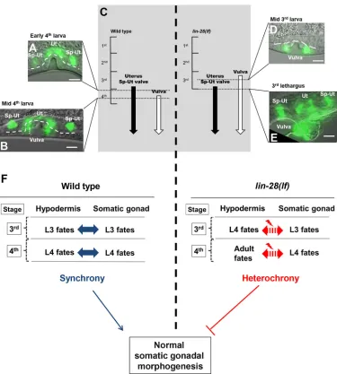
Developmental timing of hypodermal tissues and somatic gonadal tissues is discoordinated inlin-28(lf)mutants Based on the above observations indicating parallels between the control of hypodermal developmental timing and somatic gonadal morphogenesis bylin-28mutants, we investigated whether the stage specificity of somatic gonadal developmental events might be altered in lin-28(n719) hermaphrodites, in analogy to their precocious hypodermal development. As a marker to monitor the expression of stage-specific programs in the somatic gonad and hypodermis, we employedcog-1::GFP(Palmer et al., 2002), which is expressed in the wild-type dorsal uterus and Sp-Ut valve core, beginning from late-L3/early-L4 stage (Fig. 6A). cog-1::GFP is also expressed in the ventral hypodermis (vulval cell lineages), beginning in the mid L4 stage, which is after the onset of cog-1::GFPsomatic gonadal expression (Fig. 6B).
We examined whether the normal relative order ofcog-1::GFP expression in the somatic gonad and vulva is altered inlin-28(n719) mutants. Inlin-28(n719)hermaphrodites,cog-1::GFPexpression in the vulva was observed precociously in the mid-3rd larval stage (Fig. 6D), consistent with the previously described precocious vulval cell divisions of lin-28(lf ) mutants (Euling and Ambros, 1996). However, the onset of somatic gonadal expression ofcog-1:: GFP was normal, beginning from the late 3rd larval stage in both lin-28(n719) and in the wild type (Fig. 6E). Thus, the expression ofcog-1::GFPin the vulva precedes the expression in somatic gonads inlin-28(n719)mutants. These data suggest that the somatic gonadal development, at least as reflected bycog-1::GFP expression, is not precocious in lin-28(n719), despite precocious development of the hypodermis. Therefore, developmental events in hypodermal tissues and somatic gonadal tissues are temporally discoordinated inlin-28(n719)mutants (Fig. 6F). We hypothesize that this discord, between the precociously developing hypodermis and normally timed somatic gonad, causes abnormal somatic gonadal morphogenesis.
To investigate the hypothesis that the somatic gonadal defects of lin-28(n719)mutants are caused by precocious development of the hypodermis, we examined whether post-dauer development, which has been shown to suppress the precocious hypodermal development oflin-28(n719)(Euling and Ambros, 1995; Liu and Ambros, 1991), can also rescuelin-28(n719)somatic gonadal defects. We observed that after post-dauer development, lin-28(n719) adults exhibited restored fertility and embryonic viability, normal morphology of the Sp-Ut valve core, uterine lumen, and normal utse cell migration (Fig. 5A,C, Figs S7E, S4G, S5E and S5F). This suppression of
somatic gonadal defects in lin-28(n719) animals by post-dauer development supports the supposition thatlin-28acts indirectly, via downstream genes and events, to mediate normal somatic gonadal morphogenesis (Fig. 5E).
Hypodermal expression, but not somatic gonadal expression, oflin-28rescues somatic gonadal defects, embryonic lethality and reduced number of total progeny in lin-28(lf)hermaphrodites
If the somatic gonadal morphological defects of lin-28(n719) mutants originate from discoordination between the timing of hypodermal and somatic gonadal development, suppressing the precocious hypodermal development in lin-28(n719) mutants should also rescue the somatic gonadal phenotypes. To test whether lin-28 expression specifically in the hypodermis could rescue the Sp-Ut valve morphological defects oflin-28(n719), we employed Mos1-mediated single copy insertion (MosSCI) transformation to generate transgenic worms expressing lin-28:: GFP::lin-28 3′ UTR driven by tissue-specific promotors; these included lin-28 endogenous promoter sequences, a dpy-7 hypodermal promoter (Gilleard et al., 1997) and anehn-3Aearly somatic gonadal promoter (Large and Mathies, 2010). The endogenous lin-28 promoter drives lin-28::GFP expression in neurons and hypodermis, where lin-28is known to be expressed (Moss et al., 1997). Using spinning disk microscopy, we also detectedlin-28p::lin-28::GFPexpression in Z1 and Z4 cells, which are precursors of somatic gonadal tissues (Fig. S8A,B). dpy-7p::lin-28::GFPwas expressed in the hypodermis during the embryonic and L1 stages, andehn-3Ap::lin-28::GFPexpression was strongest in Z1 and Z4 from the late embryo to L1 stages (Fig. S8C,D). lin-28::GFPlevels decreased from the L2 stage in all three strains, presumably owing to repression mediated by thelin-283′UTR.
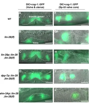
We crossed those MosSCI transgenic strains withlin-28(n719); cog-1::GFPhermaphrodites to assess the relative timing of somatic gonadal and hypodermalcog-1::GFPexpression.cog-1::GFPwas expressed in somatic gonadal tissues prior to the vulva in wild-type animals, whereas cog-1::GFP expression in the vulva was precocious in thelin-28(n719)mutants (Figs 6 and 7A,B).lin-28 expression via its endogenous promoter ( lin-28p::lin-28::GFP::lin-283′UTR) restored the normal relative timing of somatic gonadal and hypodermalcog-1::GFP expression inlin-28(n719) mutants (Fig. 7C). Similarly, indpy-7p::lin-28::GFP;lin-28(n719);cog-1:: GFPanimals,cog-1::GFPexpression in the vulva occurred at the normal time, following somatic gonadal expression. Thus, hypodermal expression of lin-28 efficiently rescues precocious hypodermal development oflin-28(n719) (Fig. 7D). By contrast, somatic gonadal expression oflin-28via theehn-3Ap::lin-28::GFP transgene did not rescue the precocious expression of hypodermal cog-1::GFPin lin-28(n719) mutants (Fig. 7E). These results are consistent with cell-intrinsic activity oflin-28in the hypodermis to control hypodermal developmental timing.
Does hypodermal expression of lin-28also rescue the somatic gonadal morphogenesis defects of lin-28(n719) hermaphrodites? Indeed,dpy-7p::lin-28::GFPdid rescue wild-type morphology of the Sp-Ut valve core inlin-28(n719), (Fig. 7F,H,I), normal uterine lumen formation (Fig. S4I) and normal utse cell migration (Fig. S5M,N). By contrast, somatic gonadallin-28expression did not rescue lin-28(n719) somatic gonadal defects (Fig. 7G,J, Figs S4J, S4K, S5O and S5P).
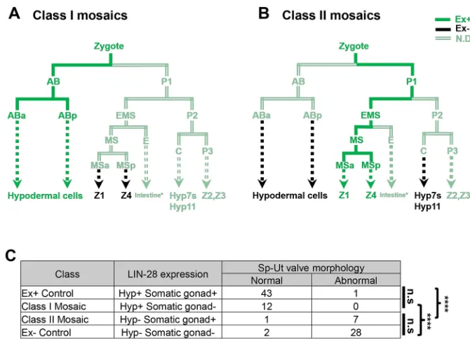
We also performed mosaic analysis to investigate whether hypodermal expression of lin-28 is necessary and sufficient for somatic gonadal morphogenesis (Yochem and Herman, 2003).
DEVEL
O
We constructed lin-28(n719);cog-1::GFP strains with an extrachromosomal transgenic array containing alin-28expression vector and asur-5::GFPplasmid as a reporter for the array. Twelve mosaic animals that completely lost the extra-chromosomal array in the Z1 and Z4 lineages, but not in hypodermal lineage, all showed wild-type Sp-Ut valve core morphology at the young adult stage (Fig. 8A, Fig. S9B). For seven out of eight mosaic animals in which sur-5::GFP was undetectable in the hypodermis, but clearly expressed in the Z1 and Z4 lineages, wild-type Sp-Ut valve core morphology was not observed (Fig. 8B, Fig. S9C). This suggests that loss of lin-28 expression in hypodermal tissues, but not in somatic gonadal tissues, is correlated with abnormal Sp-Ut valve morphogenesis (Fig. 8C, Fig. S9).
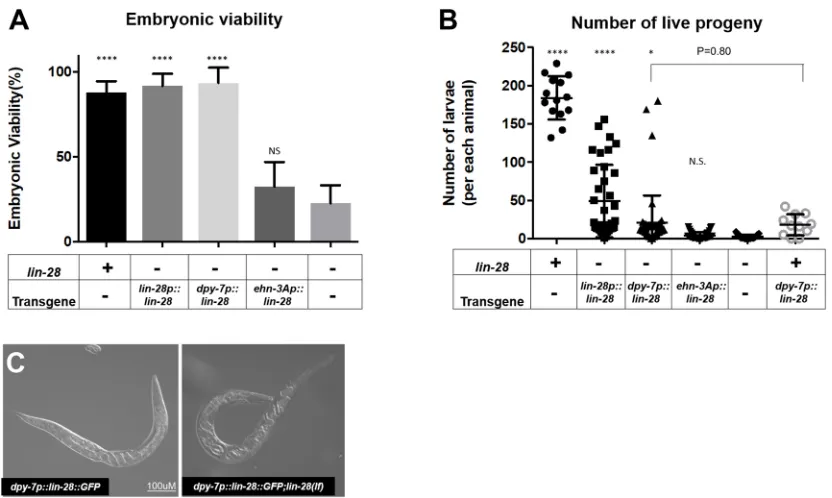
Because the hypodermal expression of lin-28 suppresses the morphological defects of the somatic gonad inlin-28(n719)mutants (Fig. 7), we investigated whether embryonic lethality and fertility defects were accordingly rescued, as would be expected. Expression of eitherlin-28p::lin-28::GFPordpy-7p::lin-28::GFPrestored the embryo viability of lin-28(n719) progeny, whereas ehn-3Ap::lin-28::GFP expression did not restore viability of lin-28(n719) progeny (Fig. 9A).
[image:8.612.119.494.54.471.2]Interestingly, although the total number of live progeny was greater for thedpy-7p::lin-28::GFP;lin-28(n719)strain than for the lin-28(n719) mutants, the number of progeny was comparable with the wild type in only∼15% of animals; the other ∼85% of animals produced fewer than 20 progeny at 25°C. In addition, the Fig. 6. Heterochronic development of the hypodermis, relative to the somatic gonad, inlin-28(lf)hermaphrodites.(A,B,D,E)cog-1::GFPexpression patterns in wild type andlin-28(n719)hermaphrodites at indicated developmental stages. The temporal order of the onset ofcog-1::GFPexpression in the somatic gonad and vulva is reversed inlin-28(lf )compared with the wild type.cog-1::GFPis expressed in both somatic gonadal tissues (uterus and Sp-Ut valve) and hypodermal tissue (vulva). The timing of expression in wild type andlin-28(n719)mutants is summarized in C. In the wild type,cog-1::GFP
expression in somatic gonadal tissues first appears during the late 3rd larval stage and is not yet apparent in the vulva at the early 4th larval stage (A). Vulval expression ofcog-1::GFPstarts at the middle of the 4th larval stage (B). Inlin-28(n719)mutants, vulval expression ofcog-1::GFPoccurs precociously in the middle of the 3rd larval stage (D), whereas somatic gonadal expression ofcog-1::GFPstarts at the normal time, in the late 3rd larval stage (E). Both vulval and somatic gonadal expression are detected at 3rd lethargus (E). (F) Model for the importance oflin-28activity for somatic gonadal development. In wild-type animals, timing of hypodermis development and somatic gonadal development are synchronized. Inlin-28(lf )mutants, hypodermal development happens precociously, whereas somatic gonadal development does not, resulting in heterochronic development between two tissues. We suggest this heterochrony causes somatic gonadal morphogenesis defects. Scale bars: 10 µm.
RESEARCH ARTICLE Development (2019) 146, dev164293. doi:10.1242/dev.164293
DEVEL
O
dpy-7p::lin-28::GFPtransgene in the wild-type background led to a similarly reduced number of progeny, indicating that the expression of dpy-7p::lin-28::GFP alone is sufficient to reduce fertility, apparently by impairing egg-laying (Fig. 9C). In contrast, somatic gonadal promoter-drivenlin-28::GFPexpression did not affect the number of progeny produced by lin-28(n719) hermaphrodites (Fig. 9B).
DISCUSSION
C. elegans lin-28(lf ) hermaphrodites exhibit a dramatic reduction in fertility, in excess of what would be expected as a simple consequence of their vulval morphogenesis defects. In principle, it is possible thatlin-28could promote fertility entirely via a germline-specific activity, analogous to the demonstrated role of mammalian Lin28 in promoting pluripotency and stem cell proliferation (Yu et al., 2007; Zhang et al., 2016). Indeed, lin-28 has been reported to regulate the germ cell pool size in mice and inC. elegans hermaphrodites (Shinoda et al., 2013; Wang et al., 2017). However, the reported effects on germ cell pool size in C. elegans, after germline knockdown oflin-28, did not include substantially reduced fertility (Wang et al., 2017). Our results suggest that the reduced fertility oflin-28(lf)hermaphrodites is caused by fertility-promoting activities of lin-28 outside the germline, i.e. within somatic cell lineages of the gonad and/or other tissues. Other examples in which lin-28controls the development of reproductive tissues other than germ cells have been reported in flies and mice. TheDrosophilaegg chamber is fused abnormally early inlin-28-null mutants, and the development of mice vaginal openings is delayed in lin-28a transgenic mice (Stratoulias et al., 2014; Zhu et al., 2010).
Here, we have investigated the somatic function of lin-28 in promotingC. eleganshermaphrodite fertility. Our results show that
lin-28 is required for the completion of certain somatic gonadal morphogenetic events, specifically, the enlargement of the uterine lumen, the migration of utse nuclei and the extension of the Sp-Ut valve core, and that these somatic gonadal morphogenesis defects likely underlie the reduced fertility oflin-28(lf )hermaphrodites. In particular, the abnormal Sp-Ut valve of lin-28(lf ) animals has a potent impact on fertility by preventing fertilized embryos from entering the uterus, thereby inhibiting ovulation and resulting in reduced embryo production.
We also found compromised eggshell integrity in lin-28(lf ) mutants, which negatively affects embryonic viability, similar to other eggshell defective mutants (Johnston et al., 2006, 2010; Maruyama et al., 2007). Moreover, we found that another spermathecal exit mutant fln-1(lf ) has eggshell-defective phenotypes similar to lin-28(lf ) (Fig. 4C), suggesting a causal link between spermathecal retention and loss of eggshell integrity. We speculate that physical damage to the eggshells may occur when the embryos are trapped in the spermatheca. Interestingly, deficiency of cbd-1, an essential component of the C. elegans eggshell, leads to pinched-off embryos reflecting incomplete spermathecal exit (Johnston et al., 2010). This suggests that damage to the eggshell of embryos lingering too long in the spermatheca could further aggravate an underlying spermathecal exit defect. Further research is needed to clarify the relationship between spermathecal exit and eggshell integrity.
[image:9.612.49.339.48.382.2]Our results point to a close coupling between the hypodermal and somatic gonadal phenotypes inlin-28(lf )mutants. First, we found that a very similar configuration of the known heterochronic genes mediates the effects of lin-28(lf ) on hypodermal developmental timing and on somatic gonadal morphogenesis (Fig. 5E). The exception is lin-41; we could not find evidence that lin-41, a
Fig. 7. Hypodermal expression oflin-28rescues developmental timing defects and Sp-Ut valve
morphogenesis inlin-28(lf)mutants.cog-1::GFPexpression in the somatic gonad and vulva (below the dashed line) in late 3rd or early 4th larval stage hermaphrodites (A-E), or in the Sp-Ut valve (outlined) in adult or 4th stage hermaphrodites (F-J), of the wild type (A,F),lin-28(lf )(B,G) andlin-28(lf ), withlin-28expression driven by various promoters (C-E,H-J).dpy-7p(D,I) is a hypodermal-specific promoter (Gilleard et al., 1997) andehn-3Ap
(E,J) is an early somatic gonadal-specific promoter (Large and Mathies, 2010). In these experiments, the promoter-drivenlin-28
transgene (C-E,H-J) is also tagged with GFP, butlin-28::GFP
expression is not detectable at these stages, so all the GFP signal shown here corresponds tocog-1::GFP. lin-28p::lin-28::GFP;lin-28(n719)(C) anddpy-7p::lin-28::GFP;lin-28(n719)(D) express
cog-1::GFPin the somatic gonad earlier than in the vulva, as in wild type (A). In contrast, bothlin-28(n719)mutants (B) andehn-3Ap::lin-28::GFP;lin-28(n719)(E) expresscog-1::GFP
precociously in the vulva. Eighty-three percent oflin-28p::lin-28:: GFP;lin-28(n719)(H) and 95% of dpy-7p::lin-28::GFP;lin-28(n719)(I) animals exhibit the Sp-Ut core dumbbell structure observed in wild type (F). However, the Sp-Ut valve core structure remains as a single lobe shape in bothehn-3Ap::lin-28::GFP; lin-28(n719)(J) andlin-28(n719)(G) animals (94% and 98%, respectively). These data suggest that hypodermal expression of
lin-28is sufficient to rescue heterochronic development (A-E) and abnormal Sp-Ut valve core morphogenesis (F-J) inlin-28(n719)
mutants, but somatic gonadal expression oflin-28cannot rescue either defect. Scale bars: 10 µm.
DEVEL
O
downstream target of let-7for hypodermal developmental timing (Slack et al., 2000), is involved in Sp-Ut valve core morphogenesis. The lin-41(RNAi) animals displayed a superficially normal dumbbell-shaped Sp-Ut valve, albeit somewhat smaller than the wild-type valve (Fig. S7C). This result may reflect either an insufficient knockdown oflin-41by RNAi in these experiments, or thatlin-41does not participate in somatic gonadal development. In the latter event, hbl-1 would appear to function as a main downstream target oflet-7in somatic gonadal morphogenesis.
Our second finding indicating a link between lin-28(lf ) hypodermal and somatic gonadal phenotypes is the observation that post-dauer development suppresses both the hypodermal and the somatic gonadal developmental defects of lin-28(lf ) hermaphrodites. This result in particular accentuates the fact that the somatic gonadal defects oflin-28(lf )do not reflect a direct role oflin-28in somatic gonadal development per se; rather, somatic gonadal morphogenesis fails in lin-28 mutants as an indirect consequence of precocious development. Consistent with this idea, another precocious mutant,lin-14(lf )(Ambros and Horvitz, 1984), showed similar Sp-Ut valve defects tolin-28(lf )mutants (Fig. S7D). Importantly, we did not find evidence for precocious development of somatic gonadal events inlin-28(lf ). In particular, we observed that the timing of the onset of expression of certain fluorescent markers of L3 and L4 somatic gonadal developmental events was not altered in lin-28(lf ), even though subsequent morphogenesis failed. Assuming that the somatic gonad and the hypodermis each has its own developmental clock, it would appear that the hypodermal developmental clock oflin-28(lf ) mutants is accelerated, whereas the somatic gonadal developmental clock runs normally, resulting in discoordination of developmental timing
between the two tissues during the L3 and L4 stages (Fig. 6F). We propose that it is this temporal discord between the accelerated hypodermis, and the normally scheduled somatic gonad, that results in failure of somatic gonadal morphogenesis.
The apparent absence of precocious development of the somatic gonad oflin-28(lf )animals suggests thatlin-28may affect somatic gonadal morphogenesis cell non-autonomously, by controlling one or more signals from the hypodermis to the somatic gonad. In strong support for this model, we found that expression of lin-28 specifically in the hypodermis could rescue the somatic gonadal developmental defects oflin-28(lf )mutants. Conversely, expression oflin-28specifically in the somatic gonadal precursor lineage did not rescue anylin-28(lf )phenotypes. Based on these observations, we propose that the principal function of lin-28 with regards to somatic gonadal development is to act within the hypodermis to specify a schedule of hypodermal events that is properly coordinated with a corresponding schedule of somatic gonadal developmental events. Apparently, normal somatic gonadal morphogenesis requires a coordination between the hypodermis and the somatic gonad during the L3 and/or L4 stages.
[image:10.612.136.471.55.297.2]By what mechanisms could hypodermal activity oflin-28regulate the development of a different tissue? There are precedents in C. elegansfor cell non-autonomous developmental signals originating from the hypodermis. A recent study showed that heterochronic genes acting in the hypodermis can modulate mTORC2 signaling in the intestine (Dowen et al., 2016). In another example, it has been reported that migration of the hermaphrodite-specific neurons and arborization of sensory neurons are regulated by hypodermal expression of the microRNA mir-79 and MNR-1/menorin, respectively (Pedersen et al., 2013; Salzberg et al., 2013). Fig. 8.lin-28(lf)mosaic analysis for Sp-Ut valve morphogenesis.(A,B) Summary of mosaic expression patterns ofsur-5::GFPin VT3884 animals where the extrachromosomal array maEx265[sur-5::GFP;lin-28p::lin-28::GFP] was lost either (A) in a precursor of Z1 and Z4 or (B) in hypodermal lineages (the AB lineage and probably also in P2 or C). (See Fig. S9 for more detailed analysis of individual mosaic animals and non-mosaic controls.) The observedsur-5::GFP
expression pattern for these two classes of mosaic animals are color coded: dark green, confirmedsur-5::GFPexpression; black, confirmed absence of
sur-5::GFPexpression; light green,sur-5::GFPexpression not determined. Dotted lines represent multiple cell divisions not shown in these abbreviated cell lineage diagrams. (Expression in the E lineage was determined in some mosaic animals, but was not relevant to Sp-Ut valve core morphogenesis; see Fig. S9B,C.) (C) Summary of the phenotypes exhibited by class I and class II mosaic animals and non-mosaic controls. Presence of wild-type Sp-Ut valve core morphology was determined in mosaic animals (A,B) as well as in the control animals, which contained or completely lost extrachromosomal arrays in both hypodermal and somatic gonadal lineages (Fig. S9A,D). Data were analyzed using a Chi-square test. n.s, not significant; ****P<0.0001.
RESEARCH ARTICLE Development (2019) 146, dev164293. doi:10.1242/dev.164293
DEVEL
O
Hypodermal glycosylated cell surface molecules or signaling cell-adhesion molecules are key downstream factors for neuronal morphogenesis in each case, implying this nonautonomous signaling may require physical contact with other tissues.
Indeed, seam cells in the hypodermis become physically connected to the utse of C. elegans hermaphrodites. The connection between these two tissues is thought to be formed during the mid to late L4 stages in wild-type animals (Newman et al., 1996). The precocious hypodermal maturation inlin-28(lf ) animals might cause this connection to be formed aberrantly, or not at all. Alternatively, the reduced number of seam cells inlin-28(lf ) mutants may alter positioning of seam cells in the hypodermis, disrupting the normal connection between seam cells and the utse. However, there is evidence that utse defects may not necessarily be associated with Sp-Ut valve abnormality.lin-29expression in the anchor cell induces signals for utse precursor cells to adopt utse fates, andlin-29(lf )mutants do not form a proper utse (Newman et al., 2000). However, loss oflin-29does not cause any detectable abnormality in Sp-Ut valve core morphology (Fig. 5D). Nevertheless, it will be interesting to examine whether the physical connection between the hypodermal seam and gonadal utse is formed properly inlin-28(lf )hermaphrodites.
It is striking that lin-28(lf )mutants exhibit defects in the final stages of morphogenesis in at least three distinct somatic gonadal structures: extension of the Sp-Ut valve core, positioning of the uterine seam cell nuclei and expansion of the lumen of the uterus. As all three of these defects are highly penetrant inlin-28(lf ), and are coordinately rescued by appropriatelin-28-expressing transgenes, we were not able to determine whether these defects are expressed
independently, or whether, for example, one of them is the primary defect that is linked to hypodermal developmental timing and the other defects are secondarily precipitated by the first. Further research is needed to identify any cause-effect relationships between utse cell migration, Sp-Ut valve morphogenesis and uterine lumen formation. Overall, our studies of lin-28 in the context of C. elegans reproductive system development provides an informative model for exploring fundamental principles of multicellular development, including how the generation of organized cellular complexity requires precise temporal coordination of events across interacting tissues. Our findings exemplify how a cell-intrinsic developmental timing program can be required not only cell-autonomously to specify temporal cell fates, but also to control cell-nonautonomous signaling that is crucial for proper development of interacting tissues. Our identification of cell non-autonomous hypodermis-to-gonad developmental signaling controlled by lin-28 and the heterochronic pathway should set the stage for future studies addressing the identity and potential evolutionary conservation of the molecular components of the downstream signal(s).
MATERIALS AND METHODS Culture ofC. elegansstrains
C. eleganswild-type (strain N2) and mutant strains (listed in Table S1) were
[image:11.612.99.515.55.304.2]grown and maintained (at 25°C unless otherwise noted) on nematode growth media (NGM) agar plates seeded withE. coli(strain HB101). A list of genotyping primers for allele confirmation can be found in Table S2. Synchronized populations of larvae at defined developmental stages were obtained as previously described (Stiernagle, 2006). Briefly, embryos were collected using 1.2% (v/v) sodium hypochlorite and 0.5 N NaOH, washed
Fig. 9. Hypodermal expression oflin-28enhances embryonic viability and number of progeny oflin-28(lf)mutants.(A) Embryonic viability of lin-28p::lin-28::GFP;lin-28(n719)anddpy-7p::lin-28::GFP;lin-28(n719)are restored to a level similar to that in wild type. However, expression ofehn-3Ap::lin-28::GFPdoes not enhance the viability oflin-28(n719)embryos. Number of animals≥15 per each assay; number of independent replicate assays=4 forlin-28(n719), 3 for all other strains. (B) The number of live larva progeny is increased inlin-28p::lin-28::GFP;lin-28(n719)(n=36) and slightly enhanced in dpy-7p::lin-28::GFP;lin-28(n719)(n=53) compared withlin-28(n719)mutants (n=25). Progeny numbers fordpy-7p::lin-28::GFPinsertional lines withoutlin-28(n719)(n=11) are similar to those ofdpy-7p::lin-28::GFP;lin-28(n719)(P=0.80), suggesting that expression ofdpy-7p::lin-28::GFPinduces fertility defects regardless oflin-28(n719). Progeny numbers forehn-3Ap::lin-28::GFP;lin-28(n719)(n=40) are similar to those oflin-28(n719)mutants. Data are mean±s.d. analyzed using an unpairedt -test compared withlin-28(lf ). N.S., not significant; *P<0.05, ****P<0.0001. (C) Embryos ofdpy-7p::lin-28::GFP(left) anddpy-7p::lin-28::GFP;lin-28(n719)(right) are trapped inside adult hermaphrodites, indicating that defective egg laying is a cause of the reduced fertility in these animals. Scale bar: 100 µm.
DEVEL
O
with M9 buffer and incubated in M9 buffer overnight at 20°C, placed on NGM plates seeded with HB101, incubated for defined lengths of time at 20°C or 25°C, and developmental stage was assessed by DIC microscopy of a sample of worms from the population (Byerly et al., 1976).
Microscopy
For DIC and fluorescence microscopy, worms were anesthetized with 0.2 mM levamisol and mounted on 2% agarose pads. All images, except the following, were obtained using a Zeiss Axiocam 503 mono: Fig. S2 (Leica DM 5500Q confocal microscopy) and Fig. S8 [3i (Intelligent Imaging Innovations) Everest spinning disk confocal microscopy]. The supplementary movies were captured using a Zeiss Axiocam 503 mono. For gonad DAPI staining, hermaphrodites were cut with a syringe needle and the extruded gonads were fixed with 95% ethanol. After washing twice with M9, the dissected gonads were incubated with 4′6′-diamidino-2-phenylindole solution (100 ng/ml) for 10 min in a humidified chamber and washed again with M9 (modified from Shaham, 2005).
RNAi
RNAi by feeding worms withE. coli-expressing double-stranded RNA was conducted as previously described (Conte et al., 2015). HT115 bacterial RNAi strains (lin-28, hbl-1, lin-29, lin-46) and an empty vector strain (L4440) from the Ahringer library were used (Kamath and Ahringer, 2003).
Analysis of fertility and embryonic viability
Individual young adult hermaphrodites were placed, one per plate, on NGM plates seeded with HB101, and the number of live progeny from each hermaphrodite was counted∼3-4 days later. To determine embryo viability, gravid adult hermaphrodites were dissected with a syringe, and the released embryos were collected and transferred to NGM plates seeded with HB101. The total number of embryos was counted immediately and, after 36 h incubation at 25°C, the number of live animals was counted. Viability was calculated as (number of live animals)/(total number of embryos seeded)×100%.
Eggshell integrity
Eggshell permeability was assessed using FM 4-64 dye (Sigma, T13320), as described previously (Johnston et al., 2006). Briefly, embryos were dissected from gravid hermaphrodites in 150 mM KCl with 30μM of FM4-64, and the proportion of embryos infiltrated by FM4-64 was measured using fluorescence microscopy.
Estimating the duration of spermathecal transit
Wild-type and lin-28(lf ) hermaphrodites containing a spermathecal GFP reporter (strains DZ325 and VT2930, respectively) were examined in the fluorescence dissecting microscope to identify worms that had not yet undergone their first ovulation event; these animals were placed individually on seeded NGM plates. Each animal was then observed every 20 min to check for the occurrence of ovulation (evidenced by an embryo surrounded by expanded fluorescent spermathecal tissue), and/or subsequent spermathecal exit (evidenced by the embryo located in the region of the uterus and adjacent to compacted fluorescent spermathecal tissue). When an animal was first observed to contain a spermathecal-resident embryo, spermathecal entry was scored as having occurred within the previous 20 min. Likewise, if a previously spermathecal-resident embryo was observed to have exited the spermatheca, the time of spermathecal exit was scored as occurring within the previous 20 min. The maximum duration of spermathecal transit was estimated for each embryo in terms of multiples of 20 min, based on the number of 20-min intervals that encompassed spermathecal entry and spermathecal exit. For animals in which ovulation and spermathecal exit had both occurred within the previous 20 min, we estimated the maximum duration of spermathecal transit for that embryo as 20 min.
Construction of plasmids
To generate transgenic strains containing tissue-specific promoters driving
lin-28::GFP, we removed the sequence between the first exon and second
exon (I:8409341-I:8410415) oflin-28ato prevent the sequence from serving as an endogenous promoter (Moss et al., 1997). GFP sequences were adapted
from XW12 (Wei et al., 2012) and were fused in frame to the C terminus of
lin-28-coding sequence. The primers used for the overlapping PCRs for these
procedures are listed in Table S2. We used Gateway Technology (Invitrogen, 12535-019) to construct transgenic vectors.lin-28::GFPwas cloned into the gateway entry vector pDONR P2P3 by BP reaction. In addition, the promoter regions ofenh-3A,lin-28anddpy-7were cloned into pDONR P4P1r by BP reaction.lin-283′UTR was cloned into pDONR P2rP3. LR reactions of these three entry vectors with pCJF150 yielded final vectors containing the following transgenes for injection: pSW40(lin-28p), pSW42(ehn-3Ap) and pSW43(dpy-7p). The sequences for all primers used in this procedure can be found in Table S2 and the maps of pSW40, pSW42 and pSW43 can be found in Fig. S10.
Generation of MosSCI transgenic lines
MosSCI single-copy insertions (into the ttTi5605Mos1 allele, near the center of chromosome II) were obtained using the protocol previously described on the‘wormbuilder’website (www.wormbuilder.org/). For each plasmid construct, MosSCI transformation was generally conducted using the direct injection approach (Frøkjær-Jensen et al., 2012), and also using the approach employing extrachromosomal array intermediates and ivermectin selection for insertion (Shirayama et al., 2012). To prepare plasmids for injection, the following plasmids were purified using a midiprep kit (Qiagen, 12143): pSW40, pSW42, PSW43, pCFJ601
(eft-3p::transposase), pMA122 (hspp::peel-1), pGH8 (rab-3p::mCherry),
pCFJ90 (myo-2p::mCherry), pCFJ104 (myo-3p::mCherry), pJL44
(hsp-16.48p∷MosTase∷glh-2 3′UTR), pCCM416 (myo-2p::avr-15) and
pRF4 [rol-6(su1006)]. For the direct injection method, injection mixtures consisted of pCFJ601 (50 ng/µl), pMA122 (10 ng/µl), pGH8 (10 ng/µl), pCFJ90 (2.5 ng/µl), pCFJ104 (5 ng/µl) and one of the transgene-containing plasmids [ pSW40, pSW42 or pSW43 (25 ng/µl)]; the mixture was injected into EG4322 hermaphrodites and injected animals were placed singly onto NGM plates seeded with HB101. Following ∼7-10 days of incubation at 25°C, cultures were heat-shocked (35°C, 1 h) to kill any worms with an extrachromosomal array and surviving animals were cloned. After allowing them to produce progeny, worms are genotyped to identify single copy transgene inserted strains. For the approach using ivermectin selection, we injected a mixture of pJL44 (50 ng/µl), pCCM416 (50 ng/µl) and pRF4 (50 ng/µl) with one of pSW40, pSW42 or pSW43 (15 ng/µl) into WM186 hermaphrodites. After allowing the progeny of injected animals to grow for multiple generations, they were heat-shocked (35°C, 1 h) to induce heat shock promoter-driven transposase expression from extra chromosomal arrays, and individual single-copy transgene-inserted strains were selected by invermectin resistance (10 ng/ml) against extrachromosomal arrays. We obtained VT3702 by the direct injection method, and VT3486 and VT3392 by the extrachromosomal array intermediate method. We crossed those strains to VT2929 to obtain VT3703, VT3517 and VT3516 (Table S1).
Mosaic analysis
A transgenic strain carrying an extrachromosomal array expressing wild-type
lin-28 and sur-5::GFP (VT3884: lin-28(n719); syIs63[cog-1::GFP];
maEx265[lin-28(+); sur-5::GFP]), was generated by injection of VT2929
with a mixture of plasmids pSW40 (20 µg/ml) and pTG96 (100 µg/ml) (Yochem et al., 1998). Populations of VT3884 animals at the 4th stage or young adult stage were screened using fluorescence microscopy to identify animals with either of two classes of mosaic patterns ofsur-5::GFPexpression (Fig. 8A,B): Class I, absence ofsur-5::GFPin Z1 and Z4, withsur-5::GFP
expression retained in the hypodermis (consistent with loss of the extrachromosomal array in a precursor of Z1 and Z4, with retention of the array at least in the AB lineage); class II, absence of detectablesur-5::GFPin the hypodermis, along withsur-5::GFPexpression retained in Z1 and Z4 (corresponding to presumed loss of the extrachromosomal array in the AB lineage, and a second loss of the array in the P2 or C blastomere). Sp-Ut valve core morphology was scored for each animal based oncog-1::GFPexpression.
Acknowledgements
We thank members of the Ambros Lab, Anna Zinovyeva (Kansas State University), Katherine McJunkin (NIH) and Yoshiki Andachi (National Institute of Genetics) RESEARCH ARTICLE Development (2019) 146, dev164293. doi:10.1242/dev.164293
DEVEL
O
for helpful discussions and critical comments about this project. We also thank Erin Cram (Northeastern University) and Anna Allen (Howard University) for sharing strains, Kang Shen (Stanford University) and Sean Ryder (University of Massachusetts Medical School) for sharing plasmids, and Amy Walker, Craig Mello and Michael Gorzyca (University of Massachusetts Medical School) for help with microscopy. We thank Wormbase and the Caenorhabditis Genetics Center for data resources and strains. The NIH had no role in study design, data collection and interpretation, or the decision to submit the work for publication.
Competing interests
The authors declare no competing or financial interests.
Author contributions
Conceptualization: S.C., V.A.; Methodology: S.C., V.A.; Validation: S.C.; Formal analysis: S.C.; Investigation: S.C.; Resources: V.A.; Data curation: S.C.; Writing - original draft: S.C.; Writing - review & editing: S.C., V.A.; Visualization: S.C.; Supervision: V.A.; Project administration: V.A.; Funding acquisition: V.A.
Funding
This work was funded by the National Institutes of Health (R01GM104904-04 and R01GM034028-32 to V.A.). Deposited in PMC for release after 12 months.
Supplementary information
Supplementary information available online at
http://dev.biologists.org/lookup/doi/10.1242/dev.164293.supplemental
References
Abbott, A. L., Alvarez-Saavedra, E., Miska, E. A., Lau, N. C., Bartel, D. P., Horvitz, H. R. and Ambros, V.(2005). The let-7 microRNA family members mir-48, mir-84, and mir-241 function together to regulate developmental timing in
caenorhabditis elegans.Dev. Cell9, 403-414.
Allen, A. K., Nesmith, J. E. and Golden, A.(2014). An RNAi-based suppressor
screen identifies interactors of the Myt1 ortholog of Caenorhabditis elegans.G34,
2329-2343.
Ambros, V.(2011). MicroRNAs and developmental timing.Curr. Opin. Genet. Dev.
21, 511-517.
Ambros, V. and Horvitz, H.(1984). Heterochronic mutants of the nematode
Caenorhabditis elegans.Science226, 409-416.
Ambros, V. and Horvitz, H. R.(1987). The lin-14 locus of Caenorhabditis elegans controls the time of expression of specific postembryonic developmental events.
Genes Dev.1, 398-414.
Byerly, L., Cassada, R. C. and Russell, R. L.(1976). The life cycle of the nematode
Caenorhabditis elegans. I. Wild-type growth and reproduction.Dev. Biol.51, 23-33.
Chang, W., Tilmann, C., Thoemke, K., Markussen, F.-H., Mathies, L. D., Kimble, J. and Zarkower, D.(2004). A forkhead protein controls sexual identity of the
C. elegans male somatic gonad.Development131, 1425-1436.
Conte, D., MacNeil, L. T., Walhout, A. J. M. and Mello, C. C.(2015). RNA
Interference in Caenorhabditis elegans.Curr. Protoc. Mol. Biol.109, 26.3.1-26.330.
Dowen, R. H., Breen, P. C., Tullius, T., Conery, A. L. and Ruvkun, G.(2016). A microRNA program in the C. elegans hypodermis couples to intestinal mTORC2/
PQM-1 signaling to modulate fat transport.Genes Dev.30, 1515-1528.
Euling, S. and Ambros, V. R.(1995). Reversal of cell fate determination in
Caenorhabditis elegans vulval development.Development122, 2507-2515.
Euling, S. and Ambros, V.(1996). Heterochronic genes control cell cycle progress and
developmental competence of C. elegans vulva precursor cells.Cell84, 667-676.
Frøkjær-Jensen, C., Davis, M. W., Ailion, M. and Jorgensen, E. M.(2012).
Improved Mos1-mediated transgenesis in C. elegans.Nat. Methods9, 117-118.
Ghosh, S. and Sternberg, P. W. (2014). Spatial and molecular cues for cell
outgrowth during C. elegans uterine development.Dev. Biol.396, 121-135.
Gilleard, J. S., Barry, J. D. and Johnstone, I. L.(1997). cis regulatory requirements for hypodermal cell-specific expression of the Caenorhabditis elegans cuticle
collagen gene dpy-7.Mol. Cell. Biol.17, 2301-2311.
Gissendanner, C. R., Kelley, K., Nguyen, T. Q., Hoener, M. C., Sluder, A. E. and Maina, C. V. (2008). The Caenorhabditis elegans NR4A nuclear receptor is
required for spermatheca morphogenesis.Dev. Biol.313, 767-786.
Hoskins, R., Hajnal, A. F., Harp, S. A. and Kim, S. K.(1996). The C. elegans vulval induction gene lin-2 encodes a member of the MAGUK family of cell junction
proteins.Development122, 97-111.
Iwasaki, K., McCarter, J., Francis, R. and Schedl, T. (1996). emo-1, a Caenorhabditis elegans Sec61p gamma homologue, is required for oocyte
development and ovulation.J. Cell Biol.134, 699-714.
Johnston, W. L., Krizus, A. and Dennis, J. W.(2006). The eggshell is required for meiotic fidelity, polar-body extrusion and polarization of the C. elegans embryo.
BMC Biol.4, 35.
Johnston, W. L., Krizus, A. and Dennis, J. W.(2010). Eggshell chitin and
chitin-interacting proteins prevent polyspermy in C. elegans. Curr. Biol. 20,
1932-1937.
Kamath, R. S. and Ahringer, J. (2003). Genome-wide RNAi screening in
Caenorhabditis elegans.Methods30, 313-321.
Kariya, K.-I., Bui, Y. K., Gao, X., Sternberg, P. W. and Kataoka, T.(2004).
Phospholipase Cepsilon regulates ovulation in Caenorhabditis elegans.Dev. Biol.
274, 201-210.
Keyte, A. L. and Smith, K. K.(2011). Heterochrony and developmental timing
mechanisms: Changing ontogenies in evolution.Semin. Cell Dev. Biol.34, 99-107.
Kimble, J. and Hirsh, D. (1979). The postembryonic cell lineages of the
hermaphrodite and male gonads in Caenorhabditis elegans.Dev. Biol.70, 396-417.
Klingenberg, C. P.(1998). Heterochrony and allometry: the analysis of evolutionary
change in ontogeny.Biol. Rev. Camb. Philos. Soc.73, 79-123.
Kovacevic, I. and Cram, E. J.(2010). FLN-1/filamin is required for maintenance of
actin and exit of fertilized oocytes from the spermatheca in C. elegans.Dev. Biol.
347, 247-257.
Large, E. E. and Mathies, L. D.(2010). hunchback and Ikaros-like zinc finger genes
control reproductive system development in Caenorhabditis elegans.Dev. Biol.
339, 51-64.
Liu, Z. and Ambros, V.(1991). Alternative temporal control systems for hypodermal
cell differentiation in Caenorhabditis elegans.Nature350, 162-165.
Maruyama, R., Velarde, N. V., Klancer, R., Gordon, S., Kadandale, P., Parry, J. M., Hang, J. S., Rubin, J., Stewart-Michaelis, A., Schweinsberg, P. et al.
(2007). EGG-3 regulates cell-surface and cortex rearrangements during egg
activation in Caenorhabditis elegans.Curr. Biol.17, 1555-1560.
McCarter, J., Bartlett, B., Dang, T. and Schedl, T.(1999). On the control of oocyte
meiotic maturation and ovulation in Caenorhabditis elegans.Dev. Biol.205, 111-128.
Moss, E. G., Lee, R. C. and Ambros, V.(1997). The cold shock domain protein LIN-28 controls developmental timing in C. elegans and is regulated by the lin-4
RNA.Cell88, 637-646.
Newman, A. P. and Sternberg, P. W.(1996). Coordinated morphogenesis of epithelia during development of the Caenorhabditis elegans uterine-vulval
connection.Proc. Natl. Acad. Sci. USA93, 9329-9333.
Newman, A. P., White, J. G. and Sternberg, P. W.(1996). Morphogenesis of the
C. elegans hermaphrodite uterus.Development122, 3617-3626.
Newman, A. P., Inoue, T., Wang, M. and Sternberg, P. W. (2000). The
Caenorhabditis elegans heterochronic gene lin-29 coordinates the vulval–
uterine–epidermal connections.Curr. Biol.10, 1479-1488.
Palmer, R. E., Inoue, T., Sherwood, D. R., Jiang, L. I. and Sternberg, P. W.(2002). Caenorhabditis elegans cog-1 locus Encodes GTX/Nkx6.1 homeodomain proteins and regulates multiple aspects of reproductive system development.
Dev. Biol.252, 202-213.
Pedersen, M. E., Snieckute, G., Kagias, K., Nehammer, C., Multhaupt, H. A. B., Couchman, J. R. and Pocock, R.(2013). An epidermal microRNA regulates
neuronal migration through control of the cellular glycosylation state.Science341,
1404-1408.
Piskounova, E., Polytarchou, C., Thornton, J. E., LaPierre, R. J., Pothoulakis, C., Hagan, J. P., Iliopoulos, D. and Gregory, R. I.(2011). Lin28A and Lin28B
inhibit let-7 microRNA biogenesis by distinct mechanisms.Cell147, 1066-1079.
Reinhart, B. J., Slack, F. J., Basson, M., Pasquinelli, A. E., Bettinger, J. C., Rougvie, a E., Horvitz, H. R. and Ruvkun, G.(2000). The 21-nucleotide let-7 RNA
regulates developmental timing in Caenorhabditis elegans.Nature403, 901-906.
Resnick, T. D., McCulloch, K. A. and Rougvie, A. E.(2010). miRNAs give worms the time of their lives: small RNAs and temporal control in Caenorhabditis elegans.
Dev. Dyn.239, 1477-1489.
Salzberg, Y., Dı́az-Balzac, C. A., Ramirez-Suarez, N. J., Attreed, M., Tecle, E., Desbois, M., Kaprielian, Z. and Bülow, H. E.(2013). Skin-derived cues control
arborization of sensory dendrites in caenorhabditis elegans.Cell155, 308-320.
Seggerson, K., Tang, L. and Moss, E. G.(2002). Two genetic circuits repress the
Caenorhabditis elegans heterochronic gene lin-28 after translation initiation.Dev.
Biol.243, 215-225.
Shaham, S.(2005).Methods in Cell Biology. WormBook.
Shinoda, G., de Soysa, T. Y., Seligson, M. T., Yabuuchi, A., Fujiwara, Y., Yi Huang, P., Hagan, J. P., Gregory, R. I., Moss, E. G. and Daley, G. Q.(2013).
Lin28a regulates germ cell pool size and fertility.Stem Cells31, 1001-1009.
Shirayama, M., Seth, M., Lee, H.-C., Gu, W., Ishidate, T., Conte, D. and Mello, C. C.(2012). piRNAs initiate an epigenetic memory of nonself RNA in the
C. elegans germline.Cell150, 65-77.
Slack, F. J., Basson, M., Liu, Z., Ambros, V., Horvitz, H. R. and Ruvkun, G.(2000). The lin-41 RBCC gene acts in the C. elegans heterochronic pathway between the
let-7 regulatory RNA and the LIN-29 transcription factor.Mol. Cell5, 659-669.
Stiernagle, T.(2006). Maintenance of C. elegans (February 11, 2006), WormBook, ed. The C. elegans Research Community, WormBook.
Stratoulias, V., Heino, T. I. and Michon, F.(2014). Lin-28 regulates oogenesis and
muscle formation in Drosophila melanogaster.PLoS ONE9, e101141.
Vadla, B., Kemper, K., Alaimo, J., Heine, C. and Moss, E. G.(2012). lin-28
controls the succession of cell fate choices via two distinct activities.PLoS Genet.
8, e1002588.
Van Wynsberghe, P. M., Kai, Z. S., Massirer, K. B., Burton, V. H., Yeo, G. W. and Pasquinelli, A. E.(2011). LIN-28 co-transcriptionally binds primary let-7 to
regulate miRNA maturation in C. elegans.Nat. Struct. Mol. Biol.18, 302-308.