Review Article
Plasticity in bilateral superior temporal cortex: Effects of deafness and
cochlear implantation on auditory and visual speech processing
Carly A. Anderson
a,b,*, Diane S. Lazard
c,d, Douglas E.H. Hartley
a,b,d,eaNational Institute for Health Research (NIHR) Nottingham Hearing Biomedical Research Unit, Ropewalk House, 113 The Ropewalk, Nottingham, NG1 5DU, United Kingdom
bOtology and Hearing Group, Division of Clinical Neuroscience, School of Medicine, University of Nottingham, Nottingham, NG7 2UH, United Kingdom cInstitut Arthur Vernes, ENT Surgery, Paris, 75006, France
dNottingham University Hospitals NHS Trust, Derby Road, Nottingham, NG7 2UH, United Kingdom
eMedical Research Council (MRC) Institute of Hearing Research, The University of Nottingham, University Park, Nottingham, NG7 2RD, United Kingdom
a r t i c l e i n f o
Article history: Received 29 March 2016 Received in revised form 20 July 2016
Accepted 25 July 2016 Available online xxx
Keywords:
Audio-visual interactions Cortical plasticity
Functional near-infrared spectroscopy Occipital cortex
Speechreading Superior temporal cortex
a b s t r a c t
While many individuals can benefit substantially from cochlear implantation, the ability to perceive and understand auditory speech with a cochlear implant (CI) remains highly variable amongst adult re-cipients. Importantly, auditory performance with a CI cannot be reliably predicted based solely on routinely obtained information regarding clinical characteristics of the CI candidate. This review argues that central factors, notably cortical function and plasticity, should also be considered as important contributors to the observed individual variability in CI outcome. Superior temporal cortex (STC), including auditory association areas, plays a crucial role in the processing of auditory and visual speech information. The current review considers evidence of cortical plasticity within bilateral STC, and how these effects may explain variability in CI outcome. Furthermore, evidence of audio-visual interactions in temporal and occipital cortices is examined, and relation to CI outcome is discussed. To date, longitudinal examination of changes in cortical function and plasticity over the period of rehabilitation with a CI has been restricted by methodological challenges. The application of functional near-infrared spectroscopy (fNIRS) in studying cortical function in CI users is becoming increasingly recognised as a potential so-lution to these problems. Here we suggest that fNIRS offers a powerful neuroimaging tool to elucidate the relationship between audio-visual interactions, cortical plasticity during deafness and following cochlear implantation, and individual variability in auditory performance with a CI.
©2016 The Authors. Published by Elsevier B.V. This is an open access article under the CC BY license (http://creativecommons.org/licenses/by/4.0/).
Contents
1. Introduction . . . 00
1.1. Variability in cochlear implant outcome . . . 00
1.2. A contributing role of cortical factors . . . 00
1.3. Aim of the review . . . 00
2. Responsiveness of superior temporal cortex to auditory speech following cochlear implantation . . . 00
3. Involvement of superior temporal cortex in visual speech processing . . . 00
4. A facilitative role of visual speech within the left superior temporal cortex and audio-visual interactions . . . 00
4.1. Maintenance of left-hemispheric specialisation . . . 00
4.2. Cooperation between the auditory and visual modality . . . 00
5. Maladaptive plasticity effects within the right superior temporal cortex . . . 00
*Corresponding author. NIHR Nottingham Hearing Biomedical Research Unit, Ropewalk House, 113 The Ropewalk, Nottingham, NG1 5DU, United Kingdom.
E-mail addresses: [email protected] (C.A. Anderson), [email protected] (D.S. Lazard), [email protected] (D.E.H. Hartley).
Contents lists available atScienceDirect
Hearing Research
jo u rn a l h o m e p a g e :w w w . e l s e v i e r . c o m / l o c a t e / h e a r e s
http://dx.doi.org/10.1016/j.heares.2016.07.013
6. Summary of existing evidence . . . 00
7. Difficulties measuring cortical plasticity following cochlear implantation . . . 00
8. The use of functional near-infrared spectroscopy (fNIRS) for measuring cortical activation in cochlear implant users . . . 00
9. Conclusions . . . 00
Acknowledgements . . . 00
References . . . 00
1. Introduction
1.1. Variability in cochlear implant outcome
Over the past few decades, continued developments in cochlear implantation have enabled many individuals with severe-to-profound sensorineural hearing loss to benefit substantially from a cochlear implant (CI). Benefits provided by a CI can include greater awareness of environmental sounds (Cooper, 2006; Summerfield and Marshall, 1995), better quality of life (Damen at al., 2007; Klop et al., 2007; UK Cochlear Implant Study Group, 2004), improved psychological well-being (Knutson et al., 1998; Olze et al., 2011; Rembar et al., 2009), and significant improve-ments in auditory speech perception (Lazard et al., 2010a;
Summerfield and Marshall, 1995; UK Cochlear Implant Study
Group, 2004). However, evidence from multiple studies consis-tently suggests that there is pronounced variability in speech perception abilities across adult CI recipients, even in quiet listening conditions (Blamey et al., 2013; Gantz et al., 1993; Holden et al., 2013; Lazard et al., 2010a; Summerfield and Marshall, 1995; Tyler et al., 1997). Specifically, both the rate and trajectory of auditory performance over time is seen to vary across individuals (Holden et al., 2013; Tyler et al., 1997), and word identification across a cohort of CI users can span the entire possible range of test scores (0e100% correct,Lazard et al., 2010a).
In some ways, the variation in speech perception is unsurpris-ing. Firstly, the CI provides an artificial sensation of hearing that is markedly different from that of normal hearing. Therefore, CI users have to acclimatize to and learn to process this novel and degraded sound signal. Ability to do so is seen to vary between individuals and as a function of time (Tyler and Summerfield., 1996). Secondly, CI recipients are commonly heterogeneous in many clinical char-acteristics that are known to influence auditory performance with a CI. These factors include, but are not limited to, the duration of deafness prior to cochlear implantation (Blamey et al., 2013; Green et al., 2007; Holden et al., 2013; Summerfield and Marshall, 1995), level of preoperative usable residual hearing (Gomaa et al., 2003; Lazard et al., 2012a), and history of hearing aid use (Lazard et al., 2012a). Device-related factors including the device brand (Lazard et al., 2012a) and the number of active electrodes (Blamey et al., 1992; Lazard et al., 2012a) have also been shown to influence CI outcome, as well as surgical factors including the positioning of the electrode array within the cochlea (Aschendorff et al., 2007; Holden et al., 2013; Skinner et al., 2007) and the depth of electrode inser-tion (Blamey et al., 1992; Finley and Skinner, 2008; Skinner et al., 2002; Yukawa et al., 2003). Multi-factor models of CI outcome have proved invaluable in establishing the relative importance of these factors in CI success, and thus in providing information that can help to guide the assessment process within the clinic and to inform patients' expectations (Blamey et al., 1992, 2013; Gantz et al., 1993; Green et al., 2007; Lazard et al., 2012a; Summerfield and Marshall, 1995). However, a large portion of variance in CI outcome remains unexplained by these models (up to 80%;Lazard et al., 2012a). Therefore, examining factors beyond these peripheral
and routine clinical characteristics may help to provide a more comprehensive understanding of individual variability in CI outcome. In this review we outline the importance of examining central factors, namely cortical function and plasticity, as de-terminants of CI outcome. Specifically, we evaluate the current evidence of deafness-related changes within the bilateral superior temporal cortex (STC).
1.2. A contributing role of cortical factors
Neuroimaging studies have indicated that the ability to recruit bilateral auditory association cortices, located within the STC, in response to auditory speech stimulation may be crucial for achieving proficient levels of auditory performance with a CI (Fujiki et al., 1999; Green et al., 2005; Mortensen et al., 2006). However, evidence of‘cross-modal’cortical plasticity has been observed in cases of profound pre-lingual deafness, whereby auditory brain regions can become more responsive to the intact senses, such as vision (Finney et al., 2001) or touch (Auer et al., 2007). Such evi-dence has generated much interest in how this cross-modal recruitment of auditory brain regions may also occur in cases of post-lingual deafness: in particular, how it may limit an individual's ability to recruit temporal brain regions in response to speech and thus understand auditory speech information with a CI (Buckley and Tobey, 2011; Doucet et al., 2006; Lee et al., 2001, 2007a; Sandmann et al., 2012).
Establishing cortical factors important to CI success could help to more reliably inform prognosis. Whether the direct measure-ment of pre-implant cortical function and plasticity, which is not currently conducted in CI clinics, could offer additional prognostic value above that of routinely available clinical information remains unknown. Furthermore, how the cortex subsequently adapts to the restoration of auditory inputs following cochlear implantation and its relation to individual variability in CI outcome is also unclear. Indeed, cochlear implantation offers a unique opportunity to study the effects of auditory deprivation, and its subsequent ameliora-tion, on cortical function and plasticity. However, longitudinal ex-aminations of this remain lacking largely due to methodological challenges.
1.3. Aim of the review
evidence pertaining to plasticity effects within the right STC, and the extent to which these may be reversible following cochlear implantation, is also evaluated (section5). Specifically, we argue that a distinction may exist between plasticity effects within the left and right STC and their relation to CI outcome.
We also highlight that examining plasticity in temporal brain regions in everyday practice, as well as functional interactions be-tween temporal and occipital brain regions, may help to better understand the variability observed in CI outcome and thus inform more accurate prognoses. However, limitations of conventional functional neuroimaging techniques for assessing cortical plasticity from pre-to post-implantation have to date constrained our un-derstanding of these issues. Therefore, after a brief summary of the currentfindings (section6), we discuss the advantages and disad-vantages of using conventional methods for imaging cortical plas-ticity in CI users (section 7). Lastly, given recent reports of the application of functional near-infrared spectroscopy (fNIRS) for measuring cortical activation in adult and paediatric CI users (Chen et al., 2015a; Lawler et al., 2015; McKay et al., 2016; Olds et al., 2016; Sevy et al., 2010), the unique opportunity that fNIRS provides for advancing thefield is discussed (section8).
2. Responsiveness of superior temporal cortex to auditory speech following cochlear implantation
Following unilateral cochlear implantation in adults with post-lingual deafness, cortical activation measured using positron emission tomography (PET) during story comprehension (Green et al., 2005) and when listening to sequential speech sentences relative to silence (Fujiki et al., 1999), white noise (Mortensen et al., 2006; Naito et al., 1995), and multi-talker babble (Mortensen et al., 2006) has been observed bilaterally within the superior temporal brain regions. This auditory speech-evoked activity has been evi-denced in experienced CI users with more than two years of CI use (Green et al., 2005) and also in relatively inexperienced CI users within thefirst few months following activation of their CI device (Green et al., 2005; Petersen et al., 2013). Such PET studies have suggested that the successful stimulation of auditory association cortices through the CI may be an important factor in achieving proficient auditory speech perception. For instance, CI users with
‘high’and‘low’levels of auditory speech comprehension have been distinguished based on their differing levels of speech-evoked activation within the STC and left inferior frontal cortex (Mortensen et al., 2006). In addition,Green et al. (2005)showed a positive correlation between the strength of activation to speech within bilateral auditory association areas and auditory perfor-mance on a sentence recognition test across inexperienced and experienced CI users. Similarly,Fujiki et al. (1999) provided evi-dence of an association between greater speech-evoked activation within the left and the right auditory association areas and greater consonant recognition score, albeit with stronger correlations for consonant, vowel and sentence recognition evident in the left temporal lobe (Fujiki et al., 1999).
These findings demonstrate that the recruitment of the STC following implantation, and perhaps the left hemisphere in particular, may be crucial in order to successfully process and un-derstand the auditory speech signal provided by the implant. However, existing evidence in post-lingually deaf unilaterally-implanted adults also suggests that a longer duration of deafness prior to implantation is associated with a lower level of auditory speech-evoked activation within bilateral auditory association cortices (Green et al., 2005), and a lower level of activation within left posterior superior temporal regions during phonological pro-cessing (Lazard et al., 2013). Therefore, in post-lingually deaf adults, it is possible that the duration of auditory deprivation experienced
before implantation may negatively impact on auditory perfor-mance with a CI, either by limiting the capability of the STC to be effectively stimulated by auditory speech, and/or due to increased responsiveness to other modalities (cross-modal plasticity).
However,Buckley and Tobey (2011)found that the amplitude of cross-modal response to visual stimulation within the right tem-poral lobe was not associated with the duration of deafness in pre-or post-lingually deaf individuals. Cross-modal activation to visual stimuli, as well as reduced auditory speech-evoked activation within temporal cortex, has also been evidenced in cases of adult-onset mild-to-moderate hearing loss (Campbell and Sharma, 2013, 2014). Animal models have further demonstrated that auditory deprivation can induce cross-modal changes, namely somatosen-sory conversion of auditory cortex, as rapidly as 16 days after the onset of deafness in the adult brain (Allman et al., 2009). Together these studies suggest that deafness-induced plasticity within temporal cortex does not necessarily require profound levels of auditory deprivation, or long durations of deafness; rather, these cortical changes can occur quite rapidly and with varying degrees of hearing loss, and are presumably associated with auditory depri-vation per se.
3. Involvement of superior temporal cortex in visual speech processing
In hearing individuals, the multi-modal propensity of the STC for speech processing has been evidenced by its responsiveness to both auditory and visual speech stimulation (Calvert et al., 1997; MacSweeney et al., 2000). More specifically, it is widely accepted that silent visual speech cues (speechreading) can elicit activations within bilateral STC (Bernstein et al., 2002; Calvert et al., 1997; Capek et al., 2008; MacSweeney et al., 2000, 2002), with greater levels of activation observed in the left STC at the group level (Calvert and Campbell, 2003; MacSweeney et al., 2002) and at the individual level (Hall et al., 2005). Furthermore, individual subject analysis of functional magnetic resonance imaging (fMRI) data has shown a positive association between individual speechreading proficiency and the level of cortical activation within the left STG that was not evident within the right hemisphere (Hall et al., 2005). This suggests a functional link between greater recruitment of the left STG for visual speech processing and speechreading ability in hearing adults (Hall et al., 2005).
Following deafness when hearing is severely degraded and unreliable on its own, individuals often come to rely on visual modes of communication such as speechreading to enable spoken communication. Unsurprisingly, adults with early-onset (Auer and Bernstein, 2007; Bernstein et al., 2000; Ellis et al., 2001) and late-onset deafness (Rouger et al., 2007) achieve a greater level of speechreading proficiency on average compared to normal-hearing listeners, reflecting a behavioural adaption to deafness. However, individual differences in speechreading ability are also observed in pre- and post-lingually deaf individuals (Auer and Bernstein, 2007; Bernstein et al., 2000; Rouger et al., 2007). Neuroimaging studies have sought to understand whether this behavioural adaptation of enhanced speechreading ability is also accompanied by modifi ca-tions at the cortical level in profoundly deaf individuals.
compared to control subjects. Importantly, such cortical modifi ca-tions during periods of profound deafness are seen to correlate with individual speechreading scores, and in this way may provide a behavioural advantage by supporting higher levels of speech-reading proficiency (Capek et al., 2008, 2010; Lee et al., 2007b; Suh et al., 2009). Furthermore, it is interesting to note that these func-tional adaptations are evident in some post-lingually deaf in-dividuals soon after the onset of deafness (Lee et al., 2007b). Therefore, long periods of auditory deprivation do not seem necessary in order for these speech-related plasticity effects to arise, and thereby presumably draw on latent multi-modal con-nections (Lee et al., 2007b).
Whilst such cortical plasticity effects may prove beneficial for communication purposes during periods of auditory deprivation, evidence suggests that cortical modifications within the temporal brain regions could have a detrimental effect on auditory rehabil-itation with a CI (Buckley and Tobey, 2011; Lazard et al., 2013, 2014; Sandmann et al., 2012). However, the effects of speechreading during deafness on temporal-lobe function and its relation to future CI outcome remains largely unexamined, yet heavily debated (Campbell et al., 2014; Lyness et al., 2013). Moreover, evidence of the existence and direction of a functional link between speech-reading proficiency during post-lingual deafness and CI outcome remains inconsistent and poorly explained (Gantz et al., 1993; Hay-McCutcheon et al., 2005; Holden et al., 2013; Summerfield and Marshall, 1995).
4. A facilitative role of visual speech within the left superior temporal cortex and audio-visual interactions
4.1. Maintenance of left-hemispheric specialisation
A large body of evidence suggests that the left hemisphere is specialised for language processing irrespective of the sensory input modality, i.e. auditory inputs for oral communication, visual inputs either used for speechreading or for communication through sign language (Corina, 1999; Hickok et al., 1998; MacSweeney et al., 2008; Neville et al., 1998; Petitto et al., 2000; Sakai et al., 2005). This left-hemispheric specialisation seems resilient to auditory depri-vation. For instance, in cases of congenital deafness, the left STC is seen to undergo plasticity whilst maintaining its linguistic function, as evidenced by the cortical processing of sign language in these regions (Cardin et al., 2013). Likewise, this left-hemispheric specialisation seems unaffected by the progressive loss of oral communication in cases of post-lingual deafness. Specifically, post-lingually deaf individuals, even after years of profound deafness, are seen to preserve the left dominance for phonology processing (Lazard et al., 2010b, 2012b). Furthermore, as the side of cochlear implantation is not seen to influence auditory performance with a CI in post-lingually deafened adults (Blamey et al., 2015), it may be hypothesized that the auditory information reaches the most relevant hemisphere depending on its content (speech or para-linguistic cues, see paragraph below for further explanation) despite unilateral stimulation with only one CI.
The left dominance for speech is thought to be attributable to intrinsic properties of the left hemisphere's macro and micro-structure, with a greater volume, wider spacing of cortical col-umns and a higher rate of myelinisation compared to that of the right hemisphere presumably allowing for faster signal analysis (Zatorre and Belin, 2001). Thus, the‘asymmetric sampling in time’
theory (Poeppel, 2003) stipulates a hemispheric dissociation be-tween a slow analysis window for non-speech acoustic stimuli and presumably the speech envelope in the right hemisphere, and a rapid analysis window for the fine acoustic structure in the left hemisphere, with fine acoustic structure being critical for
phonemic contrasts during speech perception (Lazard et al., 2012c). Consequentially, in cases when the effects of brain damage (for instance due to stroke) are too devastating, the right hemisphere is sometimes used to compensate for the deficits in speech produc-tion or comprehension. Unfortunately, this adaptive mechanism can be less efficient and even detrimental as the properties of the right hemisphere are unable to offer adequate compensations for the functions that are typically performed by the left hemisphere (Marsh and Hillis, 2006; Naeser et al., 2005; van Oers et al., 2010). Similarly, available evidence in post-lingually deaf, unilaterally-implanted adults suggests that maintaining the left-hemispheric specialisation for auditory speech processing may be an impor-tant cortical factor in CI success. For instance, greater activation to speech compared to multi-talker babble within the left STG has been observed in CI users with high levels of speech comprehen-sion; however this was not evident in CI users with low levels of speech comprehension, indicating greater functional specialisation of and ability to successfully recruit these brain regions for speech processing in rehabilitated CI users (Mortensen et al., 2006). In line with this notion,Giraud et al. (2001a)observed greater phonology-specific activation within left posterior superior temporal regions of hearing control subjects compared to post-lingually deaf, unilaterally-implanted adults, thus suggesting decreased left-hemispheric specialisation for speech processing in CI recipients. However, Giraud et al. (2001b) also provide evidence of the development of speech-specific activation with increased cochlear implant use, particularly within the left STC. From theirfindings, the authors suggest that left hemisphere language specialisation may be impacted on by deafness, yet could be subsequently rein-stated with increased CI experience (Giraud et al., 2001a, 2001b).
Indeed, maintaining the left-hemisphere specialisation for higher-level speech processing functions, such as accessing stored auditory phonological representations, has been suggested as an important factor in CI success for post-lingually deaf, unilaterally-implanted adults (Lazard et al., 2013). Using fMRI, Lazard et al. (2013)examined the functional organisation of bilateral posterior STC in post-lingually deaf CI candidates, prior to unilateral im-plantation. Cortical activations were measured during a memory task that evoked environmental sound imagery, typically right-hemisphere specialised, and a phonological task that involved accessing auditory speech representations from memory, typically left-hemisphere specialised (Hickok and Poeppel, 2007; Lazard et al., 2012c, 2014). Evidence of functional cortical reorganisation was demonstrated whereby left-hemisphere specialised phono-logical processing came to be supported by analogous contralateral regions in the right hemisphere (Lazard et al., 2013). In addition, this was accompanied by a decrease in activation to environmental sound processing within the right hemisphere. Lazard and col-leagues suggest that this contralateral reorganisation during deaf-ness may be driven by communication needs, as environmental sound processing is less important to maintain compared to the accessing of phonological representations for maintaining inter-personal communication and social interactions. Furthermore, right and left temporal reorganisations (i.e. increased neural ac-tivity for phonological processing within the left to palliate diffi -culties and greater reallocation of cognitive resources to the right) were associated with a longer duration of bilateral hearing loss, thus suggesting the effects of post-lingual deafness on central plasticity within bilateral posterior STC (Lazard et al., 2013).
success. In this way, examining pre-implant cortical measures of temporal-lobe function could help to prospectively identify those individuals at risk of poor CI outcome. Moreover, speechreading also shares a left predominant temporo-parieto-frontal network including posterior regions of the left STC in hearing (Calvert and Campbell, 2003; Hall et al., 2005) and post-lingually deaf in-dividuals (Lee et al., 2007b). Given the multi-modal rather than sensory-specific nature of these regions for language processing (Petitto et al., 2000; Straube et al., 2012), STC activations to visual speech may reflect activation elicited by amodal linguistic analysis such as phonological processing (Hall et al., 2005), a cognitive function that is implicated in successful speechreading (Andersson et al., 2001; Lyxell et al., 2003). It could therefore be hypothesized in post-lingually deafened, unilaterally-implanted individuals that recruitment of the left STC for speechreading prior to implantation could help to maintain phonological representations and left hemispheric specialisation for phonological processing. In this way, speechreading may have the potential to guard against the effects of maladaptive cortical plasticity in the left STC and promote optimal CI outcome.
4.2. Cooperation between the auditory and visual modality
Following implantation, post-lingually deafened adult CI re-cipients are seen to maintain enhanced speechreading abilities (Rouger et al., 2007) and to rely heavily on visual speech informa-tion in audio-visual condiinforma-tions (Desai et al., 2007; Rouger et al.,
2007, 2008). This apparent use of compensatory strategies is
likely due to the degraded and unreliable nature of the auditory signal provided by the CI, as well as the need for CI users to learn to match the novel auditory speech inputs onto their existing stored auditory representations and to form new associations with the corresponding visual speech cues (Strelnikov et al., 2009). Behav-ioural evidence in pre-lingually deaf paediatric CI users has indi-cated that superior speechreading abilities before implantation may benefit future auditory-only language abilities with a CI (Bergeson et al., 2005). Speechreading may provide these benefits by enabling early access to spoken language structure and the development of general linguistic skills such as phonological pro-cessing (Bergeson et al., 2005; Lachs et al., 2001). Moreover, recent behavioural and physiological evidence in early-deafened, bilater-ally-implanted ferrets has demonstrated that intermodal training (i.e. using interleaved auditory and visual cues on separate trials) can in fact improve auditory-alone localisation abilities (Isaiah et al., 2014). This intermodal training was also seen to enhance neural sensitivity to sound localisation cues within the auditory cortex (Isaiah et al., 2014). This animal model thereby suggests that vision may facilitate the restoration of auditory function following cochlear implantation, likely through modifications to the auditory cortex (Isaiah et al., 2014; Isaiah and Hartley, 2015).
For the purpose of the current review we refer to this facilitative interaction between the auditory and visual modalities as a‘ syn-ergy’. This audio-visual synergy has been evidenced at the cortical level in post-lingually deafened, unilaterally-implanted adults. For instance, evidence suggests that CI users with proficient auditory speech perception abilities may capitalise on enhanced levels of visual cortex activity to compensate for the degraded auditory input provided by the implant (Doucet et al., 2006; Giraud et al.,
2001c; Strelnikov et al., 2010, 2013). Giraud et al. (2001c)
observed greater activation within the visual cortex (BA18/V2) in post-lingually deaf, unilaterally-implanted adults compared to hearing controls, when listening to meaningful sounds including speech. This visual activity elicited by auditory speech appeared to be behaviourally relevant as it was positively related to speech-reading proficiency, and was also seen to increase with longer
duration of CI use (Giraud et al., 2001c). The authors propose that this recruitment of the visual cortex for auditory perception reflects an enhanced synergy between the two modalities in CI users, and that a progressive refinement in coupling between auditory and visual speech representations with increased CI use could offer a facilitative mechanism for the rehabilitation of auditory speech perception abilities.
In an analogous way, it is possible that greater temporal-lobe response to visual speech may represent a close coupling be-tween the two modalities at the cortical level that in turn could offer a compensatory mechanism and aid recovery of auditory speech perception (as discussed byRouger et al., 2012). Indeed, it has been suggested that activation of the STG by visual speech may reflect indirect activations due to the inherent link between audi-tory and visual speech information (Hall et al., 2005; MacSweeney et al., 2000). For instance, viewing someone silently talking may elicit activation through auditory verbal imagery (J€ancke and Shah, 2004; Shergill et al., 2001) and anticipation of hearing the talker's voice, as suggested by Pekkola et al. (2005). In this way, greater temporal-lobe activation to visual speech in CI users may reflect more strongly formed associations between the two modalities.
Emerging evidence has indicated that a synergy between the modalities within the left temporal lobe may be an important neural correlate of CI outcome. Firstly, Rouger et al. (2012) demonstrated activations to visual speech within posterior por-tions of the left STC at an early stage following unilateral implan-tation (approximately one week on average) that were still seen to remain after approximately eight months of CI use. From this, the importance of left temporal brain regions (as part of a temporo-parieto-frontal network, i.e. the dorsal route) for phonological processing and mapping of visual onto auditory speech represen-tations following implantation was suggested (Rouger et al., 2012). Subsequent studies have indicated a tight coupling between left temporal and occipital-lobe activations in relatively experienced and proficient unilateral CI users (Strelnikov et al., 2015).Strelnikov et al. (2015)interpret thesefindings as evidence of a synergistic neural mechanism enabling the recovery of auditory speech perception following implantation. However, these studies did not examine whether a functional link existed between cortical acti-vation and auditory performance with a CI. Therefore, the role of visual speech-evoked activation within the left temporal lobe, and temporo-occipital coupling, in determining CI outcome remains largely unknown.
5. Maladaptive plasticity effects within the right superior temporal cortex
In addition to the functional changes that can occur within cortex following deafness, neuroimaging studies have demon-strated that peripheral hearing loss is also associated with struc-tural changes. In older adults, age-related hearing loss, and thus a decline in the peripheral auditory system, is seen to have central effects on the morphology of the primary auditory cortex (Eckert et al., 2012; Peelle et al., 2011).Peelle et al. (2011)demonstrated a significant inverse relationship between level of hearing loss and gray matter volume in the right primary auditory cortex, whilst a non-significant trend existed in the left auditory cortex. Further to this, accelerated whole brain and temporal lobe volume decline has been indicated in individuals with hearing loss compared to normal-hearing controls, when adjusted for risk factors including age (Lin et al., 2014). Specifically, these effects were more pro-nounced within the right temporal lobe, including the STG, compared to the left temporal lobe (Lin et al., 2014).
individuals, as evidenced by activation to low-level ‘basic’visual stimuli (Dewey and Hartley, 2015; Finney et al., 2001; Shiell et al., 2014). Furthermore, existing evidence in individuals following unilateral and bilateral implantation suggests that the activation of right temporal lobe by basic visual stimuli may be detrimental to CI outcome, perhaps through precluding or limiting auditory responsiveness (Sandmann et al., 2012). However, whether or not the involvement of the right STC for processing higher-level visual speech cues during periods of deafness similarly exerts a negative influence on future CI outcome remains poorly understood.
Within posterior regions of the right STC, there is no clear evi-dence of a functional link between cortical activation to visual speech and speechreading ability, either in hearing individuals (Hall et al., 2005), nor in post-lingually deaf CI users (Lee et al., 2007b; Rouger et al., 2012). Thus, unlike that of the left STC, greater activation to visual speech within the right STC does not seem to provide a behavioural advantage for oral communication (Lee et al., 2007b; outlined in section4). This is in line with the notion that deafness-related plasticity effects observed in the left STC may be of linguistic origin, whereas plasticity effects in the right STC are thought to be of a sensory nature (Cardin et al., 2013). This right-hemisphere plasticity, suspected to be driven by auditory deprivation rather than visual language use (Cardin et al., 2013; Fine et al., 2005), presumably draws on the right-hemispheric propensity for visual motion processing in pre-lingually deafened individuals (Fine et al., 2005; Finney et al., 2001).
Preliminary evidence from unilaterally-implanted, post-lingually deaf CI users has initially suggested possible maladaptive speech-related plasticity effects within the right STC. Specifically, Strelnikov et al. (2013)demonstrated that greater cortical activa-tion to visual speech within middle poractiva-tions of the right STG/su-perior temporal sulcus (STS), measured using PET soon after cochlear implantation (approximately one week on average), was associated with poorer auditory word recognition scores obtained between four and seven months after implantation. Such evidence alludes to a detrimental link between visual speech processing within the right STC and later restoration of auditory abilities. These findings also suggest the potential value of such a cortical measure in helping to predict likely future CI outcome at an early stage after implantation. In addition, the level of activation to visual speech within the anterior portions of the right STS of adult CI users, an auditory region in normal-hearing controls that is implicated in voice recognition, has been seen to decrease from an early to late stage post-implantation, nearing the levels of activation observed in hearing control subjects (Rouger et al., 2012).Rouger et al. (2012) suggest that this reduction in speechreading activation may be indicative of a reversal of cross-modal plasticity within anterior regions of the right STC, which may enable the recovery of auditory function with increased CI use. However, we note that thesefi nd-ings should be interpreted with caution as the lack of corre-sponding repeated measurement data from hearing control subjects in this study means that it is not possible to attribute changes in cortical activation over time solely to the CI process.
Together the available evidence suggests that recruitment of the anterior-to-posterior portions of the right STC for basic visual processing in unilaterally- and bilaterally-implanted adults (Sandmann et al., 2012), as well as for visual speech processing (Rouger et al., 2012; Strelnikov et al., 2013) and phonological pro-cessing (Lazard et al., 2013) in unilaterally-implanted post-lingually deafened adults, may indicate a profound change in function of the right STC that may limit success in restoring proficient levels of auditory speech perception with a CI. However, from the evidence to date, it is not clear whether these plasticity effects are fully or even partially reversible over time following implantation. Neither is it understood whether the reversal of visually-evoked activations
within the right STC corresponds to increased responsiveness to auditory speech cues and restoration of auditory speech percep-tion. As discussed byChen et al. (2015a), it seems possible that the variability observed in CI outcome may result from individual dif-ferences in the brain's ability to adapt to increased auditory inputs provided by cochlear implantation, and to lose the compensatory effects developed during deafness. Yet this remains largely specu-lative due to the scarcity of longitudinal follow-ups of temporal auditory rewiring, as well as audio-visual and temporo-occipital interactions in the same individuals.
6. Summary of existing evidence
Following cochlear implantation, the ability of the superior temporal cortices to respond effectively to the re-instated auditory input seems an important factor in achieving proficient levels of auditory speech perception (Fujiki et al., 1999; Green et al., 2005; Mortensen et al., 2006). However, higher-order cortices such as these auditory association regions are thought to be particularly susceptible to cortical modifications during deafness, perhaps due to their multi-modal propensity (Dormal and Collignon, 2011; Kral et al., 2003; Lomber et al., 2010; Meredith et al., 2011). In particular, cross-modal plasticity can take place whereby superior temporal brain regions can become more responsive to visual cues when auditory inputs are deprived (Capek et al., 2008, 2010; Lazard et al., 2014; Lee et al., 2007b). Such cortical plasticity effects may underlie compensatory behavioural benefits during deafness (Lee et al., 2007b; Suh et al., 2009). However, emerging evidence suggests that deafness-related plasticity can impact the typical functional organisation of bilateral superior temporal brain regions (Lazard et al., 2013), and could prove to be maladaptive for auditory speech processing abilities when auditory input is later reinstated by cochlear implantation (Lazard et al., 2013; Strelnikov et al., 2013).
7. Difficulties measuring cortical plasticity following cochlear implantation
Auditory performance in CI users is seen to improve over the first 3.5 years following implantation, with the steepest slope in auditory improvement observed in thefirst year of CI use (Blamey et al., 2013). Longitudinal studies have also provided evidence of a critical ‘learning phase’over the first six months (Lenarz et al., 2012; Rouger et al., 2007), with evidence of auditory performance rising to 90% of thefinal level achieved two years post-CI by 6.9 months of CI use, and reaching a stable plateau by 6.3 months (Holden et al., 2013). This learning curve observable in CI users soon after implantation may reflect central plasticity effects. Indeed, plasticity of the language networks can be seen to occur rapidly following the onset of deafness (Lee et al., 2007b), receipt of a CI (Rouger et al., 2012), as well as in other cases of neurorehabilitation such as recovery from aphasia after stroke (Thulborn et al., 1999). Therefore, the ability to repeatedly measure cortical plasticity both prior to and during this initial period of auditory recovery may be crucial in order to fully understand the role of the cortex in variable CI outcomes.
It proves notoriously challenging to obtain measures of cortical function repeatedly, and safely, over a short time period in CI users due to various methodological limitations of conventional neuro-imaging modalities. For instance, fMRI involves the use of a high magneticfield and therefore is not compatible with the magnetic component of the CI device. While it is possible for CI users to safely undergo MRI scanning at low-power 1.5 T if the CI device is tightly bound (Crane et al., 2010), large artefacts are generated ipsilateral to the implant, thus preventing imaging of large regions of tem-poral cortex (Crane et al., 2010; Kim et al., 2015). The size of the artefact can be reduced by surgically removing the implanted magnet (Kim et al., 2015). However, not only does this create risk and inconvenience to the CI user that would be unjustified for most research purposes, but it also precludes auditory stimulation through the CI and consequently imaging of auditory cortical function. Even with the use of protective head bandages during low power MRI scanning, adverse events are still experienced. These include pain and discomfort due to heating of the implanted elec-trical component as well as magnet displacement and demagnet-isation (Kim et al., 2015). In this way, fMRI is not deemed fully compatible or safe for use in such research with CI users.
Alternatively, neuroimaging techniques such as electroenceph-alography (EEG) and magnetoencephelectroenceph-alography (MEG) are safe for use with CI users. However, EEG and MEG respectively involve the measurement of electrical currents and magneticfields produced by cortical activity. Therefore, evoked potentials acquired using such techniques are often affected by electric and magnetic arte-facts produced by the electronic and magnetic components of the CI device. For instance, in the case of EEG, electrical stimulation from the CI generates electrical artefacts in the EEG signal that are thus time-locked to the presentation of the auditory stimulus (Debener et al., 2008; Gilley et al., 2006). In order to minimise these artefacts, the duration of auditory stimulation is often limited in the order of hundreds of milliseconds (e.g.Chen et al., 2015b; Debener et al., 2008; Gilley et al., 2006; Sharma et al., 2002). As a conse-quence, the presentation of spoken words or sentences is pre-cluded, meaning such techniques are unable to provide information about the cortical processing of speech at the level of word iden-tification and sentence comprehension.
Signal processing techniques such as independent component analysis have been deemed effective in recovering auditory evoked potentials (AEPs) elicited by tones and white noise (Debener et al., 2008), environmental sounds (Viola et al., 2011), and synthesized syllables (Gilley et al., 2006) from CI-related artefacts. However,
Martin (2007)found that such techniques were not successful in removing the CI-related artefact from EAPs elicited by synthesized vowel stimulation. This highlights that these issues can prove problematic in CI users. Moreover, even in cases where the CI artefact during speech stimulation can be successfully removed from the EEG signal, the integrity of the resultant AEPs and pres-ervation of important information about the speech-evoked neural response in CI users is largely unknown.
In contrast to the above methods, PET can be used to measure changes in cerebral blood flow and metabolism as a result of neuronal activity. As PET does not rely on electrical or magnetic cortical signal, it enables artefact-free functional imaging in CI users and is seen to be the most commonly used neuroimaging technique in CI research (Giraud et al., 2001b). However, one major drawback of PET is its invasive nature, involving the injection of a radioactive tracer isotope into the bloodstream (for example, fl uorodeox-yglucose). This requires a high level of tolerance from the subject and means that its use is restricted to approximately one exposure per year, unless the dose, and consequentially the number of sampling sessions, is reduced (Giraud et al., 2001b). Indeed, exist-ing PET studies that have conducted more than one post-operative scan over thefirst eight months of CI use have noted that the design of stimulus presentation paradigms, specifically the number of stimulation conditions, can be somewhat constrained due to limi-tations set by European legislation (Strelnikov et al., 2015). More-over, its use is forbidden in the European community in paediatric populations and is very restrictive for women in their childbearing years. Therefore, the repeated use of PET within the same indi-vidual is constrained and can limit the adequate assessment of cortical changes that can occur rapidly over a short period of time.
8. The use of functional near-infrared spectroscopy (fNIRS) for measuring cortical activation in cochlear implant users
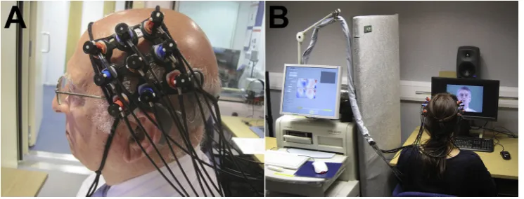
Near-infrared spectroscopy is a non-invasive optical neuro-imaging technique that offers a promising solution to the issues associated with performing brain imaging with CI recipients highlighted above (Lawler et al., 2015; Saliba et al., 2016; Sevy et al., 2010). As a functional neuroimaging technique, fNIRS is used to measure functional haemodynamic responses in both the infant and adult brain. Specifically, fNIRS is based on the principle of neurovascular coupling whereby an increase in neuronal activation and metabolic demand of the cells provokes an increase in local cerebral bloodflow (CBF) and oxygen delivery for consumption by the active cells (Villringer and Chance, 1997). This can be likened to fMRI that also measures cerebral haemodynamics, namely the blood oxygenated level-dependent (BOLD) signal, which has been shown to correlate well with the temporo-spatial characteristics of the fNIRS signal (Steinbrink et al., 2006; Toronov et al., 2007). In this way, fNIRS does not provide a direct measure of neuronal activa-tion: rather it measures the consequential haemodynamic response comprising stimulus-evoked changes in levels of oxygenated hae-moglobin (HbO) and deoxygenated haehae-moglobin (HbR) in the blood. In order to measure these changes in HbO and HbR con-centrations,fibre-optic bundles named optodes are placed in con-tact with the subject's scalp (Fig. 1A) and cortical measurements are acquired throughout the stimulus presentation paradigm (Fig. 1B). For more comprehensive information regarding the principles, available analysis techniques, and technological developments in fNIRS, which is outside the scope of the current review, we refer the reader to some key existing reports in the literature (Boas et al., 2004; Ferrari and Quaresima, 2012; Huppert et al., 2009; Lloyd-Fox et al., 2010; Scholkmann et al., 2014; Tak and Ye, 2014).
well-established neuroimaging techniques (Lloyd-Fox et al., 2010). For instance, in comparison to methods such as EEG with a high temporal resolution, fNIRS is limited somewhat by the sluggish nature of the haemodynamic response. However, in comparison to the temporal resolution of fMRI, fNIRS has a higher sampling rate and can therefore acquire measurements more rapidly and provide a more comprehensive picture of the haemodynamic response (Huppert et al., 2006; Lloyd-Fox et al., 2010). The spatial sensitivity of fNIRS is limited by the depth penetration of light into the cortex (Toronov et al., 2007), as well as light absorption and scattering by the skull and the cerebrospinal fluid (Okada and Delpy, 2003). Furthermore, fNIRS does not permit imaging of the underlying brain anatomy, meaning that it is not possible to localise the precise anatomical origin of cortical activations, or to account for individual differences. However, ongoing technological advancements that address such drawbacks, including the use of 3D digitizers and spatial-registration software, now enable more accurate estimation of the anatomical source of cortical activation measured using fNIRS and help to reduce variability across subjects (Singh et al., 2005; Tak and Ye, 2014; Tsuzuki et al., 2012; Tsuzuki and Dan, 2014).
In addition to these temporo-spatial characteristics, fNIRS offers several pertinent advantages for use in both adult and paediatric populations. Firstly, fNIRS is intrinsically quiet, making it particu-larly adept and ecologically valid when studying auditory function. Secondly, by its non-invasive and portable nature, fNIRS requires the lowest level of subject tolerance compared to alternative neu-roimaging techniques such as PET and fMRI as the injection of a radioactive tracer and horizontal placement into a constrained scanning environment is not required. The technique also offers a relatively high level of resilience to subjects' head and body movements (Piper et al., 2014). Thus fNIRS offers the opportunity for safe andflexible repeated testing across a variety of subject populations.
As such, fNIRS has generated much interest in the field of developmental neuroscience due to its ability to measure brain activity in children and infants without the need for natural sleep or sedation. For instance fNIRS has been applied to the study of paediatric sensory function and perceptual development (Taga et al., 2003; Watanabe et al., 2013), language acquisition (Gervain et al., 2008; Homae et al., 2011), social-cognitive function (Csibra et al., 2004; Minagawa-Kawait et al., 2009; Southgate et al., 2014) and the assessment and early identification of neuro-developmental disorders (Lloyd-Fox et al., 2013; Negoro et al., 2010; Zhu et al., 2014). Moreover, these advantageous features of
fNIRS have enabled bed-side monitoring of cerebral haemody-namics and the assessment of clinical outcomes in a range offields including, but not limited to, psychiatry, oncology, and clinical neurology (Ehlis et al., 2014; Hirsch et al., 2009; Jean-Pierre, 2014; Obrig, 2014).
Pertinent to this review, it has been demonstrated that fNIRS is fully compatible with a CI.Sevy et al. (2010) provided the first report of the successful use of fNIRS in paediatric CI recipients. Using a four-channel fNIRS system, cortical activation within bilateral temporal brain regions was measured during presentation of speech stimuli comprising 20 s segments of vignettes from a children's story. The study demonstrated that fNIRS was able to detect significant levels of auditory speech-evoked activation in a group of paediatric CI recipients on the day of CI activation, and in another group with more than four months of CI use. Crucially, these auditory speech-evoked fNIRS recordings were free from any CI-generated artefacts that are known to contaminate EEG re-cordings. Overall,Sevy et al.'s (2010)findings indicated the suit-ability of fNIRS for examining speech-evoked cortical activation in paediatric CI users without the need to limit the stimulation paradigm or to employ aggressive artefact removal techniques that are often required by techniques such as EEG.
SinceSevy et al.'s (2010)report, fNIRS has been applied in adult CI users to study brain activity during low-level visual stimulation, as well as linguistic and non-linguistic auditory stimulation (Bisconti et al., 2015; Chen et al., 2015a; Olds et al., 2016). However, to date, little is known about the functional link between auditory speech-evoked cortical activation measured using fNIRS and auditory performance: we were only able to identify two recent studies that had sought to understand these relationships in normal-hearing adults (Pollonini et al., 2014) and adult CI users (Olds et al., 2016).Pollonini et al. (2014)examined whether fNIRS could detect differences in speech-evoked activation when normal-hearing adults listened to clear speech or to speech that was degraded in order to simulate listening through a cochlear implant. The results demonstrated that clear speech elicited a significantly larger area of temporal-lobe activation in both hemispheres compared to degraded speech. This study provides initial evidence to suggest that fNIRS can detect differences in cortical activations elicited by speech stimuli of varying levels of intelligibility in a group of normal-hearing adults.
[image:8.595.108.481.66.208.2]The study demonstrated that the extent of speech-evoked activa-tion within the lateral temporal lobe/STG in each hemisphere, expressed as an activation area ratio for normal to scrambled speech, was positively correlated with auditory word and sentence recognition scores (Olds et al., 2016). Although further work is imperative, thefindings to date are encouraging for the future use of fNIRS to objectively assess the quality of the speech signal delivered by the implant to auditory brain regions and for under-standing the neural substrates of variable CI outcome (Olds et al., 2016; Pollonini et al., 2014).
Emerging evidence should be highlighted when considering the practical applicability of fNIRS for assessing and monitoring cortical plasticity and audio-visual interactions both before and after im-plantation (Dewey and Hartley, 2015; Chen et al., 2015a; Wiggins and Hartley, 2015). These studies provide thefirst demonstrations of the ability of fNIRS to measure audio-visual interactions within the occipital cortex of hearing adults (Wiggins and Hartley, 2015), cross-modal responses within the temporal brain regions of deaf individuals (Dewey and Hartley, 2015), and cross-modal responses in both the occipital and temporal lobes of CI users (Chen et al., 2015a). Specifically,Dewey and Hartley (2015)reported enhanced cortical activation to a visual motion stimulus within the right temporal lobe of profoundly deaf individuals compared to hearing controls. This illustrates the feasibility of fNIRS for detecting cross-modal activation during deafness that has previously been reported in fMRI studies (Fine et al., 2005; Finney et al., 2001). In post-lingually deaf, unilaterally-implanted adults, Chen et al. (2015a) observed cross-modal activations in temporal and occipital brain regions using fNIRS. Chen and colleagues identified that a greater level of cross-modal activation to basic visual stimulation within the left auditory cortex (‘maladaptive reorganisation’), relative to the level of cross-modal activation to auditory speech within the left visual cortex (‘beneficial reorganisation’), was related to poorer auditory performance on a speech in noise task. The authors conclude that reorganisation within temporal and occipital brain regions may jointly influence auditory performance with a CI, and that an optimal balance between beneficial and maladaptive plas-ticity effects may be an important factor in determining CI outcome. Furthermore, the authors recognise the need for longitudinal studies in order to understand the impact of cortical plasticity that takes place both before and after cochlear implantation on CI outcome (Chen et al., 2015a).
Certainly, the current review suggests that longitudinal mea-surements acquired within the same individuals from pre-to post-implantation are vital if the relationship between cortical plasticity and CI outcome is to be fully elucidated. However, the measure-ments must be sufficiently reproducible in order to reliably attri-bute changes in cortical activation to the CI rehabilitation process. Existing studies that have examined the test-retest reliability of fNIRS measurements in adults during basic visual stimulation (Plichta et al., 2006), verbalfluency tasks (Kakimoto et al., 2009; Schecklmann et al., 2008), and during auditory and visual social stimulation in infants (Blasi et al., 2014), have reported good reproducibility of fNIRS measurements across test sessions when responses are assessed at the group level and averaged over a small number of channels overlying a cortical region of interest.
Wiggins et al. (2016) have more recently provided the first known assessment of the test-retest reliability of speech-evoked, temporal-lobe fNIRS responses in normal-hearing adults. The re-sults showed that highly reproducible temporal-lobe responses to auditory speech (with or without corresponding visual speech cues) could be obtained at the group level over a retest interval of three months, so long as suitable signal processing was used to reduce the influence of systemic physiological interference. These findings indicate that fNIRS is potentially well-suited for the
longitudinal study of speech-evoked auditory responses in adults. However, it should be noted that the reliability of fNIRS measure-ments at an individual level has typically been shown to be highly variable between people (Blasi et al., 2014; Kakimoto et al., 2009; Plichta et al., 2006; Schecklmann et al., 2008; Wiggins et al., 2016). As discussed by Plichta et al. (2006), reasons for limited reliability at the single-subject level remain largely unknown. Po-tential limiting factors could include inconsistency across in-dividuals and across testing sessions in the positioning of optodes in relation to surface landmarks (Plichta et al., 2006). Anatomical factors such as differences in scalp and skull thickness, as well as individual variability in head size, may also contribute to the vari-ance at a single-subject level (Huppert et al., 2006).
With the aforementioned in mind, it is important to consider that the applicability of fNIRS in assessing cortical plasticity in in-dividual CI recipients could potentially be limited by various fac-tors. Possible practical limitations include variability across individuals in the surgical placement of the CI receiver. For instance, in cases where the device is placed in a more anterior position, it is possible that fNIRS measurement acquisition from posterior temporal regions of interest could be precluded by the overlap of optodes with the external component of the device. However, standardisation of CI placement across surgeons within the same CI clinics likely limits this issue. Furthermore, light attenuation may be greater for individuals with darker and thicker hair, which could result in inadequate levels of light intensity being transmitted and received between source-detector pairs. The development of hair-penetrating brush optodes is anticipated to help improve this situation in the future (Khan et al., 2012). These potentially limiting factors should therefore be taken into account when considering the applicability of fNIRS as a clinical tool for monitoring individual outcomes after implantation.
9. Conclusions
Although cochlear implantation can provide significant im-provements in speech understanding to profoundly deaf in-dividuals, the success rate is highly variable and remains largely unpredictable (Lazard et al., 2010a, 2012a). Various clinical char-acteristics are known to influence CI outcome, yet from these fac-tors alone we are currently not able to reliably predict how well an individual will perform with a CI (Blamey et al., 2013; Lazard et al., 2012a; Summerfield and Marshall, 1995). A better understanding of the factors and mechanisms underlying variability in CI outcome is of clinical importance as sensitive prognostic tools are needed to help more accurately predict clinical outcomes of individual cochlear implant recipients in order to set and counsel their ex-pectations most effectively. A growing body of evidence suggests that cortical plasticity within temporal and temporo-occipital brain regions could be an important factor in understanding and pre-dicting how much benefit an individual will receive from their CI (Buckley and Tobey, 2011; Chen et al., 2015a; Doucet et al., 2006; Giraud et al., 2001a, 2001b, 2001c; Sandmann et al., 2012; Strelnikov et al., 2013, 2015). From the available evidence, we suggest that a distinction may need to be made between possible facilitative and maladaptive plasticity effects within the left and the right temporal cortices, although this is not currently fully under-stood. Furthermore, a synergistic relationship between the auditory and visual modality and temporo-occipital interactions have been suggested as facilitative mechanisms during the restoring of audi-tory function in CI users (Giraud et al., 2001b, 2001c; Strelnikov et al., 2013, 2015).
repeated cortical imaging in CI recipients proves challenging: well-established neuroimaging techniques, including fMRI, EEG, MEG and PET, are all either limited in their compatibility with CI devices or are generally not well suited to longitudinal research for safety reasons (Gilley et al., 2006; Giraud et al., 2001b; Kim et al., 2015). Unlike these techniques, fNIRS is unaffected by CI-generated arte-facts (Sevy et al., 2010) and is well-suited to unrestricted longitu-dinal functional imaging in CI users, whatever their age (Lawler et al., 2015; Saliba et al., 2016; Sevy et al., 2010). Recent reports also attest to the exciting opportunity that fNIRS provides for the study of cortical function and plasticity in deaf adults and CI users (Chen et al., 2015a; Dewey and Hartley, 2015; Olds et al., 2016), as well as audio-visual interactions in adults (Wiggins and Hartley, 2015) and infants (Watanabe et al., 2013). Therefore, in conjunc-tion with behavioural measures of speech percepconjunc-tion, fNIRS offers a powerful tool to identify cortical predictors and correlates of vari-able CI outcome.
Acknowledgements
C.A.A is funded by the National Institute for Health Research (NIHR) Biomedical Research Unit Program, and was also funded by Cochlear Europe Ltd. during the writing of this article. The views expressed are those of the authors and not necessarily those of the NHS, the NIHR, or the Department of Health. The authors thank Dr Ian Wiggins for his comments on earlier versions of the manuscript. The authors also thank the two anonymous reviewers for their helpful comments and suggestions.
References
Allman, B.L., Keniston, L.P., Meredith, M.A., 2009. Adult deafness induces somato-sensory conversion of ferret auditory cortex. Proc. Natl. Acad. Sci. 106, 5925e5930.
Andersson, U., Lyxell, B., R€onnberg, J., Spens, K.-E., 2001. Cognitive correlates of visual speech understanding in hearing-impaired individuals. J. Deaf Stud. Deaf Educ. 6, 103e116.
Aschendorff, A., Kromeier, J., Klenzner, T., Laszig, R., 2007. Quality control after insertion of the nucleus contour and contour advance electrode in adults. Ear Hear 28, 75Se79S.
Auer Jr., E.T., Bernstein, L.E., Sungkarat, W., Singh, M., 2007. Vibrotactile activation of the auditory cortices in deaf versus hearing adults. Neuroreport 18, 645. Auer, E.T., Bernstein, L.E., 2007. Enhanced visual speech perception in individuals
with early-onset hearing impairment. J. Speech Lang. Hear. Res. 50, 1157e1165.
Bergeson, T.R., Pisoni, D.B., Davis, R.A.O., 2005. Development of audiovisual comprehension skills in prelingually deaf children with cochlear implants. Ear Hear 26, 149e164.
Bernstein, L.E., Auer, E.T., Moore, J.K., Ponton, C.W., D, M., Singh, M., 2002. Visual speech perception without primary auditory cortex activation. NeuroReport 13, 311e315.
Bernstein, L.E., Tucker, P.E., Demorest, M.E., 2000. Speech perception without hearing. Percept. Psychophys. 62, 233e252.
Bisconti, S., Shulkin, M., Hu, X., Basura, G.J., Kileny, P.R., Kovelman, I., 2016. Func-tional near-infrared spectroscopy brain imaging investigation of phonological awareness and passage comprehension abilities in adult recipients of cochlear implants. J. Speech Lang. Hear. Res. 59, 239e253.
Blamey, P., Artieres, F., Baskent, D., Bergeron, F., Beynon, A., Burke, E., Dillier, N., Dowell, R., Fraysse, B., Gallego, S., Govaerts, P.J., Green, K., Huber, A.M., Kleine-Punte, A., Maat, B., Marx, M., Mawman, D., Mosnier, I., O'Connor, A.F., O'Leary, S., Rousset, A., Schauwers, K., Skarzynski, H., Skarzynski, P.H., Sterkers, O., Terranti, A., Truy, E., Van de Heyning, P., Venail, F., Vincent, C., Lazard, D.S., 2013. Factors affecting auditory performance of postlinguistically deaf adults using cochlear implants: an update with 2251 patients. Audiol. Neurootol. 18, 36e47.
Blamey, P.J., Maat, B., Baskent, D., Mawman, D., Burke, E., Dillier, N., Beynon, A., Kleine-Punte, A., Govaerts, P.J., Skarzynski, P.H., 2015. A retrospective multi-center study comparing speech perception outcomes for bilateral implantation and bimodal rehabilitation. Ear Hear 36, 408e416.
Blamey, P.J., Pyman, B.C., Clark, G.M., Dowell, R.C., Gordon, M., Brown, A.M., Hollow, R.D., 1992. Factors predicting postoperative sentence scores in post-linguistically deaf adult cochlear implant patients. Ann. Otol. Rhinol. Laryngol. 101, 342e348.
Blasi, A., Lloyd-Fox, S., Johnson, M.H., Elwell, C., 2014. Testeretest reliability of
functional near infrared spectroscopy in infants. Neurow 1, 025005e025005.
Boas, D.A., Dale, A.M., Franceschini, M.A., 2004. Diffuse optical imaging of brain activation: approaches to optimizing image sensitivity, resolution, and
accuracy. NeuroImage 23, S275eS288.
Buckley, K.A., Tobey, E.A., 2011. Cross-modal plasticity and speech perception in pre-and postlingually deaf cochlear implant users. Ear Hear 32, 2e15.
Calvert, G.A., Bullmore, E.T., Brammer, M.J., Campbell, R., Williams, S.C.R., McGuire, P.K., Woodruff, P.W.R., Iversen, S.D., David, A.S., 1997. Activation of auditory cortex during silent lipreading. Science 276, 593e596.
Calvert, G.A., Campbell, R., 2003. Reading speech from still and moving faces: the neural substrates of visible speech. J. Cognit. Neurosci. 15, 57e70.
Campbell, J., Sharma, A., 2013. Compensatory changes in cortical resource allocation in adults with hearing loss. Front. Syst. Neurosci. 7, 1e9.
Campbell, J., Sharma, A., 2014. Cross-modal re-organization in adults with early stage hearing loss. PLoS One 9, e90594.
Campbell, R., MacSweeney, M., Woll, B., 2014. Cochlear implantation (CI) for pre-lingual deafness: the relevance of studies of brain organization and the role of first language acquisition in considering outcome success. Front. Hum. Neuro-sci. 8, 834.
Capek, C.M., Macsweeney, M., Woll, B., Waters, D., McGuire, P.K., David, A.S., Brammer, M.J., Campbell, R., 2008. Cortical circuits for silent speechreading in deaf and hearing people. Neuropsychologia 46, 1233e1241.
Capek, C.M., Woll, B., MacSweeney, M., Waters, D., McGuire, P.K., David, A.S., Brammer, M.J., Campbell, R., 2010. Superior temporal activation as a function of linguistic knowledge: insights from deaf native signers who speechread. Brain Lang. 112, 129e134.
Cardin, V., Orfanidou, E., Ronnberg, J., Capek, C.M., Rudner, M., Woll, B., 2013. Dissociating cognitive and sensory neural plasticity in human superior tem-poral cortex. Nat. Commun. 4, 1473.
Chen, L.-C., Sandmann, P., Thorne, J.D., Bleichner, M.G., Debener, S., 2015a. Cross-modal functional reorganization of visual and auditory cortex in adult cochlear implant users identified with fNIRS. Neural Plast. 2015.
Chen, L.-C., Sandmann, P., Thorne, J.D., Herrmann, C.S., Debener, S., 2015b. Associ-ation of concurrent fNIRS and EEG signatures in response to auditory and visual stimuli. Brain Topogr. 1e16.
Cooper, H., 2006. Selection criteria and prediction of outcomes. In: Cooper, H., Craddock, L. (Eds.), Cochlear Implants: a Practical Guide, second ed. John Wiley &Sons, Chichester, England, pp. 132e150.
Corina, D.P., 1999. On the nature of left hemisphere specialization for signed lan-guage. Brain Lang. 69, 230e240.
Crane, B.T., Gottschalk, B., Kraut, M., Aygun, N., Niparko, J.K., 2010. Magnetic reso-nance imaging at 1.5 T After cochlear implantation. Otol. Neurotol. 31, 1215e1220.
Csibra, G., Henty, J., Volein,A., Elwell, C., Tucker, L., Meek, J., Johnson, M.H., 2004. Near infrared spectroscopy reveals neural activation during face perception in infants and adults. J. Pediatr. Neurol. 2, 85e89.
Damen, G.W.J.A., Beynon, A.J., Krabbe, P.F.M., Mulder, J.J.S., Mylanus, E.A.M., 2007. Cochlear implantation and quality of life in postlingually deaf adults: long-term follow-up. Otolaryngol. Head Neck Surg. 136, 597e604.
Debener, S., Hine, J., Bleeck, S., Eyles, J., 2008. Source localization of auditory evoked potentials after cochlear implantation. Psychophysiology 45, 20e24.
Desai, S., Stickney, G., Zeng, F.G., 2007. Auditory-visual speech perception in normal-hearing and cochlear-implant listeners. J. Acoust. Soc. Am. 123, 428e440.
Dewey, R.S., Hartley, D.E.H., 2015. Cortical cross-modal plasticity following deafness measured using functional near-infrared spectroscopy. Hear Res. 325, 55e63.
Dormal, G., Collignon, O., 2011. Functional selectivity in sensory-deprived cortices. J. Neurophysiol. 105, 2627e2630.
Doucet, M.E., Bergeron, F., Lassonde, M., Ferron, P., Lepore, F., 2006. Cross-modal reorganization and speech perception in cochlear implant users. Brain 129, 3376e3383.
Eckert, M.A., Cute, S.L., Vaden, K.I., Kuchinsky, S.E., Dubno, J.R., 2012. Auditory cortex signs of age-related hearing loss. J. Assoc. Res. Otolaryngol. 13, 703e713.
Ehlis, A.-C., Schneider, S., Dresler, T., Fallgatter, A.J., 2014. Application of functional near-infrared spectroscopy in psychiatry. NeuroImage 85 (Part 1), 478e488.
Ellis, T., MacSweeney, M., Dodd, B., Campbell, R., 2001. TAS: a new test of adult speechreading-deaf people really can be better speechreaders. In: AVSP 2001-International Conference on Auditory-visual Speech Processing.
Ferrari, M., Quaresima, V., 2012. A brief review on the history of human functional near-infrared spectroscopy (fNIRS) development and fields of application. NeuroImage 63, 921e935.
Fine, I., Finney, E.M., Boynton, G.M., Dobkins, K.R., 2005. Comparing the effects of auditory deprivation and sign language within the auditory and visual cortex. J. Cognit. Neurosci. 17, 1621e1637.
Finley, C.C., Skinner, M.W., 2008. Role of electrode placement as a contributor to variability in cochlear implant outcomes. Otology& neurotology : official publication of the American Otological Society. Am. Neurotol. Soc. Eur. Acad. Otol. Neurotol. 29, 920e928.
Finney, E.M., Fine, I., Dobkins, K.R., 2001. Visual stimuli activate auditory cortex in the deaf. Nat. Neurosci. 4, 1171e1173.
Fujiki, N., Naito, Y., Hirano, S., Kojima, H., Shiomi, Y., Nishizawa, S., Konishi, J., Honjo, I., 1999. Correlation between rCBF and speech perception in cochlear implant users. Auris Nasus Larynx 26, 229e236.
Gantz, B.J., Woodworth, G.G., Knutson, J.F., Abbas, P.J., Tyler, R.S., 1993. Multivariate predictors of audiological success with multichannel cochlear implants. Ann. Otol. Rhinol. Laryngol. 102, 909e916.
Gervain, J., Macagno, F., Cogoi, S., Pena, M., Mehler, J., 2008. The neonate brain detects speech structure. Proc. Natl. Acad. Sci. U. S. A. 105, 14222e14227.