Redetermination of the crystal structure
of
b
-zinc molybdate from single-crystal
X-ray diffraction data
Olfa Mtioui-Sghaier,aRafael Mendoza-Meron˜o,aLilia Ktari,bMohamed Dammakband Santiago Garcı´a-Grandaa*
a
Departamento de Quı´mica Fı´sica y Analı´tica, Facultad de Quı´mica, Universidad de Oviedo-CINN, C/ Julia´n Claverı´a, 8, 33006 Oviedo, Spain, andbLaboratoire de Chimie Inorganique, Faculte´ des Sciences de Sfax, Route de Soukra, 3000 Sfax, Tunisia. *Correspondence e-mail: [email protected]
Received 8 May 2015; accepted 21 June 2015
Edited by M. Weil, Vienna University of Technology, Austria
The crystal structure of the-polymorph of ZnMoO4was re-determined on the basis of single-crystal X-ray diffraction data. In comparison with previous powder X-ray diffraction studies [Katikaneani & Arunachalam (2005). Eur. J. Inorg. Chem. pp. 3080–3087; Cavalcanteet al.(2013).Polyhedron,54, 13–25], all atoms were refined with anisotropic displacement parameters, leading to a higher precision with respect to bond lengths and angles. -ZnMoO4 adopts the wolframite structure type and is composed of distorted ZnO6and MoO6 octahedra, both with point group symmetry 2. The distortion of the octahedra is reflected by variation of bond lengths and angles from 2.002 (3)–2.274 (4) A˚ , 80.63 (11)–108.8 (2) for
equatorial and 158.4 (2)– 162.81 (14)for axial angles (ZnO6),
and of 1.769 (3)–2.171 (3) A˚ , 73.39 (16)–104.7 (2), 150.8 (2)– 164.89 (15)(MoO6), respectively. In the crystal structure, the
same type of MO6 octahedra share edges to built up zigzag chains extending parallel to [001]. The two types of chains are condensed by common vertices into a framework structure. The crystal structure can alternatively be described as derived from a distorted hexagonally closed packed arrangement of the O atoms, with Zn and Mo in half of the octahedral voids.
Keywords:crystal structure; redetermination;-ZnMoO4; hydrothermal synthesis; wolframite structure type.
CCDC reference:1408028
1. Related literature
Most molybdates of divalent cations crystallize either in the scheelite-type or in the wolframite-type (Macavei & Schulz,
1993). Zinc molybdate (ZnMoO4) is an inorganic semi-conductor. It adopts the wolframite-type of structure (Keeling, 1957) and is dimorphic. The two phases, referred to as -(triclinc symmetry) and - (monoclinic symmetry), can be selectively obtained by controlling the synthetic conditions (Abrahams et al., 1967; Zhang et al., 2010). Previous crystal structure refinements of-ZnMoO4, based on X-ray powder diffraction data, were reported by Cavalcanteet al.(2013) and Katikaneani & Arunachalam (2005). For structure refinement of ZnWO4, isotypic with the title compound, see: Trotset al.
(2009).
2. Experimental
2.1. Crystal data
ZnMoO4 Mr= 225.31 Monoclinic,P2=c a= 4.6980 (3) A˚ b= 5.7380 (4) A˚ c= 4.8960 (4) A˚ = 90.311 (7)
V= 131.98 (2) A˚3 Z= 2
MoKradiation = 13.62 mm 1 T= 293 K
0.080.060.03 mm
2.2. Data collection
Oxford Diffraction Xcalibur CCD diffractometer
Absorption correction: multi-scan (CrysAlis PRO; Oxford Diffraction, 2014) Tmin= 0.905,Tmax= 1.000
1207 measured reflections 405 independent reflections 358 reflections withI> 2(I) Rint= 0.036
2.3. Refinement
R[F2> 2(F2)] = 0.028 wR(F2) = 0.068 S= 1.10 405 reflections
29 parameters
max= 1.20 e A˚ 3
min= 1.17 e A˚ 3
Data collection: CrysAlis CCD (Oxford Diffraction, 2014); cell refinement: CrysAlis RED (Oxford Diffraction, 2014); data reduc-tion: CrysAlis RED; program(s) used to solve structure: SIR2011
(Burla et al., 2012); program(s) used to refine structure:
SHELXL2014 (Sheldrick, 2015); molecular graphics: DIAMOND
(Brandenburg & Putz, 1999); software used to prepare material for publication:WinGX(Farrugia, 2012), publCIF(Westrip, 2010) and
PARST(Nardelli, 1995).
Acknowledgements
We acknowledge financial support from the Spanish Minis-terio de Economı´a y Competitividad (MAT2013–40950-R), Gobierno del Principado de Asturias (GRUPIN14–060) and ERDF.
Supporting information for this paper is available from the IUCr electronic archives (Reference: WM5159).
data reports
i6
Mtioui-Sghaieret al. doi:10.1107/S205698901501186X Acta Cryst.(2015).E71, i6–i7References
Abrahams, S. C. (1967).J. Chem. Phys.46, 2052–2063.
Brandenburg, K. & Putz, H. (1999).DIAMOND. Crystal Impact GbR, Bonn, Allemagne.
Burla, M. C., Caliandro, R., Camalli, M., Carrozzini, B., Cascarano, G. L., Giacovazzo, C., Mallamo, M., Mazzone, A., Polidori, G. & Spagna, R. (2012).J. Appl. Cryst.45, 357–361.
Cavalcante, L. S., Moraes, E., Almeida, M. A. P., Dalmaschio, C. J., Batista, N. C., Varela, J. A., Longo, E., Siu Li, M., Andre´s, J. & Beltra´n, A. (2013). Polyhedron,54, 13–25.
Farrugia, L. J. (2012).J. Appl. Cryst.45, 849–854.
Katikaneani, P. & Arunachalam, R. (2005).Eur. J. Inorg. Chem.pp. 3080– 3087.
Keeling, R. O. (1957).Acta Cryst.10, 209–213.
Macavei, J. & Schulz, H. (1993).Z. Kristallogr.207, 193–208. Nardelli, M. (1995).J. Appl. Cryst.28, 659.
Oxford Diffraction (2014).CrysAlis PRO,CrysAlis CCDandCrysAlis RED. Oxford Diffraction Ltd., Abingdon, England.
Sheldrick, G. M. (2015).Acta Cryst.C71, 3–8.
Trots, D. M., Senyshyn, A., Vasylechko, L., Niewa, R., Vad, T., Mikhailik, V. B. & Kraus, H. (2009).J. Phys. Condens. Matter,21, 325402.
Westrip, S. P. (2010).J. Appl. Cryst.43, 920–925.
supporting information
sup-1
Acta Cryst. (2015). E71, i6–i7
supporting information
Acta Cryst. (2015). E71, i6–i7 [doi:10.1107/S205698901501186X]
Redetermination of the crystal structure of
β
-zinc molybdate from single-crystal
X-ray diffraction data
Olfa Mtioui-Sghaier, Rafael Mendoza-Mero
ñ
o, Lilia Ktari, Mohamed Dammak and Santiago
Garc
í
a-Granda
S1. Synthesis and crystallization
Reagents were used as commercial sources with no further purification. An aqueous solution was prepared by a mixture
of 0.047 g 2,2′-bipyridine, 0.015 g of molybdenum trioxide and 0.043 g of zinc acetate in 10 ml water. The reaction
mixture was stirred at room temperature to homogeneity, then transferred into a teflon-lined stainless steel vessel (40 ml)
and heated to 453 K for 48 h under autogenous pressure and after-wards cooled slowly to room temperature. The
resulting material was obtained as colorless single-crystals without side products. The solid was filtered off, washed
thoroughly with distilled water, and finally air-dried at room temperature.
S2. Refinement
The remaining maximum and minimum electron densities were found 0.77 Å and 0.90 Å, respectively, from the O1 atom.
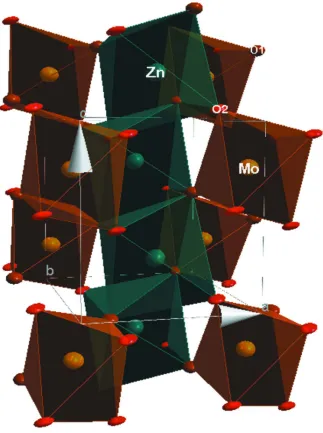
Figure 1
A view of the crystal structure of β-ZnMoO4. Anisotropic displacement parameters are drawn at the 50% probability
level.
β-Zinc molybdate
Crystal data
MoO4Zn
Mr = 225.31
Monoclinic, P2/c
a = 4.6980 (3) Å
b = 5.7380 (4) Å
c = 4.8960 (4) Å
β = 90.311 (7)°
V = 131.98 (2) Å3
Z = 2
F(000) = 208
Dx = 5.670 Mg m−3
Mo Kα radiation, λ = 0.71073 Å
Cell parameters from 570 reflections
θ = 3.6–31.1°
µ = 13.62 mm−1
T = 293 K
supporting information
sup-3
Acta Cryst. (2015). E71, i6–i7 Data collection
Oxford Diffraction Xcalibur CCD diffractometer
Radiation source: Enhance (Mo) X-ray Source
Detector resolution: 10.2673 pixels mm-1
ω– and φ–scans
Absorption correction: multi-scan
(CrysAlis PRO; Oxford Diffraction, 2014)
Tmin = 0.905, Tmax = 1.000
1207 measured reflections 405 independent reflections 358 reflections with I > 2σ(I)
Rint = 0.036
θmax = 31.3°, θmin = 3.6°
h = −6→6
k = −8→8
l = −6→6
Refinement
Refinement on F2
Least-squares matrix: full
R[F2 > 2σ(F2)] = 0.028
wR(F2) = 0.068
S = 1.10
405 reflections 29 parameters
0 restraints
w = 1/[σ2(F
o2) + (0.0209P)2 + 0.5403P]
where P = (Fo2 + 2Fc2)/3
(Δ/σ)max < 0.001
Δρmax = 1.20 e Å−3
Δρmin = −1.17 e Å−3
Special details
Geometry. All e.s.d.'s (except the e.s.d. in the dihedral angle between two l.s. planes) are estimated using the full covariance matrix. The cell e.s.d.'s are taken into account individually in the estimation of e.s.d.'s in distances, angles and torsion angles; correlations between e.s.d.'s in cell parameters are only used when they are defined by crystal symmetry. An approximate (isotropic) treatment of cell e.s.d.'s is used for estimating e.s.d.'s involving l.s. planes.
Fractional atomic coordinates and isotropic or equivalent isotropic displacement parameters (Å2)
x y z Uiso*/Ueq
Mo1 1.0000 0.81190 (10) 0.2500 0.00507 (18)
Zn1 1.5000 0.69182 (15) 0.7500 0.0092 (2)
O1 1.2538 (7) 0.6236 (6) 0.4014 (7) 0.0080 (7)
O2 0.7835 (7) 0.8950 (6) 0.5603 (7) 0.0058 (7)
Atomic displacement parameters (Å2)
U11 U22 U33 U12 U13 U23
Mo1 0.0065 (3) 0.0045 (3) 0.0041 (3) 0.000 −0.0002 (2) 0.000
Zn1 0.0087 (4) 0.0116 (4) 0.0073 (4) 0.000 0.0009 (3) 0.000
O1 0.0089 (17) 0.0110 (16) 0.0041 (17) 0.0006 (14) 0.0003 (13) −0.0004 (14)
O2 0.0088 (16) 0.0064 (14) 0.0021 (16) −0.0005 (13) 0.0016 (12) −0.0010 (13)
Geometric parameters (Å, º)
Mo1—O1i 1.769 (3) Zn1—O2iv 2.002 (3)
Mo1—O1 1.769 (3) Zn1—O2v 2.002 (3)
Mo1—O2 1.894 (3) Zn1—O1vi 2.094 (3)
Mo1—O2i 1.894 (3) Zn1—O1 2.094 (3)
Mo1—O2ii 2.171 (3) Zn1—O1vii 2.274 (4)
Mo1—O2iii 2.171 (3) Zn1—O1viii 2.274 (4)
O1i—Mo1—O2 97.25 (15) O2v—Zn1—O1 96.96 (14)
O1—Mo1—O2 100.46 (15) O1vi—Zn1—O1 158.4 (2)
O1i—Mo1—O2i 100.46 (15) O2iv—Zn1—O1vii 162.81 (14)
O1—Mo1—O2i 97.25 (15) O2v—Zn1—O1vii 88.37 (13)
O2—Mo1—O2i 150.8 (2) O1vi—Zn1—O1vii 82.25 (14)
O1i—Mo1—O2ii 164.89 (14) O1—Zn1—O1vii 80.63 (11)
O1—Mo1—O2ii 88.90 (14) O2iv—Zn1—O1viii 88.37 (13)
O2—Mo1—O2ii 73.39 (16) O2v—Zn1—O1viii 162.81 (14)
O2i—Mo1—O2ii 84.01 (11) O1vi—Zn1—O1viii 80.63 (11)
O1i—Mo1—O2iii 88.90 (14) O1—Zn1—O1viii 82.24 (14)
O1—Mo1—O2iii 164.89 (15) O1vii—Zn1—O1viii 74.53 (18)
O2—Mo1—O2iii 84.01 (11) Mo1—O1—Zn1 126.54 (19)
O2i—Mo1—O2iii 73.39 (16) Mo1—O1—Zn1viii 133.83 (18)
O2ii—Mo1—O2iii 78.49 (18) Zn1—O1—Zn1viii 97.75 (14)
O2iv—Zn1—O2v 108.8 (2) Mo1—O2—Zn1ix 125.93 (18)
O2iv—Zn1—O1vi 96.96 (14) Mo1—O2—Mo1ii 106.61 (16)
O2v—Zn1—O1vi 95.54 (14) Zn1ix—O2—Mo1ii 124.32 (17)
Symmetry codes: (i) −x+2, y, −z+1/2; (ii) −x+2, −y+2, −z+1; (iii) x, −y+2, z−1/2; (iv) x+1, y, z; (v) −x+2, y, −z+3/2; (vi) −x+3, y, −z+3/2; (vii) x, −y+1,