STRUCTURE AND FUNCTION IN THE NEMATODES:
INTERNAL PRESSURE AND CUTICULAR
STRUCTURE IN ASCARIS
BY J. E. HARRIS AND H. D. CROFTON
Department of Zoology, University of Bristol
(Received 14 November 1956)
No one who has made even a casual study of the Nematoda can fail to have been struck by the extraordinary similarity in form and organization between the very large number of species, genera, orders and families which constitute this class of the very diversiform phylum Aschelminthea. Though there are species which show some variation from the normal 'nematode fades', they are few in number and merely seem to emphasize still further the great uniformity of the group as a whole. The characteristic features of the nematodes are present in fresh-water, marine and parasitic forms; they are largely independent of size, of diet and of stage of development.
This uniformity can hardly be ascribed to simplicity of organization. Well below the level of complexity of the nematodes, the protozoa, sponges and coelenterates show a rich variety of adult forms and of larval stages within comparatively restricted taxonomic or ecological groupings. The platyhelminths, otherwise similar in their degree of organization so far as the presence of specialized tissues and organs are concerned, show a far greater diversity of form and structure; the elementary student may be forgiven at times for thinking that there is only one nematode but that the model comes in different sizes and with a great variety of life histories.
Among the factors which act as powerful determinants of organic form and structure, we owe to D'Arcy Thompson the idea that physical and perhaps more particularly mechanical factors are highly significant. Even in man-made struc-tures this is equally true. A bridge, a ship, an aeroplane are recognizable at once because their design is based necessarily and largely on the mechanical forces which play such a predominant part in their economy; the decorative and even to some extent the functional variations which may be added to the structure are severely limited by the need to satisfy the rigorous requirements of mechanical strength and efficiency. Is it in such limitations that we may find the key to the underlying uni-formity in the nematodes?
functioning of any system of muscles must provide for their contraction against a suitable antagonistic force, in the absence of circular musculature there must be an elastic return mechanism which could be provided by an internal hydrostatic pressure. The extent to which this system will be a determining factor in the organization of the nematode body will clearly depend on the magnitude and dis-tribution of the forces involved.
The present paper, the first of a series in which the general biology of the nematode body is considered in mechanical terms, is concerned with the determina-tion of the internal turgor pressure, its reladetermina-tionship to the structure of the cuticle and to the force exerted by the longitudinal musculature.
MATERIAL AND METHODS
The present studies have been made on adult female specimens of Ascaris lumbri-coides obtained from freshly killed pigs from a Bristol slaughterhouse. The worms were brought to the laboratory in a limited volume of pig intestinal fluid contained
in a previously warmed wide-mouthed vacuum flask. Experiments were carried out in the medium recommended by Hobson (1948)—30% sea water—in which the worms remain active for long periods. Not only do whole worms survive well in this artificial medium, but portions of worms such as those used in some of the additional experiments, reported below, survive and maintain their characteristic activity for several days. All experiments were, however, carried out as soon as possible after the arrival of a fresh batch of worms, usually within 6 hr. of obtaining the specimens.
Two methods of recording the turgor pressure have been employed—a direct method using a pressure gauge connected with a cannula inserted into the body cavity, and an indirect method based on the distortion of the body wall when external pressure is applied.
The open end was connected through one arm of a T-tube by a continuous water column to the source of pressure to be measured; all connexions were made with glass tubes jointed with cement or sealing wax to reduce changes in internal volume of the instrument to a minimum. It was found convenient to cement the base of a hypodermic syringe on to the pressure tip so that various types of cannula modified from ordinary hypodermic needles could readily be attached and replaced. The second arm of the T-tube led through a simple tap to a vertical water column providing a pressure head. This was used to flush out the cannula or to indicate if it was blocked, and also, when the opening of the cannula was sealed off, to calibrate the gauge.
Fig. i. The use of the glass helix pressure gauge. Inset (lower right), an enlarged view of the cannula. The arrow points to the lateral opening.
To the closed end of the helix was fixed a light black glass pointer, some 15 cm. in length, which moved over a millimetre scale. Calibration showed that the move-ment was linearly related to the pressure over a wide range, a deflexion of 10 mm. corresponding to a pressure of 55 mm. of mercury. The natural period of oscilla-tion of the system was less than 0-5 sec. and the response was quite sufficiently rapid for the preparations used; reasonable shielding of the needle from stray air currents was all that was required.
Considerable trouble was encountered in finding a suitable form of cannula, which required to be sharp in order to penetrate the tough cuticle of the animal, but steeply tapered in order to seal the puncture successfully. Best results were obtained by soldering a small blob of metal around the base of a hypodermic needle which was then ground down to a length of 1-2 mm. The metal blob was then trimmed to a conical shape with an apical angle of about 450. The hole in the tip was at the side, to reduce the chance of blocking during insertion, and was about 0-2 mm. in diameter.
A slightly wider vertical hole was drilled in the top of the block to meet the hori-zontal channel, and gave access to the tip of the pressure cannula. The block was carried on the arm of a simple dissecting microscope, whose rackwork served to raise and lower it as required. The block, its support and the syringe tip of the pressure apparatus were immersed in a bath containing 30% sea water thermo-statically controlled at 38-390 C. (see Fig. 1).
[image:4.451.91.363.274.389.2]Continuous recording of the changes in pressure was made possible by a simple slit camera. A multi-speed Palmer kymograph was mounted with the shaft hori-zontal. The shaft carried a 12 in. drum enclosed in a light-tight box; in the roof of the box was a slit, \ mm. in width. An image of the needle of the pressure gauge was thrown on the slit by a 6 in. photographic lens used at a magnification of (x 2); illumination was provided by a strip light about 50 cm. above the needle. Time marking was accomplished by a 6 V. flash-lamp bulb, mounted 50 cm. above the slit, and switched on at half-minute intervals by a time clock.
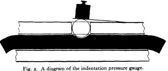
Fig. 2. A diagram of the indentation pressure gauge.
120 J. E. HARRIS AND H. D. CROFTON
calibration curve was not linear, but the method provided a simple and rapid indica-tion of the approximate extent of the internal pressure changes in an intact and active worm.
RESULTS
(i) Measurement of turgor pressure
Whichever form of pressure gauge was used, its successful functioning could readily be tested by applying finger pressure to the body of the worm. The immediate and reversible response which was obtained (except when the needle was blocked) showed that there was normally free transmission of the internal pressure through-out the body. It would appear that the fluid-filled spaces in the pseudocoelomic cavity are in open communication. Occasionally, particularly when the worm had been threaded through a narrow tube, there was clear evidence of a block in trans-mission of pressure across the constricted region, but this has not been encountered in an unconstricted worm. The recorded pressures may therefore be taken to be the true internal turgor pressure.
Table i . Pressures measured by the glass helix gauge in adult female Ascaris
Wonn A B C D E F G H I Notes Fresh worm
Three successive teats: tail region tail region
vulval region (varying) Three successive tests (maximum values)
Same worm cooling down
Ditto with warm saline applied to head region (two tests)
Three successive tests with little variation
An active worm showing great variations in pressure (max. and min. in four tests)
Worm kept in saline 4 hr. after collection Ditto, another specimen
Ditto, 6 hr. after collection
Same worm immediately after a defaecation Worm 6 hr. after collection
Pressure
cm. of water
*35 45 45 37- 83 142 172 172 30- 37 83 75
6 7 - 8 3 67- 75 67 15-127 105—142 37-135 -150 6 0 52 52 2 2 37 mm. Hg ICO 33 33 27- 60 1 0 4 1 2 7 127 22- 27 6 0 55 50- 60 50- 55 5° 11- 94 77-104 27-100 - n o
44 38 38 16 27
The indentation pressure gauge in its present form is not suited to instantaneous determinations of internal pressure, though a modification is being designed for con-tinuous recording. It was used on intact worms by adding a constant weight and observing the proportion of the total time during which the internal pressure was high enough to raise the cover-glass above the Perspex base. Using the values for internal pressure obtained from the method of calibration described above, it was found that the pressure only rarely fell below 25 mm. Hg, but that for 37 % of the time it was above 65 mm. Values which were above 100 mm. were found to occur for a small portion of the total time of observation.
180
120
60
| °
180
120
[image:6.451.53.403.202.438.2]60
Fig. 3. Tracing from a photographic record of pressure changes made with a glass helix pressure gauge. The two halves of the record are consecutive. Time marks are at 30 sec. intervals.
The highest values which have been recorded in the present series of studies were obtained from a worm in which the head region was tightly coiled in front of the point of penetration of the cannula, and the tail was at the same time strongly contracted. For about 15 sec. the worm maintained a pressure greater than 180 mm. Hg, rising for a few seconds to a maximum of 225 mm. Hg (306 cm. of water), equivalent to nearly a third of an atmosphere.
These values are almost an order of magnitude higher than those obtained by Picken (1936) for Potamobius fluviatilis (10-5-18 mm. Hg), for Carcinus moenas (3-5—19 mm.), for Peripatopsis (2-15 mm.) and also than those obtained by Chap-man & Newell (1947) for Aremcola (10-20 mm., mean values of resting and maxi-mum pressure), by Chapman (1949) for CalUactis parasitica (0-10 mm.) and by Newell (1950) for Lumbricus terrestris (1-5-21 mm.).
122 J. E. HARRIS AND H. D. CROFTON
There is evidence of a rhythmical rise and fall in pressure at intervals of about 30 sec. in the early part of the record. The recorded pressures vary from 49 to 180 mm. Hg with a mean value of 95 mm., substantially similar to the values noted above.
Further confirmation of these high values for the internal pressure has been obtained from two other lines of experimental investigation.
The half worm, ligatured to a cannula placed in connexion with a water mano-meter, usually lay completely inert at low pressures—up to 15 mm. Hg. At 45 mm. occasional contractions were recorded, and from 60 to 100 mm. regular and con-tinuous bursts of rhythmical activity occurred at intervals of about 20 sec. Above 150 mm. the activity was irregular; the tail became extended in length, and at pressures of 200 mm. and above failed to show contractions. The pressure con-ditions in these experiments are of course not strictly comparable with those in the intact worm, since in the isolated halves contraction is isotonic, i.e. against a con-stant manometer pressure; as we shall see later, contraction in the intact worm is accompanied by a rise in pressure.
As a final check on these observations, if we assume that the pressure is generated by the tension exerted by the longitudinal body musculature we can calculate its value from the experimentally observed values for this tension. Miss C. E. Bradley, working in this laboratory, has found that isolated 'anterior preparations' of Ascaris as used by Baldwin (1943) will show rhythmical contractions against a load of 10—20 g., but fail to do so against loads of 25-30 g. Assuming that the cross-sectional area of the preparation is 15 sq.mm. a tension of 10-20 g. corresponds to an internal pressure of 75-150 mm. Hg, which is consistent with the observed values.
(2) The structure of the cuticle
There is a common impression that the cuticle of nematodes is stiff and unyield-ing. Hyman (1951, p. 399) says that' because of their stiff cuticle... the nematodes possess poor powers of locomotion'. Stauffer (1924), in his study on locomotion in the nematodes, says that the cuticle, in conjunction with the internal body pressure, helps to maintain a constant body length, and compares the system with the verte-bral column or notochord in a vertebrate.
In actual fact the cuticle of Ascaris is extensible and capable of much more extension than is perhaps generally realized. We have recorded changes in length (of half worms artificially inflated as well as of the corresponding portions of intact worms) of the order of 10-15 % for pressure differences within the normal physio-logical range; similar alterations can occur in the diameter.
In these changes in length and diameter of the body the fine structure of the cuticle appears to play an important part. Prominent among the structural com-ponents of the cuticle of Ascaris and of most nematodes is a set of diagonal crossed fibres, readily visible in fixed specimens. Three such fibre layers together form a spiral basketwork, incorrectly illustrated by Chitwood & Chitwood (1950).
seems reasonable to regard them as practically inextensible in the living animal. Picken et al. have commented on the value of this arrangement in permitting anisometric extension of the cuticle.
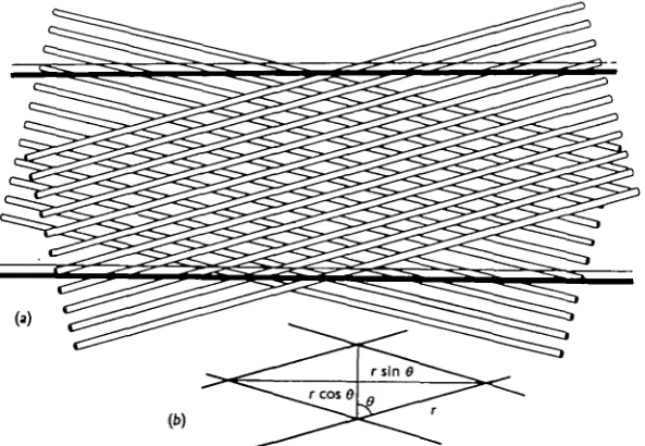
The two sets of fibres may be considered to enclose between them a system of minute parallelograms (Fig. 4) which, even though the lengths of their sides remain constant, may be distorted by shearing with a consequent change in length of their diagonals. As these diagonals lie, in relation to the nematode, longitudinally and transversely, the distortion of the structure will lead to the worm becoming longer and thinner or shorter and thicker—the changes in length and diameter will be strictly interrelated as in a set of 'lazy tongs'.
[image:8.451.79.376.213.418.2](b)
Fig. 4. A schematic representation of the basketwork structure of fibrils in the cuticle of Ascaris.
(a) General diagram illustrating the orientation of two of the fibril layers in relation to two of the
transverse external annulations. (b) The equivalent unit parallelogram.
In such a structure there will exist one critical shape of the unit parallelogram from which any small distortion will change the length, /, and diameter, a, of the worm in such a way that the volume (oc/a2) remains constant.
If r is the length of any side of the parallelogram and 8 is the acute angle between any side and the longitudinal axis of the worm, then
/ocrcosfl and accrsinO,
so that V, the volume, ocr3 cos 0 sin2 8. The condition that Kshall be unchanged by a small change in 8 is provided by dVjdd = o, i.e.
yx (cos 8 sina 6) = o, do
or
from which
124 J- E. HARRIS AND H. D. CROFTON
If the angle 6 is larger than this (i.e. if the fibres lie more nearly in the transverse plane), an increase in length will more than compensate for decrease in diameter so that the worm will increase in volume on extension; if 9 < 550 the reverse will be true and lengthening of the worm will decrease its volume.
Measurements on a number of fixed specimens gave a mean value for 9 of 750 30', rather greater than the 700 suggested by Picken et al. (1947), who quoted a (possibly approximate) figure. In any case it is clear that for such an angle, length and volume changes will be directly related; if the worm increases in volume it will do so by extending and growing thinner. As it swells in this way the angle 6 will decrease, but an extension of well over 50 % would be required before the critical angle of 550 was reached; this extension appears to be far beyond the capabilities of the normal animal.
It was possible to make an approximate test of this hypothetical behaviour of the cuticle by measuring the change in length for a given change in volume.
A normal worm was threaded through a rubber stopper, bored with a hole sufficiently small to prevent it moving through. The stopper was thrust into a tube (containing the tail end of the worm) which was equipped with a capillary side tube to serve as a dilatometer. A series of simultaneous measurements of tail length and tail volume was made over a period in which no fluid was passing through the gut, but the worm was showing periodic tail contractions. For nineteen such contrac-tions, the mean shortening was 8-72 mm. for a mean decrease in volume of 144-5 nun.3. The length of the tail portion was 11 cm. and the diameter (over most of this length) 5 mm. A simple calculation (based on the assumption that the worm is a cylinder) showed that for a decrease in volume of this amount the diameter must have become larger by 0-032 mm.
It can readily be shown that for a small change in length J T - = tan2 9, from which tan # = 3-323 and # = 73° 15'. The agreement with the observed value of 750 30' is astonishingly close, particularly when we take into account the slight shrinkage in length of the worms when they are fixed, which might well increase the value of 8 in the observations on fixed specimens.
It is worth noting that the basketwork structure suggested above does not in itself provide an elastic component; a framework of this type could, however, function as an elastic structure so long as (a) distortion changes its volume, i.e. 6 #= 550, and (b) it is equipped with suitable muscles. If 9 > 550 such muscle fibres must be longitudinal, while if #<55° the muscle fibres must be transverse or circular.
pressures recorded in Fig. 3 are in fact due entirely to the elastic force of the cuticle itself; increases above this resting level will then mark the activity of some part of the body musculature.
Such a system will clearly be capable of coming into static equilibrium over an appreciable range of body volume, thereby providing for the maintenance of the characteristic internal pressure (or the cycle of pressure changes) even if additional fluid is being taken into the gut, or after defaecation has released a certain volume of gut contents. Similarly, osmotic changes, which must be a normal feature of the environment of a gut parasite, can readily be dealt with if transfer of water can occur without seriously affecting the mechanical stability of the system.
In contrast to the state of affairs in a worm with an isotropic elastic cuticle, or in one in which circular and longitudinal muscle layers are present, there is in this arrangement a strict mathematical relationship between length and diameter; the internal volume will completely determine the length of the animal. At this length, the muscles can then generate pressures and provide the elasticity for the working of the system in a co-ordinated fashion. Any true elastic component of the cuticle itself will merely serve to limit the extent of increase of volume and length without otherwise affecting the argument.
A comparison of the situation in Ascaris with that described by Cowey (1952) in the nemertine shows an interesting and fundamental difference. So far as we have been able to discover, the internal hydrostatic pressure in an active Ascaris is always considerable; the cross-section of the body does not depart significantly from the circular shape and some part of the longitudinal musculature is always in a state of tonus. The angle between the fibrils and the longitudinal axis is therefore sensibly constant, as is the total length of the animal. This is a necessary consequence of the relatively free transmission of hydrostatic pressure throughout the pseudocoelomic cavity. Local shortenings in length, brought about by contraction of muscles in a particular region, are reflected in similar extensions in other parts against the elastic (or active) force exerted by the musculature in these distant regions; substantial variations in total length are found only when there are similar changes in total volume—during feeding and defaecation.
It must be realized that this picture of a basketwork with a theoretically uniform mesh over the whole of the animal is an oversimplification. It will be fairly true over a restricted length, but in a body as long as this it is clearly possible for the shape of the unit parallelogram to change gradually from one region to another. Local contraction of the tail, for example, will shorten that region by displacing fluid into the head end, where lengthening of the body will occur. The total length of the body will remain relatively unchanged, but only as a result of compensating changes in different regions of the animal. By such changes the movement of different parts of the body is largely co-ordinated, as we hope to show in a later paper; in this way a pattern of organized activity can be achieved in terms of responses which are largely local in character, i.e. which do not require an elaborate central nervous machinery of co-ordination.
by contraction of the muscles. The graph of Fig. 5 shows that, over a period in which internal pressure and tail length were simultaneously recorded, there was a clear correlation between tail shortening and increase of pressure. In a later paper, which will be concerned with feeding and volume transfer between the head and tail regions, we hope to publish one of our records which shows (among other facts irrelevant to this present account) that volume transfer follows upon the shortening of the tail with a slight delay. This indicates that the sequence is: muscle contraction ->tail shortening ->• decrease of tail volume and increase of pressure-> volume transfer->increase of head volume and pressure-*head lengthening.
o—oPositlon of tail tip
•—•Internal pressure
30
0 1 2 0 1 2 3
Time (min.)
Fig. 5. The internal pressure and the position of the tail tip plotted against time for two different worms. Each worm was fixed in the region of the vulva, so that changes in the position of the tail tip represent actual changes in the length of the whole post-vulval region.
DISCUSSION AND CONCLUSIONS
In the introduction it was suggested that physical and mechanical forces determined the general body structure of nematodes and that the limitations imposed by the particular mechanical system accounted for the uniformity of the group.
system of inextensible diagonal or spiral fibres, and the other is elastic. The body musculature consists entirely of longitudinal fibres; the only other muscles to be found are the so-called ' sphincters' at the anus and vulva, and the specially arranged muscles in the reproductive system and in the pharynx. It is to be noted that the ' sphincters' are almost invariably ' dilators' and that the only circular muscles to be found are in the ducts of the reproductive system.
The nervous system is simple, consisting of a cephalic and caudal concentration of nerve cells linked by a few longitudinal nerve cords containing few if any nerve cells. The gut consists of a powerful pumping triradiate pharynx and a simple straight intestine composed of a single layer of cells. The body cavity is a pseudo-coelom which is open and not divided serially into segments. The excretory system is basically a special system of internally closed intracellular ducts which open to the exterior. Cilia are absent; the spermatozoa are never truly flagellate and usually move in amoeboid fashion. The characteristic type of movement is by wave-like undulations of the body.
Many of the characteristics given above appear to be closely correlated with the mechanical features established by the work described in the present paper.
We have demonstrated the presence, in Ascaris, of a system in which the longi-tudinally arranged muscles act not against other antagonistic muscles but against forces exerted by the internal pressure on the cuticle. The success of this unique system depends on the presence of a spiral basketwork of inextensible fibres which permit anisometric extension and shortening of the cuticle. If body movements such as those concerned in locomotion are produced by these longitudinal muscles, it follows that the strength and efficiency of these movements can be increased only if the internal pressure of the body fluid increases also.
Such a system will be even more efficient if the cuticle contains, in addition to the main fibre network, a true elastic component. This component may be assumed to play a significant role when the meshwork is considerably extended by great elongation of the animal. Under such circumstances the elastic force will reinforce the action of the muscles, will therefore reduce the tension which must be maintained by them, and make the contraction of such muscles even more effective. This is true for the longitudinal elastic component; the circular elastic component has a corresponding role at low internal pressures, for it will, by resisting transverse expansion of the meshwork, maintain a slight longitudinal extension even when the internal pressure is negligible—keeping the muscle fibres in that state of slight stretch which is desirable for their effective action. The system is in fact designed to function not only at a high internal turgor pressure but over a wide range of pressures.
128 J. E. H A R R I S AND H. D. C R O F T O N
The possibility of co-ordinating the movement of different parts of the body by the transfer of fluid resulting from local changes in volume has already been referred to; with such mechanical co-ordination the need for local reflex networks does not exist. The simplicity of the nervous system is probably related to this feature.
Because of this possibility of local changes in length, the alimentary canal must be freely extensible. Such a tube will necessarily collapse under the internal pressure and can only be filled by some pumping mechanism capable of producing an even higher pressure. This is provided, as we shall show in a later paper, by the special structure of the triradiate pharynx; ciliary mechanisms would be completely in-capable of generating the pressures required even if the walls of the canal were in some way distended to allow them to beat freely.
The presence of dilator muscles, e.g. the so-called 'depressor ani', is also a con-comitant of the high internal pressure. The gut contents would escape, forced out by the internal pressure, if the pharynx and anus were not provided with 'self-sealing ' devices requiring muscular activity only to open them.
The excretory and reproductive systems show special features equally related to the internal pressure. In the common type of excretory system found in Ascaris the very long excretory tubes are embedded in the lateral chords and will conse-quently not be collapsed by the pressure; this will in fact provide effective filtration of the excretory product even against high internal colloid osmotic pressure. In the alternative ' renette-cell' type of excretory organ the duct is free in the body cavity but very short. Here it seems likely that the cell itself is the excretory organ and, like the mammalian salivary gland, may generate pressures in its duct higher than those in the surrounding medium.
The reproductive system is the only one in the body which possesses a peristaltic musculature and true sphincter muscles. Here, however, the mechanical conditions are markedly different from those in the gut or the excretory system. There is no controllable pump like the pharynx to fill the system, and the escape of eggs in the female which would take place with a simple vulval dilator muscle would need to be limited to those which were fertilized and equipped with their shells and egg mem-branes. Such controlled emission can best be produced by a combination of peristaltic movement—which pinches off a selected group of ova—and a sphincter muscle (derived possibly from the same circular and oblique fibres) which releases at the appropriate stage in the peristaltic cycle. Since the ova before fertilization are amorphous and tightly packed, amoeboid spermatozoa injected at high pressure by a special male copulatory apparatus will be necessary for successful fertilization—the absence of true flagella in the sperm may, however, also be connected with the absence of cilia elsewhere.
the muscles, it follows that in nematodes of widely different size, if the proportion of the cross-section occupied by muscles is similar, the internal pressure will also be the same. Furthermore, since the tangential stress in a cylinder subjected to internal pressure is proportional to the diameter, the thickness of the nematode cuticle required to withstand this stress can be proportional to the diameter of the animal. The mechanical factors which determine the general features of the nematode will thus be independent of the scale on which the model is built.
SUMMARY
1. Experimental determinations of the hydrostatic pressure in the pseudocoel of living Ascaris himbricoides were made by a direct method, using a glass helix pressure gauge and by an indirect method using an indentation gauge, both of which are described.
2. The mean value of this pressure was 70 mm. Hg (95 cm. of water), and showed wide and often rhythmical variations from 16 mm. to as high as 225 mm. Hg. Observations on the behaviour of artificially distended worms and of the tension developed by the muscles confirm these results.
3. The mechanical structure of the cuticle, with its inextensible spiral fibrils, forming a basketwork at an angle of 75° to the longitudinal axis, provides for an anisometric expansion and contraction under the action of the longitudinal muscles which is closely in accordance with the observed changes in volume and length.
4. A discussion of the significance of this mechanism and of the high internal pressure suggests that the great similarity of form among nematodes is determined to a considerable extent by mechanical factors.
We are glad to acknowledge the help of Mr M. Gillett in the construction of the apparatus described in the paper.
REFERENCES
BALDWIN, E. (1943). An in vitro method for the chemotherapeutic investigation of anthelminthic potency. Parasitology, 35, 89-1 n .
BAYLIS, H. A. (1924). The systematic position of the nematoda. Ann. Mag. Nat. Hist. ser. 9, 13,
CHAPMAN, G. (1949). The mechanism of opening and closing of Calliactis paratitica. J. Mar. Biol.
Ass. U.K. 38, 641-9.
CHAPMAN, G. & NEWELL, G. E. (1947). The role of the body fluid in relation to movement in soft-bodied invertebrates. I. The burrowing of Arenicola. Proc. Roy. Soc. B, 134, 431-45. CHITWOOD, B. G. & CHITWOOD, M. B. (1933). The characters of a protonematode. J. Parasit. 20,
13°-CHITWOOD, B. G. & 13°-CHITWOOD, M. B. (1950). An Introduction to Nematology, rev. ed. Baltimore (Md.)
COWEY, J. B. (1952). The structure and function of the basement membrane muscle system in
Amphiporus lactifloreus (Nemertea). Quart. J. Micr. Set. 93, 1.
GRAY, J. (1951). Undulating propulsion in small organisms. Nature, Land., 168, 929.
HOBSON, A. D. (1948). The physiology and cultivation in artificial media of nematodes parasitic in the alimentary tract of animals. Parasitology, 38, 183-227.
HYMAN, L. H. (195 I). The Invertebrates. III. Acanthocephala, Aschelminthes and Entoprocta. Pp. vii + 572. New York.
NEWELL, G. E. (1950). The role of the coelomic fluid in the movements of earthworms. J. Exp. Biol.
27, 110-21.
PICKEN, L. E. R. (1936). The mechanism of urine formation in invertebrates. I. The excretion mechanism in certain arthropods. J. Exp. Biol. 13, 309-28.
PICKEN, L. E. R., PRYOR, M. G. M. & SWANN, M. M. (1947). Orientation of fibrils in natural
membranes. Nature, Lond., 159, 434.
SEURAT, L. G. (1920). Histoire naturelle det nhnatodes de la Bcrberic. I. Morphologie, diveloppement,
ithologie et affinitit da nimatodes. 221 pp. Algiers.