Characterization and expression pattern analysis of the
facilitative glucose transporter 10 gene (slc2a10) in Danio rerio
NICOLA CHIARELLI, MARCO RITELLI, NICOLETTA ZOPPI, ANNA BENINI, GIUSEPPE BORSANI,
SERGIO BARLATI and MARINA COLOMBI*
Division of Biology and Genetics, Department of Biomedical Sciences and Biotechnology, Medical Faculty, University of Brescia, Brescia, Italy.
ABSTRACT The SLC2A10 gene located on chromosome 20q13.1 encodes the facilitative glucose transporter 10 (GLUT10), a class III member of the SLC2A facilitative glucose transporter family. Mutations in the human SLC2A10 gene cause arterial tortuosity syndrome (ATS), a rare autosomal recessive connective tissue disorder. In this work, we report the characterization of the slc2a10 ortholog gene in zebrafish (Danio rerio) and its expression pattern during embryonic development and in adult tissues. The slc2a10 gene consists of 5 exons, spanning 8 kb and mapping to a region on chromosome 11 that exhibits conserved synteny with human chromo-some 20. The gene encodes Glut10, a 513 amino acid protein that maintains the 12 transmembrane domain structure typical of the GLUTs family, and shares the specific functional motifs involved in sugar transport with the vertebrate GLUT10. RT-PCR analysis showed that two specific splice variants, both including the 5’-UTR region, were expressed during embryogenesis and in different adult zebrafish tissues and organs. In situ hybridization analyses demonstrated a maternal origin of the total slc2a10 mRNA and its ubiquitous distribution until the early somitogenesis stage. In later embryonic stages, slc2a10 mRNA was detected in the otic vesicles, hatching gland cells, pectoral fin, posterior tectum and swim bladder. Overall, these results suggest a wide role of slc2a10 during zebrafish development.
KEY WORDS:
slc2a10, Glut10, facilitative glucose transporter, arterial tortuosity syndrome
The transport of glucose and hexoses across the plasma mem-brane is mediated by a family of facilitative glucose transporters (SLC2A gene, GLUT protein). In the mammalian genome, 14 different GLUT isoforms with distinct kinetic properties of trans-port and tissue/cell-specific expression patterns have been de-scribed (Zhao and Keating, 2007). These sugar transporters are integral membrane proteins containing 12 transmembrane hydro-phobic segments, which are connected by 6 extracellular and 5 intracellular loops with both amino and carboxyl-termini exposed in the cytosol. On the basis of amino acid sequence similarities and transport characteristics, the mammalian GLUTs are grouped into three classes: class I, (GLUT1-4 and GLUT14), class II (GLUT5-7-9 and 11) and class III (GLUT6, 8, 10, 12 and the H+ -coupled myo-inositol transporter, HMIT) (Joost and Thorens, 2001; Joost et al., 2002; Uldry and Thorens, 2004). Human
BIOLOGY
www.intjdevbiol.com*Address correspondence to: Marina Colombi. Division of Biology and Genetics, Department of Biomedical Sciences and Biotechnology, University of Brescia, Viale Europa 11, 25123 Brescia, Italy. Fax: +39-030-370-1157. e-mail: [email protected]
web: http://www.unibs.it/on-line/med/Home/Docenti/scheda99.html
Supplementary Material (a Table) for this paper is available at: http://dx.doi.org/10.1387/ijdb.103179nc
Accepted: 15 February 2011. Final author corrected PDF published online: 31 March 2011.
ISSN: Online 1696-3547, Print 0214-6282
© 2011 UBC Press Printed in Spain
GLUT10 has been shown to facilitate D-glucose, D-galactose and 2-deoxy-D-glucose uptake when expressed in Xenopus laevis
oocytes (Dawson et al., 2001). GLUT10 mRNA is expressed at
highest levels in the liver and pancreas, but is also expressed in the heart, lung, brain, skeletal muscle, placenta and adipose tissue (Dawson et al., 2001; McVie-Wylie et al., 2001). In vitro,
SLC2A10 is expressed by cultured vascular smooth muscle cells (VSMC) and skin fibroblasts, which localize the protein either to the plasma membrane or the perinuclear envelope (Coucke et al.,
2006). Recently, it has been proposed that GLUT10 may trans-port ascorbic acid (AA), a cofactor for collagen and elastin synthesis, to the secretory pathway (Segade, 2010). In particular, the author demonstrated that GLUT10 localizes to the endoplas-mic reticulum. Moreover, Lee et al. (2010) showed that GLUT10 also localizes to the mitochondria of smooth muscle cells, facili-tating transport and uptake of AA into these organelles, which in turn protects cells against oxidative stress.
Loss-of-function mutations in the SLC2A10 gene lead to arterial tortuosity syndrome (ATS) (OMIM #208050) (Coucke et al., 2006; Drera et al., 2007, Callewaert et al., 2008a, Faiyaz-Ul-Haque et al., 2009; Zaidi et al., 2009; Ritelli et al., 2009), a rare autosomal recessive connective tissue disorder characterized by tortuosity and elongation of the large and medium sized arteries, a propensity for aneurysm formation, vascular dissection, and pulmonary artery stenosis. Two mouse models for ATS were proposed, but they did not fully mimic the human syndrome, and the reported phenotypes of these mice remain inconclusive with respect to GLUT10 function (Callewaert et al., 2008b, Cheng et al., 2009). In fact, the role of this transporter in the etiology of ATS is still controversial (Coucke et al., 2006; Akhurst, 2006; Segade, 2010; Lee et al., 2010).
The teleost fish Danio rerio has become an important animal model for studies of vertebrate development as well as the molecular bases of human diseases (Parant et al., 2010). Re-cently, 18 members of the SLC2A family were identified in this experimental model, including the zebrafish ortholog of the hu-man SLC2A10 gene (Tseng et al., 2009).
In this study, we report a more detailed characterization of the
slc2a10 gene; in particular, we identified two different slc2a10
splice variants, defined their spatiotemporal expression pattern during embryogenesis by quantitative real-time PCR and whole-mount in situ hybridization (ISH) and characterized the specific distribution of slc2a10 mRNA in several adult zebrafish tissues by RT-PCR.
Results
Characterization of slc2a10 in zebrafish
To identify the zebrafish ortholog of the human SLC2A10
gene, the sequence of human GLUT10 protein was used as a query in BLAT searchesagainstthe Zebrafish genome sequence using the UCSC Genome Browser. This analysis led to the identification of the genomic locus LOC560546, predicted to
Fig. 1. Graphical representation of conserved synteny neighboring the SLC2A10 locus between D. rerio chromosome 11 (Dre11) and H. sapiens chromosome 20 (Hsa20).The gene trace representation was generated using the Synteny DB. Genes are drawn as squares with their order but without preservation of their physical location. Lines connecting squares between the two clusters represent orthologous gene pairs. Lines of the same color, referred to as subclusters, generally indicate strong local conservation. The analysis was performed on D. rerio Sanger Zv7 and H. sapiens NCBI v36 genome assemblies with a sliding window size of 100 genes.
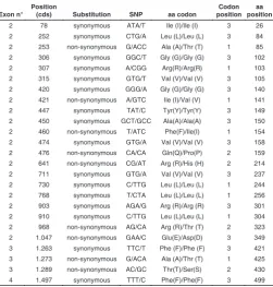
Exon n° Position
(cds) Substitution SNP aa codon
Codon position
aa position
2 78 synonymous ATA/T Ile (I)/Ile (I) 3 26 2 252 synonymous CTG/A Leu (L)/Leu (L) 3 84 2 253 non-synonymous G/ACC Ala (A)/Thr (T) 1 85 2 306 synonymous GGC/T Gly (G)/Gly (G) 3 102 2 307 synonymous A/CGG Arg(R)/Arg(R) 1 103 2 315 synonymous GTG/T Val (V)/Val (V) 3 105 2 420 synonymous GGG/A Gly (G)/Gly (G) 3 140 2 421 non-synonymous A/GTC Ile (I)/Val (V) 1 141 2 447 synonymous TAT/C Tyr(Y)/Tyr(Y) 3 149 2 450 synonymous GCT/GCC Ala(A)/Ala(A) 3 150 2 460 non-synonymous T/ATC Phe(F)/Ile(I) 1 154 2 474 synonymous GTG/A Val (V)/Val (V) 3 158 2 476 non-synonymous CA/CA Gln(Q)/Pro(P) 2 159 2 641 non-synonymous CG/AT Arg (R)/His (H) 2 214 2 711 synonymous GTG/A Val (V)/Val (V) 3 237 2 730 synonymous C/TTG Leu (L)/Leu (L) 1 244 2 768 synonymous T/CTA Leu (L)/Leu (L) 1 256 2 903 synonymous AGA/G Arg (R)/Arg (R) 3 301 2 910 synonymous C/TTG Leu (L)/Leu (L) 1 304 2 968 non-synonymous AG/CA Arg (R)/Thr (T) 2 323 2 1.047 non-synonymous GAA/C Glu(E)/Asp(D) 3 349 3 1.263 synonymous TTC/T Phe (F)/Phe (F) 3 421 3 1.273 non-synonymous G/ACA Ala (A)/Thr (T) 1 425 3 1.289 non-synonymous AC/GC Thr(T)/Ser(S) 2 430 4 1.497 synonymous TTT/C Phe(F)/Phe(F) 3 499
TABLE 1
SNPs IDENTIFIED IN THE slc2a10 CODING REGION
Allsynonymous and non-synonymous substitutions are defined using as reference sequence the
slc2a10 coding sequence (accession nos.: HM560591 and HM560592) obtained in this study, compared to those present in GenBank (accession nos.: XM_679477.2 and NM_001111163). The nucleotides are numbered starting from the first base of the start codon (ATG) of the slc2a10
encode for a protein similar to human GLUT10, on chromosome 11 of the zebrafish genome. This putative encoded protein shows a high degree of amino acid sequence similarity (43%) with human GLUT10. Furthermore, analysis of an approximately 5 Mb genomic region surrounding the SLC2A10 gene in zebrafish and in humans showed the presence of conserved synteny between this region of Danio rerio chromosome 11 and Homosapiens
chromosome 20q13.12. In both species, the gene is flanked on its 5’ side by the TP53RK gene and more distantly by the ACOT8, MYBL2, IFT52 and SFRS6 genes. On the 3’ side, the CSE1L and
TMEM189 genes can be identified (Fig. 1). These data highly suggest that the genomic locus LOC560546 contains the ortholog of the human SLC2A10 gene, which is named slc2a10 (protein Glut10)according to the Zebrafish Nomenclature Guidelines.
Using the BLAST algorithm on the dbEST database, we identified several entries corresponding to slc2a10 gene tran-scripts. We selected two putative full-length IMAGE clones, no. 5601920 and no. 7225584, which were completely sequenced. This analysis revealed a 24-bp insertion that contains a stop codon in IMAGE clone 7225584, whereas clone no. 5601920 contains an entire ORF (1.542 bp, 513 aa). Furthermore, se-quence analysis showed that these two clones differ in their 5’ UTR region, suggesting the presence of two splice variants. Recently, Tseng et al. (2009) identified eighteen members of glucose transporters in zebrafish, including slc2a10 (deposited mRNA sequence NM_001111163). Using the GenBank entry XM_679477.2 generated using a gene prediction method and the sequence of IMAGE clone no. 5601920, we performed multiple amino acid alignments that led to the identification of seven amino
acid differences among the three sequences (not shown). Evolu-tionary conservation analyses, performed by multiple ClustalW alignment of several GLUT10 orthologs (H. sapiens, M. muscu-lus, R. norvegicus, G. gallus and X. tropicalis) suggested that six out of these seven missense substitutions should be considered polymorphic variants. The highly conserved lysine at position 284 in the transmembrane domain 8 (TMD8) of the protein was the exception, having changed to a glutamic acid in the IMAGE clone (not shown). In order to clarify these discrepancies, we decided to re-clone and sequence the slc2a10 cds using an RT-PCR strat-egy performed on adult zebrafish RNA. This nucleotide sequence was used as internal reference sequence and deposited in the GenBank database (accession nos.: HM560591 and HM560592). In addition, to validate this sequence, the cds was amplified and sequenced from total RNAs purified from different adult fishes and a pool of embryos. The ClustalW multiple sequence alignment of all these sequences and those deposited in GenBank (accession nos: XM_679477 and NM_001111163) revealed a total of 25 SNPs in the slc2a10 coding region, nine of which are missense substitutions (Table 1). These nucleotide differences are likely due to high genetic variation existing in the different zebrafish strains (Guryev et al., 2006). Moreover, we did not find the p.K284E variant identified in clone no. 5601920. This substitution could be a mutational event, which probably arose during cloning, similar to the 24-bp insertion identified in clone no. 7225584.
The 5’ and 3’ UTRs of slc2a10 were obtained using a combined strategy based on computational analyses and sequencing of IMAGE clones and RT-PCR fragments of slc2a10 cDNAs. As suggested by the sequencing of the IMAGE clones, we identified two mRNA splice variants in the 5’-UTR, which were expressed both during embryogenesis and in adult zebrafish tissues (Fig. 2A and 2C). In particular, we characterized a longer isoform with a Sequence at exon–intron junction
Exon n°
Exon size
(bp) 5’ splice donor 3’ splice acceptor
Intron size (bp)
Amino acid interrupted
1 >400 AAAATGGgtaagttg tgctccagGTTGTTCT 2.391 Gly-2 2 1.185 GGACCAAgtaagtgg gtttgacagTGACGTG 1.286 Met-397 3 123 ATAATCGgtgtgcac tcttctcagATGTCATT 93 Asp-438 4 136 AGACACGgtaagagg ttctttcagAATGATC 1.950 Arg-483 5 202
TABLE 2
EXON-INTRON ORGANIZATION OF THE slc2a10 GENE
Exon sequences are indicated by uppercase letters and intron sequences by lowercase letters. Splice donor and acceptor sites are underlined. Exon and intron size are reported as base pairs. The corresponding interrupted amino acid residue in the Glut10 protein is also reported.
B
C
A
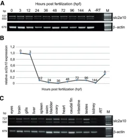
Fig. 2. Temporal expression pattern analysis of slc2a10 during development and in adult zebrafish tissues. RNA samples were purified from different embryonic and larval stages and from adult fishes and amplified with specific primer pairs of slc2a10. RT-PCR analysis (A)
177-nucleotide stretch and a shorter variant without this nucleotide sequence. Both splice variants were deposited in GenBank with the accession numbers: HM560591 (+ 177 splice form) and HM560592 (-177 splice form).
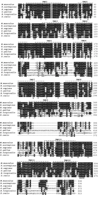
The slc2a10 gene is distributed among five exons, which is the same as the human counter-part, and the intron-exon splice junctions are conserved within the two species (Table 2). The Glut10 protein was compared to vertebrate pro-teins from H. sapiens, M. musculus, R. norvegicus, G. gallus and X. tropicalis (Fig. 3). Multiple sequence alignment revealed that zebrafish Glut10 is 45% identical to human GLUT10, 44% identical to M. musculus and R. norvegicus orthologs and shares 48% sequence identity to G. gallus and X. tropicalis GLUT10. Furthermore, Glut10 maintains the typical 12 hydrophobic TMD structure and structural mo-tifs involved in glucose transport activity (Fig. 3). In particular, zebrafish Glut10, as in the human and the mouse counterparts, retains the tryp-tophan residue W399 in TMD10 and the glutamic acid E404 and glycine G412 residues in intracel-lular loop 5 (Fig. 3). These residues are impor-tant for substrate recognition and sugar trans-port specificity (Barrett et al., 1999; McVie-Wylie et al., 2001; Dawson et al., 2001). Inter-estingly, zebrafish Glut10 does not have the PESPR motif in loop 6 present in all GLUTs, which may contribute to its low-rate saturable glucose transport activity (Dawson et al., 2001; Zhao and Keating, 2007).
Temporal expression pattern of the slc2a10 gene
To investigate the temporal expression pat-tern of slc2a10 during zebrafish development, we performed both RT-PCR and quantitative PCR (Q-PCR) analyses on cDNA obtained from
Fig. 3. Multiple sequence alignment and amino acids sequence identity of Glut10 protein with GLUT10 orthologous proteins in various species.
different developmental stages. In particular, to study the expres-sion of two slc2a10 mRNA splice variants, a primer pair was designed to specifically amplify these isoforms. Two distinct PCR products, 727 and 550 bp, were detected at all developmental stages analyzed, including the zygote, indicating that slc2a10 is also maternally expressed (Fig. 2A). The expression level of
slc2a10 transcripts was maintained throughout gastrulation, embryo segmentation (6-somite stage) and pharyngula periods (24-48 hpf). The slc2a10 gene was also detected during the larval period, the protruding-mouth (72 hpf), the 4-day-old and 6-day-old larval stage up to the adult fish.
Using Q-PCR, we quantified the expression levels of slc2a10
mRNAs during embryonic development (Fig. 2B). Simultaneous detection of ef-1 gene expression was used as the reference gene to normalize the expression level of slc2a10. Quantified mRNA values were reported for slc2a10 relative to the 1-cell stage expression level. Quantitative analysis revealed the pres-ence of slc2a10 maternal transcripts. The expression level of
slc2a10 mRNA was high at the one-cell stage and at the onset of mid-blastula transition (1k-cell stage, 3 hpf); starting from this stage, the transcript levels rapidly decreased untill the 6-somite stage and remained at lower levels during subsequent stages of embryonic development (Fig. 2B).
In the adult fishes, the slc2a10 tissue-specific distribution was analyzed by conventional RT-PCR analysis of total RNA samples purified from several tissues and organs (Fig. 2C). Both splice variants were observed in all adult samples analyzed: liver, heart, gills, eyes, brain, spleen, swim bladder, ovary, caudal fin, testes, intestine and kidney. To assess RNA integrity, the transcript for the -actin gene was amplified by RT-PCR from all tissues analyzed.
Spatiotemporal expression of slc2a10 during development
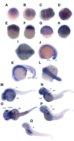
The spatiotemporal expression pattern of slc2a10 in embryos from the one-cell stage to 4 dpf larvae was investigated by whole-mount ISH using a DIG-UTP-labeled probe, which recognizes both slc2a10 mRNA splice variants. A sense probe was used in parallel control experiments that did not produce any signal (data not shown). According to RT-PCR results, ISH experiments confirmed that slc2a10 is expressed during early embryonic development. High levels of slc2a10 mRNA were expressed in equal distributions from the 1-cell stage to the 1k-cell stage (Fig. 4 A,F). During the gastrula period (50% epiboly and shield stage), the embryos showed the ubiquitous distribution of the slc2a10
transcripts (Fig. 4 G,H). In the mid-segmentation period (15 somites), transcript appeared in the otic placode and in the notochord (Fig. 4 I). At the 20-25 somite stage, a hybridization signal was detected in the yolk syncytial layer and in the notocord (Fig. 4 J,K,L). In 24 hpf embryos, slc2a10 mRNA was largely diffused in the cephalic region; an intense signal was also ob-served in the hatching gland cells and in the otic vesicles, whereas the staining in the notochord disappeared (Fig. 4M). At the long-pec stage (48 hpf), slc2a10 transcript was mainly restricted to the pectoral fin buds, hatching gland cells, otic vesicles, lens and to the posterior tectum (Fig. 4 N,O). During the larval period at the protruding mouth stage (72 hpf), the signal persisted in the otic vesicles, in the pectoral fin buds, and at a low level in the posterior tectum (Fig. 4P), which is in agreement with previous reports (Tseng et al., 2009). In the swimming larva at 4 dpf, the signal
Fig. 4. Whole-mount in situ hybridization analysis of slc2a10 expres-sion during zebrafish development.slc2a10 mRNA was ubiquitously distributed from the 1-cell stage to the 1k-cell stage (A,F) and during the gastrula period (G,H). At the 15-somite stage, a hybridization signal was localized in the otic placode and notochord (I); at the 20-25 somites stage, slc2a10 transcript was also expressed in the YSL region and in the notochord (J,K,L). During the pharyngula period (24-hpf), an intense signal was largely diffused in the cephalic region (M). In 48-hpf embryos
(N,O), the slc2a10 mRNA was detected in the lens, posterior tectum, otic vesicles, hatching gland cells and pectoral fin buds. During the larval stage in 72-hpf embryos (P), slc2a10 expression was restricted to the otic vesicles and pectoral fins; in 4-dpf larvae (Q), the staining was observed in the swim bladder structure. Abbreviations: hgc, hatching gland cells; nt, notochord; pfb, pectoral fin buds; op, otic placode; ov, otic vesicles; sb, swim bladder; YSL, yolk syncytial layer.
G
O
B
C
D
E
F
H
I
J
K
L
P
A
M
N
appeared in the swim bladder structure (Fig. 4Q). During zebrafish embryogenesis, ISH analyses did not show a specific expression of slc2a10 mRNA in blood vessels or in the cardiovascular system.
Discussion
In the present study, we characterized the zebrafish ortholog of the human SLC2A10 gene and its mRNA expression pattern during development and in adult tissues.
Bioinformatics analysis indicated the presence of a unique
slc2a10 ortholog gene located on chromosome 11 of the zebrafish genome in a region that shows conserved synteny with the H. sapiens chromosome 20q13.12. From an evolutionary perspec-tive, the SLC2A10 gene structure has been remarkably con-served from fish to mammals. The zebrafish slc2a10 is com-posed, like the human counterpart, of five exons with the transla-tion initiatransla-tion codon in exon 1. The deduced amino acid sequence of the Glut10 protein adopts the characteristic twelve transmem-brane domain structure and contains most of the sugar/polyol transporter signatures (Joost et al., 2002; Tseng et al., 2009). Sequencing of the Glut10 coding region disclosed several non-synonymous strain-specific polymorphisms. Multiple alignment of orthologous proteins underlined that these amino acid substi-tutions did not involve residues that are either evolutionarily conserved or essential for sugar transport. In contrast, all human mutations involved functionally relevant amino acids or highly conserved residues (Coucke et al., 2006; Drera et al., 2007, Callewaert et al., 2008a, Faiyaz-Ul-Haque et al., 2009; Zaidi et al.,
2009; Ritelli et al., 2009).
Unlike the human SLC2A10 gene, the slc2a10 primary tran-script in zebrafish gives rise to two specific mRNA splice variants differing in the 5’-UTR for a 177-nucleotide stretch; both encode the same polypeptide. The presence of these isoforms, not previously identified (Tseng et al., 2009), suggests the possible existence of a differential processing of zebrafish slc2a10 pre-mRNA. Bioinformatics analysis indicated the presence in the 177-nucleotide stretch of motifs involved in the transcriptional and post-transcriptional gene expression regulation, i.e., IRES se-quences and the K-box motif. Therefore, these two splice variants could provide a potential regulatory mechanism for slc2a10 gene expression at both transcriptional and post-transcriptional levels, even though their function is still unknown and requires further investigation.
During embryogenesis, both slc2a10 mRNA splice variants were detectable in newly fertilized eggs and in adult zebrafish. Because the slc2a10 transcripts showed a maternal contribution as well as zygotic expression, they could be required in the early stages of zebrafish development. ISH revealed that, in early embryogenesis, slc2a10 was homogeneously distributed. During the segmentation stage, gene expression was restricted to the otic placode and to the notochord. In particular, at the 20-25 somite stage the slc2a10 transcript was clearly observed in the YSL, suggesting a role for slc2a10 in sugar transport in yolk stores. At 24 hpf, slc2a10 showed localized expression corre-sponding to the encephalic region, lens, otic vesicles, posterior tectum, pectoral fin buds and hatching gland cells. The slc2a10
mRNA detected in the otic vesicle was also observed for its murine ortholog. In particular, De Silva et al. (2006) reported that
Slc2a10 is involved in the differentiation pathway of hair cell lineage and in the development of the cochlea. These data suggest that in both animal models the gene is required for sugar transport in the cochlear tissue and could be implicated in the morphogenesis of the inner ear. The presence of slc2a10 mRNA in the zebrafish brain could be suggestive of the existence of glucose sensing areas involved specifically in glucose homeosta-sis, as described in mouse (Dahlin et al., 2009). Our ISH results do not show a specific expression of slc2a10 in the developing blood vessels of zebrafish embryos, which is probably due to a low abundance of slc2a10 mRNA. Likewise, there has been no report of specific blood vessel expression of Slc2a10 in mouse embryos as evidenced by ISH data generated by the Eurexpress Consor-tium.
In the adult zebrafish, both slc2a10 splicing variants were detected and showed a tissue-specific expression pattern (liver, heart, gills, eyes, brain, spleen, swim bladder, ovary, caudal fin, testes, intestine and kidney), as previously described for its human and mouse orthologs (Dawson et al., 2001; Wood et al.,
2003; De Silva et al., 2006). In particular, Glut10 mRNA was expressed in tissues in which this transporter is known to play a functional role in glucose homeostasis in mammals, such as the liver and kidney (Uldry and Thorens, 2004). In addition, slc2a10
expression in the zebrafish testes could be related to a possible requirement of glucose as an energy substrate for spermatozoa, and its expression in ovary could be related to a possible require-ment in oogenesis. In agreerequire-ment with the knowledge that in mammals GLUT10 is abundantly expressed in smooth muscle-rich organs (Wu et al., 2009), slc2a10 was expressed in zebrafish intestine, heart and swim bladder.
Regarding GLUT10 function in ATS etiopathogenesis, two mouse models have been proposed (Callewaert et al., 2008b; Cheng et al., 2009). Both Slc2a10 mutant mice do not show the pathological features of the human disorder and therefore remain inconclusive with respect to GLUT10 function.
Recently, a new hypothesis regarding the role of GLUT10 in ATS etiology was proposed (Segade, 2010). GLUT10 may be a transporter of ascorbic acid (AA), a cofactor of the hydroxylases that modify collagen and elastin. From this perspective, the loss of GLUT10 should result in defective synthesis and abnormal assembly of these proteins. This assumption is supported by previous results (Siman et al., 1997; Maeda et al., 2000; Coucke
et al., 2006). Moreover, Maeda et al.,showed abnormalities of the arterial walls in knockout mice for the L-gulono--lactone oxidase (Gulo), the key enzyme involved in AA synthesis. A recent study has further validated Segade’s hypothesis that GLUT10 is a transporter of ascorbate (Lee et al., 2010). Indeed, Lee and colleagues demonstrated that GLUT10 primarily localizes to mitochondria in smooth muscle cells and adipocytes, facilitates transport of the oxidized form of vitamin C (DHA) into mitochon-dria and also increases cellular uptake of DHA. These authors also showed an increase of intracellular reactive oxygen species (ROS) levels that may lead to abnormal elastogenesis in the aortic smooth muscle cells of an ATS mouse model (Lee et al., 2010). This new functional role for GLUT10 may help to explain why the mutant mice do not fully mimic the human syndrome (Callewaert
fed a diet supplemented with AA because of the lack of a functional gulo gene. Glut10 expression in zebrafish organs and tissues observed in this study could therefore be needed for AA transport to reduce oxidative stress, particularly in muscle cells. With this taken into consideration, slc2a10 morpholino-mediated knockdown might disclose arterial abnormalities due to defective intracellular AA transport and improve our knowledge of the human syndrome.
Materials and Methods
Bioinformatics analysis
Nucleotide sequence assembly was performed using Sequencher version 4.9 software (www.genecodes.com). Zebrafish genomic se-quences were analyzed using the UCSC Genome Browser (www.genome.ucsc.edu/) and the Ensembl Genome Browsers (www.ensembl.org). Multiple sequence alignments and phylogenetic analyses were performed using the ClustalW algorithm (www.ebi.ac.uk/ clustalw/), and the formatting of the alignment was generated using the GeneDoc program. Zebrafish Glut10 sequence was compared to those of
H. sapiens (accession no. NP_110404), M. musculus (accession no. NP_569718), R. norvegicus (accession no. NP_001102433), G. gallus
(accession no. XP_417383.2) and X. tropicalis (accession no. AA_22090). The presence of synteny between human and zebrafish genomic regions was evaluated using the Synteny DB (www.teleost.cs.uoregon.edu/ synteny_db/) (Catchen et al., 2009). Nucleotide and amino acid se-quences were compared to the non-redundant sequence databases present at the NCBI using the BLAST algorithm. Putative TMDs were defined using the Prediction of Transmembrane helices in proteins, TMHMM server version 2.0 (www.cbs.dtu.dk/services/TMHMM-2.0). The DNA sequences at the exon/intron boundaries were determined using the Splice Site Prediction by Neural Network (www.fruitfly.org/seq_tools/ splice.html) program.
Identification of zebrafish slc2a10 cDNA clones
I.M.A.G.E (Integrated Molecular Analysis of Gene Expression) Con-sortium cDNA clones no. 5601920 (Genbank accession no. BM860136, 5’ sequence and no. BM859903, 3’ sequence) and no. 7225584 (GenBank accession no. CN015886, 5’ sequence), encoding the putative zebrafish Glut10, were purchased from Geneservice (U.K.). Both cDNA clones were completely sequenced using an ABI Prism 3100 automated se-quencer (Applied Biosystems, Foster City, CA, USA) and vector and gene specific primers.
Fish breeding and embryos/tissues collection
The AB wild-type strain of zebrafish was used for all experiments. The fishes were maintained at 28.5C in 14:10 h light/dark cycle following a standard procedure (Westerfield,2000). The fertilized eggs were col-lected in Petri dishes and examined until the required developmental stage was reached according to established embryonic stages (Kimmel
et al., 1995). For RNA extraction, zebrafish embryos and larvae were rinsed in PBS solution, frozen immediately in liquid nitrogen and stored at -80C until use. For tissue dissections from adult animals, the fishes were killed by an excess of ethyl 3-aminobenzoate methanesulfonate salt solution (Tricaine, Sigma Aldrich St. Louis, MO, USA). The tissues were quickly removed, soaked in liquid nitrogen and stored at -80C until RNA extraction.
RNA preparation, RT-PCR analysis and cloning of the full length Glut10 coding sequence
Total RNA was extracted from zebrafish embryos at different develop-mental stages and from several organs and tissues of adult fishes using TRIzol® protocol (Invitrogen, Carlsbad, CA, USA). The RNA concentra-tion was determined using the NanoDrop® ND-1000 spectrophotometer
(NanoDrop Technologies, Germany). cDNA was prepared from 3 g of total RNA, using 200 U/l of M-MLV reverse transcriptase (Invitrogen) in the presence of random hexamer oligonucleotides, following the manufacturer’s instructions. Amplification was performed for 34 cycles (60C annealing) following the instructions for GoTaq DNA polymerase (Promega, Madison, WI, USA) and with specific primer pairs for the
slc2a10 gene (Supplementary Table S1). In particular, to study the expression of two slc2a10 mRNA splice isoforms, the forward primer was designed upstream of the splice donor site in the 5'-UTR and the reverse primer was located in the coding region of slc2a10. To assess the RNA integrity, a 679-bp fragment of the zebrafish -actin gene (GenBank accession no. AF057040) was amplified. The PCR reactions were per-formed for 28 cycles using the following -actin specific primers: -actin-for: (5'-GTCCCTGTACGCCTCTGGTCG-3') and -actin-rev: (5'-GCCGGACTCATCGTACTCCTG-3'). To isolate the complete Glut10 coding sequence, RT-PCR-based amplification of a 1.549-bp fragment was performed on a total RNA sample from an adult AB zebrafish.
The following forward and reverse primers were used:
slc2a10-ATG-F (5’- CATCAAAATGGGTTGTTCTGTCCTG-3’) slc2a10 -TGA-R (5’-CTTCATCAGGTTGCTGTACTCGTCA-3’). A 250-ng sample of total RNA was retrotranscribed and amplified with the SuperScript III One-Step RT-PCR System (Invitrogen) using the cycling parameters provided by the manufacturer. The purified PCR product was directly cloned into the pcDNA3.1/CT-GFP-TOPO™ vector (GFP TA Cloning® system, Invitrogen) and sequenced on both strands using Big Dye terminator v3.1 protocol on an ABI PRISM 3100 Genetic Analyzer (Applied Biosystems).
Quantitative real-time RT-PCR
Q-PCR was performed using primers designed with the Primer Ex-press™ software (Applied Biosystems). The primer sequences were:
slc2a10 gene, forward: 5’-CGAAGGACATCCGAGGAAGA-3’, reverse: AGACAGGCCAAATGACATCGAT-3’, TaqMan probe: 5’-TCATCAACTGCTTCAATGTGGGCGC-3’; ef-1 gene: forward: CGATTCCACCGCATTTGTAGA-3’, reverse: CCACGTCGACTCCGGAAA-3’, TaqMan probe: 5’-TCCACCACCACCGGCCATCTG-3’. The TaqMan probes (Eurofins MWG Technologies, Ebersberg, Germany) were labeled at the 5’ end with the reporter dye 6-carboxy-fluorescein and at the 3’ end with the quencher dye 6-carboxytetramethyl rhodamine. Amplification was performed with the ABI Prism 7500 sequence detection system (Applied Biosystems). The PCR was carried out with TaqMan Universal PCR Master Mix, and thermal cycling was performed according to the manufacturer’s protocol (Applied Biosystems). The relative quantification (RQ) of gene expres-sion was determined by the equation 2-(Ct). All data analysis was performed with the 7500 system SDS software, version 1.3.1 (Applied Biosystems). Threshold cycles (Ct) were standardized by Ct for the endogenous control ef-1 mRNA.
Whole-mount in situ hybridization
Antisense and sense riboprobes were synthesized from slc2a10
Acknowledgements
This work was supported by Fondazione Cariplo “ZebraGene” Grant (2006).
References
AKHURST, R.J., (2006). A sweet link between TGFBeta and vascular disease? Nat Genet 38: 400–401.
BARRETT, M.P., WALMSLEY, A.R., GOULD, G.W. (1999). Structure and function of facilitative sugar transporters. Curr Opin Cell Biol 11: 496-502.
CALLEWAERT, B.L., WILLAERT, A., KERSTJENS-FREDERIKSE, W.S., DE BACKER, J., DEVRIENDT, K., ALBRECHT, B., RAMOS-ARROYO, M.A., DOCO-FENZY, M., HENNEKAM, R.C., PYERITZ, R.E., KROGMANN, O.N., GILLESSEN-KAESBACH, G., WAKELING, E.L., NIK-ZAINAL, S., FRANCANNET, C., MAURAN, P., BOOTH, C., BARROW, M., DEKENS, R., LOEYS, B.L., COUCKE, P.J., DE PAEPE, A.M. (2008a). Arterial tortuosity syndrome: clinical and molecular findings in 12 newly identified families. Hum Mutat 29: 150-158.
CALLEWAERT, B.L., LOEYS, B.L., CASTELEYN, C., WILLAERT, A., DEWINT, P., DE BACKER, J., SEDLMEIER, R., SIMOENS, P., DE PAEPE, A.M., COUCKE, P.J. (2008b). Absence of arterial phenotype in mice with homozygous slc2a10
missense substitutions. Genesis 46: 385–389.
CATCHEN, J.M., CONERY, J.S., POSTLETHWAIT, J.H. (2009). Automated iden-tification of conserved synteny after whole genome duplication. Genome Re-search 19: 1497-1505.
CHENG, C.H., KIKUCHI, T., CHEN, Y.H., SABBAGHA, N.G., LEE, Y.C., PAN, H.J., CHANG, C., CHEN, Y.T. (2009). Mutations in the SLC2A10 gene cause arterial abnormalities in mice. Cardiovascular Research 81: 381–388.
COUCKE, P.J., WILLAERT, A., WESSELS, M.W., CALLEWAERT, B., ZOPPI, N., DE BACKER, J., FOX, E., MANCINI, G.M., KAMBOURIS, M., GARDELLA, R., FACCHETTI, F., WILLEMS, P.J., FORSYTH, R., DIETZ, H.C., BARLATI, S., COLOMBI, M., LOEYS, B., DE PAEPE, A. (2006). Mutations in the facilitative glucose transporter GLUT10 alter angiogenesis and cause arterial tortuosity syndrome. Nat Genet 38: 452-457.
DAHLIN, A., ROYALL, J., HOHMANN, J.G., WANG, J. (2009). Expression Profiling of the Solute Carrier Gene Family in the Mouse Brain. J Pharmacol Exp Ther
329: 558–570.
DAWSON, P.A., MYCHALECKYJ, J.C., FOSSEY, S.C., MIHIC, S.J., CRADDOCK, A.L., BOWDEN, D.W. (2001). Sequence and functional analysis of GLUT10: a glucose transporter in the Type 2 diabetes-linked region of chromosome 20q12-13.1. Mol Genet Metab 74: 186-199.
DE SILVA, M.G., HILDEBRAND, M.S., CHRISTOPOULOS, H., NEWMAN, M.R., BELL, K., RITCHIE, M., SMYTH, G.K., DAHL, H.H. (2006). Gene expression changes during step-wise differentiation of embryonic stem cells along the inner ear hair cell pathway. Acta Otolaryngol 126: 1148-1157.
DRERA, B., GUALA, A., ZOPPI, N., GARDELLA, R., FRANCESCHINI, P., BARLATI, S., COLOMBI, M. (2007). Two novel SLC2A10/GLUT10 mutations in a patient with arterial tortuosity syndrome. Am J Med Genet 143: 216-218.
FAIYAZ-UL-HAQUE, M., ZAIDI, S.H., AL-SANNA, N., ALSWAID, A., MOMENAH, T., KAYA, N., AL-DAYEL, F., BOUHOAIGAH, I., SALIEM, M., TSUI, L.C., TEEBI, A.S. (2009). A novel missense and a recurrent mutation in SLC2A10
gene of patients affected with arterial tortuosity syndrome. Atherosclerosis 203: 466-471.
GURYEV, V., KOUDIJS, M.J., BEREZIKOV, E., JOHNSON, S.L., PLASTERK, R.H.A., VAN EEDEN, F.J.M., CUPPEN, E. (2006). Genetic variation in the zebrafish. Genome Research 16: 491-497.
JOOST, H.G., BELL, G.I., BEST, J.D., BIRNBAUM, M.J., CHARRON, M.J., CHEN, Y.T., DOEGE, H., JAMES, D.E., LODISH, H.F., MOLEY, K.H., MOLEY, J.F., MUECKLER, M., ROGERS, S., SCHÜRMANN, A., SEINO, S., THORENS, B. (2002). Nomenclature of the GLUT/SLC2A family of sugar/polyol transport facilitators. Am J Physiol Endocrinol Metab 282: 974-976.
JOOST, H.G. and THORENS, B. (2001). The extended GLUT-family of sugar/ polyol transport facilitators: nomenclature, sequence characteristics, and po-tential function of its novel members. Mol Membr Biol 18: 247-256.
KIMMEL, C.B., BALLARD, W.W., KIMMEL, S.R., ULLMANN, B., SCHILLING, T.F. (1995). Stages of embryonic development of the zebrafish. Dev Dynam 203: 253-310.
LEE, Y.C., HUANG, H.Y., CHANG. C.J., CHENG, C.H., CHEN, Y.T. (2010). Mitochondrial GLUT10 facilitates dehydroascorbic acid import and protects cells against oxidative stress: mechanistic insight into arterial tortuosity syn-drome. Hum Mol Genet 19: 3721-3733.
MAEDA, N., HAGIHARA, H., NAKATA, Y., HILLER, S., WILDER, J., REDDICK, R. (2000). Aortic wall damage in mice unable to synthesize ascorbic acid. Proc Natl Acad Sci. USA 97: 841–846.
MCVIE-WYLIE, A.J., LAMSON, D.R., CHEN, Y.T. (2001). Molecular cloning of a novel member of the GLUT family of transporters, SLC2A10 (GLUT10), local-ized on chromosome 20q13.1: a candidate gene for NIDDM susceptibility.
Genomics 72: 113-117.
PARANT, J.M., GEORGE, S.A., HOLDEN, J.A., YOST, H.J. (2010). Genetic modeling of Li-Fraumeni syndrome in zebrafish. Dis Model Mech 3: 45-56. RITELLI, M., DRERA, B., VICCHIO, M., PUPPINI, G., BIBAN, P., PILATI, M.,
PRIOLI, M.A., BARLATI, S., COLOMBI, M. (2009). Arterial tortuosity syndrome in two Italian paedriatric patients. Orphanet J Rare Dis 4: 20.
SEGADE, F. (2010). Glucose transporter 10 and arterial tortuosity syndrome: The vitamin C connection. FEBS letters 584: 2990-2994.
SIMAN, C.M. and ERIKSSON, U.J. (1997). Vitamin C supplementation of the maternal diet reduces the rate of malformation in the offspring of diabetic rats.
Diabetologia 40: 1416–1424.
THISSE, C. and THISSE, B. (2008). High-resolution in situ hybridization to whole-mount zebrafish embryos. Nature Prot 3: 59-69.
TSENG, Y.C., CHEN, R.D., LEE, J.R., LIU, S.T., LEE, S.J., HWANG, P.P. (2009). Specific Expression and Regulation of Glucose Transporters in Zebrafish Ionocytes. Am J Physiol Regul Integr Comp Physiol 297: 275-290.
ULDRY, M. and THORENS, B. (2004). The SLC2 family of facilitated hexose and polyol transporters. Eur J Physiol 447: 480–489.
WESTERFIELD, M. (2000). The zebrafish book: A guide for the laboratory use of zebrafish. University of Oregon Press, Eugene.
WOOD, I.S., HUNTER, L., TRAYHURN, P. (2003). Expression of Class III facilita-tive glucose transporter genes (GLUT-10 and GLUT-12) in mouse and human adipose tissues. Biochem Biophys Res Commun 308: 43–49.
WU, C., OROZCO, C., BOYER, J., LEGLISE, M., GOODALE, J., BATALOV, S., HODGE, C.L., HAASE, J., JANES, J., HUSS, J.W. 3rd, SU, A.I. (2009). BioGPS:
an extensible and customizable portal for querying and organizing gene annotation resources. Genome Biology 10: R130.
ZAIDI, S.H., MEYER, S., PELTEKOVA, V.D., LINDINGER, A., TEEBI, A.S., FAIYAZ-UL-HAQUE, M. (2009). A novel non-sense mutation in the SLC2A10
gene of an arterial tortuosity syndrome patient of Kurdish origin. Eur J Pediatr
168: 867-870.
Further Related Reading, published previously in the
Int. J. Dev. Biol.
Xenopus glucose transporter 1 (xGLUT1) is required for gastrulation movement in Xenopus laevis
Keiko Suzawa, Akira Yukita, Tadayoshi Hayata, Toshiyasu Goto, Hiroki Danno, Tatsuo Michiue, Ken W. Cho and Makoto Asashima Int. J. Dev. Biol. (2007) 51: 183-190
TBX1, a DiGeorge syndrome candidate gene, is inhibited by retinoic acid Lifeng Zhang, Tao Zhong, Yuexiang Wang, Qiu Jiang, Houyan Song and Yonghao Gui Int. J. Dev. Biol. (2006) 50: 55-61
Cdx2 specifies the differentiation of morphological as well as functional absorptive enterocytes of the small intestine
Hiroyuki Mutoh, Kiichi Satoh, Hiroto Kita, Hirotsugu Sakamoto, Hiroko Hayakawa, Hironori Yamamoto, Norio Isoda, Kiichi Tamada, Kenichi Ido and Kentaro Sugano
Int. J. Dev. Biol. (2005) 49: 867-871
Embryonic expression of Xenopus SGLT-1L, a novel member of the solute carrier family 5 (SLC5), is confined to tubules of the pronephric kidney
Samer R Eid, Anne Terrettaz, Katsumi Nagata and André W Brändli Int. J. Dev. Biol. (2002) 46: 177-184
Astroglia-microvessel relationship in the developing human telencephalon
D Virgintino, E Maiorano, M Errede, A Vimercati, P Greco, L Selvaggi, L Roncali and M Bertossi Int. J. Dev. Biol. (1998) 42: 1165-118