Drug Design, Development and Therapy
Dove
press
O r i g i n a l r e s e a r c h
open access to scientific and medical research
Open access Full Text article
Development of antiproliferative long-circulating
liposomes co-encapsulating doxorubicin and
curcumin, through the use of a quality-by-design
approach
lucia ruxandra Tefas1
Bianca sylvester1
ioan Tomuta1
alina sesarman2,3
emilia licarete2,3
Manuela Banciu2,3
alina Porfire1
1Department of Pharmaceutical
Technology and Biopharmaceutics, Faculty of Pharmacy, University of Medicine and Pharmacy “iuliu
Haţieganu”, 2Department of
Molecular Biology and Biotechnology, Faculty of Biology and geology,
3Molecular Biology centre, institute
for interdisciplinary research in
Bio-Nano-Sciences, Babeş-Bolyai
University, cluj-napoca, romania
Abstract: The aim of this work was to use the quality-by-design (QbD) approach in the devel-opment of long-circulating liposomes co-loaded with curcumin (CUR) and doxorubicin (DOX) and to evaluate the cytotoxic potential of these liposomes in vitro using C26 murine colon carcinoma cell line. Based on a risk assessment, six parameters, namely the phospholipid, CUR and DOX concentrations, the phospholipid:cholesterol molar ratio, the temperature during the evaporation and hydration steps and the pH of the phosphate buffer, were identified as potential risk factors for the quality of the final product. The influence of these variables on the critical quality attributes of the co-loaded liposomal CUR and DOX was investigated: particle size, zeta potential, drug loading and entrapment efficiency. For this, a 26-2 factorial design was employed
to establish a proper regression model and to generate the contour plots for the responses. The obtained data served to establish the design space for which different combinations of variables yielded liposomes with characteristics within predefined specifications. The validation of the model was carried out by preparing two liposomal formulations corresponding to the robust set point from within the design space and one outside the design space and calculating the percent-age bias between the predicted and actual experimental results. The in vitro antiproliferative test showed that at higher CUR concentrations, the liposomes co-encapsulating CUR and DOX had a greater cytotoxic effect than DOX-loaded liposomes. Overall, this study showed that QbD is a useful instrument for controlling and optimizing the manufacturing process of liposomes co-loaded with CUR and DOX and that this nanoparticulate system possesses a great potential for use in colon cancer therapy.
Keywords: doxorubicin, curcumin, co-loaded long-circulating liposomes, quality by design, design of experiments
Introduction
The World Health Organization has stated that cancer is one of the major leading causes of mortality around the world.1 However, in the past decades, advances in
cancer treatment have led to an increase in the survival rate of cancer patients.2 In spite
of the indisputable benefits of cancer treatment, and even with the development of new anticancerous agents, often cancer treatment regimens are unsuccessful.3 Failure
in cancer treatment mostly occurs as a result of drug-induced toxicity and resistance of cancer cells.4 Because of these drawbacks, it is obvious that chemotherapy alone
faces challenges, and a new approach is needed in order to achieve a satisfactory outcome in cancer patients. Therefore, combination therapy has emerged as a new
correspondence: ioan Tomuta Department of Pharmaceutical Technology and Biopharmaceutics, Faculty of Pharmacy, University of
Medicine and Pharmacy “Iuliu Haţieganu”, 41 Victor Babeş Street, 400012
Cluj-napoca, romania Tel/fax +40 264 59 5770 email [email protected]
Journal name: Drug Design, Development and Therapy Article Designation: Original Research
Year: 2017 Volume: 11
Running head verso: Tefas et al
Running head recto: Antiproliferative liposomes co-encapsulating DOX and CUR DOI: http://dx.doi.org/10.2147/DDDT.S129008
Drug Design, Development and Therapy downloaded from https://www.dovepress.com/ by 118.70.13.36 on 21-Aug-2020
For personal use only.
Number of times this article has been viewed
This article was published in the following Dove Press journal: Drug Design, Development and Therapy
Dovepress
Tefas et al
and effective strategy capable of overcoming the limitations of chemotherapy.5 It is suggested that the association of
common chemotherapeutic agents with existing anticancer agents, new ones or even natural products, could maximize the chemotherapy efficacy and reduce the toxic effects to normal cells. Recently, there has been growing interest in the scientific field toward the use of natural products.6 Natural
products with potential anticancer activity are mostly dietary compounds which can be found in various foods like fruits, vegetables and other plants. They could be good candidates for combination therapy because aside from the fact that they have shown efficacy toward a wide range of cancerous diseases, their side effects and toxicity are low.7
Doxorubicin (DOX), an anthracycline antibiotic, is one of the efficient chemotherapeutic agents commonly used in the treatment of solid tumors, acute leukemia and malignant lymphoma.8,9 Its main mechanisms of action involve
topoi-somerase II inhibition, DNA intercalation and free radical formation.10 DOX can inhibit the replication of tumor cells
by intercalating in the base pairs of DNA.9 Also, it induces
apoptosis in cancerous cells as a result of oxidative dam-age to DNA.11 However, despite the fact that it is a very
potent chemotherapeutic agent, its clinical use is limited by its induced toxicity and drug resistance.8 DOX can induce
severe cytotoxic effects in normal cells, mostly leading to cardiotoxicity and myelosupression.9 Incorporating DOX
into PEGylated liposomes reduces the toxicity associated with DOX use and prolongs the circulation time in the bloodstream, overall improving the accumulation at the tumor site through the enhanced permeability and retention effect.12 Liposomal DOX is currently marketed as various
products, namely Doxil®/Caelyx® (Johnson & Johnson)
and Lipo-Dox® (Taiwan Liposome), for the treatment of
Kaposi’s sarcoma, ovarian cancer, breast cancer and multiple myeloma.13 Doxil (in the US) or Caelyx (in Europe) was the
first DOX-loaded liposomal formulation to be approved in 1995 by the regulatory authorities.12,14 Preclinical and
clini-cal studies have shown that liposomal DOX has a smaller distribution volume, an increased circulation time and a decreased clearance compared to free DOX. Also, due to the small size of the particles, liposomal DOX preferentially accumulates within the tumor tissue with altered vasculature, which accounts for the lower toxicity and side effects, such as cardiotoxicity and myelosupression.15
Curcumin (CUR; 1,7-bis(4-hydroxy 3-methoxy phenyl)-1,6-heptadiene-3,5-dione), also called diferuloylmethane, is a diphenolic compound extracted from the rhizome of the perennial herb Curcuma longa (turmeric), a plant which
belongs to the Zingiberaceae family.16–18 CUR is a yellow
pigment extensively used as a spice, coloring agent and flavoring agent, but also in traditional Asian medicine to treat different types of diseases.6 There are many reports
which state that CUR has a plethora of biological activities, including antioxidant, anti-inflammatory, antimicrobial, anticarcinogenic, hepatoprotective and neuroprotective properties.17,19,20 Of late, it has attracted considerable
atten-tion in cancer prevenatten-tion and therapeutics. The underlying molecular mechanisms of action through which CUR exerts its anticancerous effect are various and possibly involve the modulation of different intracellular signaling pathways, such as those involved in proliferation, inflammation and apoptosis.21 CUR has been shown to inhibit the activation of
NF-κB6 and also to downregulate the intracellular levels of
P-glycoprotein and MRP1, which play an important role in multidrug resistance, including chemotherapy.22 Moreover, in
vivo studies have demonstrated that CUR possesses potential cardioprotective effects.23 Besides its potential beneficial
effects, the use of CUR in cancer chemoprevention and ther-apy is of interest due to its safety profile and lack of toxicity in animals or humans.16 CUR has been reported to be an ideal
chemosensitizer capable of reversing multidrug resistance in cancer cells.24 Recent research suggests that the association
of a chemotherapeutic agent with a chemosensitizer is much more efficient than a traditional combination of two or several cytotoxic drugs.8 Also, it has been demonstrated that CUR
has synergistic effects with DOX when associated.24 In spite
of its low toxicity and multiple biological properties, the clinical use of CUR is limited by its low solubility in water and poor bioavailability.25
The co-delivery of two or several drugs via nanopar-ticulate drug delivery systems, for chemotherapy, shows multiple advantages such as synergism of the pharmaco-logical effect, decrease of the effective dose, reduction of side effects and overcoming of multidrug resistance.26
Up to the present, several studies have emphasized the superior anticancer efficacy of the DOX-CUR association co-delivered by various nanoparticulate systems, including lipid nanoparticles,8 polymeric nanoparticles,27 polymeric or
lipidic micelles24,26,28–30 and liposomes,25 to hepatic, brain and
breast cancer, leukemia and melanoma. Therefore, develop-ing a drug delivery system such as liposomes can provide a solution to the aforementioned challenges, by decreasing the toxicity of DOX, enhancing the solubility of CUR and overall improving the stability of both the drugs. Even though the co-encapsulation of DOX and CUR in liposomes has been previously reported in another study, to the best of our
Drug Design, Development and Therapy downloaded from https://www.dovepress.com/ by 118.70.13.36 on 21-Aug-2020
Dovepress antiproliferative liposomes co-encapsulating DOX and cUr
knowledge, no study with liposomal DOX and CUR has been performed in colon cancer.
The current necessity in drug development is an approach like quality-by-design (QbD). QbD is a concept used to ensure a predefined quality of a product by assisting the design and development of the manufacturing process.31 As mentioned
in the ICH guideline Q8(R2), a QbD study includes the fol-lowing elements of the pharmaceutical development: 1) defin-ing the quality target product profile (QTPP), 2) identifydefin-ing the critical quality attributes (CQAs) of a drug product and the critical process parameters (CPPs), 3) performing a risk assessment in order to identify which formulation or process parameters can potentially influence the product’s CQAs and 4) establishing the design space that ensures desired product specifications.32,33 In order to study the relation between the
CPPs and the CQAs, often the design of experiments (DOE) strategy is used in the formulation and process design.34
The present study was aimed at developing liposomes co-encapsulated with CUR and DOX with long-circulating properties (CUR-DOX-LCL) and evaluating their antiprolif-erative activity in vitro using C26 murine colon carcinoma cells. The QbD approach used for liposomal development allowed us to evaluate the influence of several critical for-mulation and process parameters on the CUR-DOX-LCL characteristics and to establish the design space, such as the combination of process parameters and material attributes providing the assurance of quality.
Materials and methods
Materials
1,2-Dipalmitoyl-sn-glycero-3-phosphocholine (DPPC),
N
-(carbonyl-methoxypolyethyleneglycol-2000)-1,2-distearoyl-sn-glycero-3-phosphoethanolamine (PEG-2000-DSPE) and sodium salt were purchased from Lipoid GmbH (Ludwigshafen, Germany). Cholesterol, CUR and DOX hydrochloride were purchased from Sigma-Aldrich (St Louis, MO, USA). Potassium dihydrogen phosphate was purchased from Chemical Company (Iasi, Romania). Sodium hydroxide was purchased from EMD Millipore (Billerica, MA, USA). Ethanol was purchased from Chemical Com-pany, and methanol and acetonitrile were purchased from LGC Standards GmbH (Wesel, Germany). Murine colon carcinoma cells, C26, were obtained from Cell Line Services (Baden-Eppelheim, Germany). RPMI 1640 cell-culturing media (Lonza) containing l-glutamine, HEPES, antibiotics and 10% fetal calf serum was purchased from Sigma-Aldrich. The Cell Proliferation ELISA kit, BrdU, was obtained from Roche Applied Science (Penzberg, Germany).
Methods
Preparation of cUr-DOX-lcl and
DOX-lcl
CUR-DOX-LCL were prepared by the film hydration method.35 According to this method, DPPC,
PEG-2000-DSPE, cholesterol and CUR were dissolved in 10 mL ethanol. The obtained solution was subjected to rotary evaporation (Heidolph, Schwabach, Germany) at a specific temperature and 80 rpm, for 30 minutes. After the organic solvent had evaporated, the remaining lipid film was hydrated with 5 mL of DOX solution in phosphate buffer saline, in a water bath, at the same temperature as for the evaporation step, at 100 rpm for 30 minutes. DOX-loaded liposomes (DOX-LCL) were prepared according to the same method, but without the addition of CUR in the ethanolic solution of lipids.
In both the cases, the obtained liposomal dispersion was extruded through polycarbonate membranes (Whatman International Ltd, Maidstone, UK) using a LiposoFast LF-50 extruder (Avestin Europe GmbH, Mannheim, Germany). Each sample was passed thrice through membranes with a specific porosity of 0.8, 0.6, 0.4 and 0.2 μm, respectively. After size reduction, non-entrapped CUR and DOX were removed by the dialysis method using Silde-A-Lyzer®
Dialysis Cassettes (Thermo Fisher Scientific, Waltham, MA, USA) with a 10 kDa molecular weight cut-off (MWCO), in phosphate buffer saline, for 24 hours. The study was carried out on mouse cells, and did not require ethical permission.
Particle size analysis
The particle size and particle size distribution of liposomes were determined by dynamic light scattering using a Zetasizer Nano ZS (Malvern Instruments, Malvern, UK) apparatus. After dilution with distilled water (1:100), the samples were analyzed at an angle of 90°, at 25°C. All measurements were carried out in triplicate and are expressed as mean ± standard deviation.
Zeta potential analysis
The zeta potential of liposomal samples was measured by laser Doppler electrophoresis, with the Zetasizer Nano ZS, after appropriately diluting the samples with distilled water. The determination was performed at 25°C, and results are expressed as an average of three consecutive measurements ± standard deviation.
ee of cUr and DOX
The amounts of CUR and DOX entrapped in the liposomes were determined by high-performance liquid chromatography
Drug Design, Development and Therapy downloaded from https://www.dovepress.com/ by 118.70.13.36 on 21-Aug-2020
Dovepress
Tefas et al
(HPLC) analysis with fluorescence detection. Briefly, the lipo-somal samples were dissolved in methanol, and the resulting solution was further diluted with a mixture of acetonitrile and water (20:80, v/v), and then 2 μL of sample was injected into the HPLC system. The chromatographic separation was conducted on an Agilent 1100 Series HPLC system (Agi-lent Technologies, Santa Clara, CA, USA) equipped with a Zorbax C18 column (3.5 μm) and a fluorescence detector. The mobile phase was composed of 0.2% formic acid and acetonitrile and had a flow rate of 1 mL/min. A gradient elution was used so that after the first minute of analysis, the percentage of organic solvent increased abruptly from 18% to 50% which was held over a period of 5 minutes, and then decreased back to 18% which remained until the end of the run. The excitation and emission wavelengths were set at 490 and 560 nm for DOX and 425 and 533 nm for CUR, respectively. All measurements were performed at 30°C and in triplicate. The entrapment efficiency (EE) was calculated according to the following equation:
EE % Entrapped drug concentration Total drug concentratio ( )=
nn ×100
QbD approach
identifying the cQas of liposomal cUr and DOX by risk assessment
A risk assessment was carried out for CUR-DOX-LCL in order to define the CQAs of the product and identify the potential risk factors with the highest influence on the quality of the final product (or QTPP). Based on scientific literature information and on preliminary formulation studies, the size, surface charge, drug loading and EE were chosen as CQAs of our liposomal product. Potential risk factors which are likely to influence the CQAs have been identified through risk analysis. At the end of this step, six risk variables were selected to be further investigated by means of experimental design.
DOe
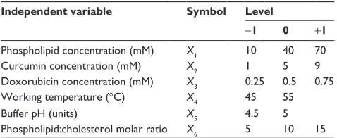
A screening study was conducted in order to find the fac-tors with the most relevant influence on CUR-DOX-LCL characteristics (or CQAs). The experiment was designed and the data were statistically analyzed using Modde 11 Pro software (Umetrics, Malmö, Sweden). Six variables were established as independent factors to be investigated, namely the phospholipid concentration, the phospholipid:cholesterol molar ratio, the CUR concentration, the DOX concentration, the working temperature and the pH of the buffer. Each
independent factor was assigned two levels, low and high, which were represented by the values of -1 and +1, respec-tively (Table 1). Taking into account the number of experi-mental factors and their levels, a 26-2 fractional factorial design
resolution IV was generated, resulting in 19 experiments. The experimental setup is given in Table 2. The investigated responses (dependent variables) were the CQAs of the CUR-DOX-LCL, such as the encapsulated CUR concentration (Y1), the encapsulated DOX concentration (Y2), the EE for CUR (Y3) and DOX (Y4) and the size (Y5) and zeta potential (Y6) of the liposomes. The data were fitted by means of partial least squares using the statistical module of Modde 11 Pro software. The experiments were performed in a random order to reduce the experimental variability.
establishment of the design space
The design space was established with Modde 11 Pro soft-ware, as the combination of factors for which the target
Table 1 independent variables and their levels
Independent variable Symbol Level
-1 0 +1
Phospholipid concentration (mM) X1 10 40 70
curcumin concentration (mM) X2 1 5 9
Doxorubicin concentration (mM) X3 0.25 0.5 0.75
Working temperature (°c) X4 45 55
Buffer ph (units) X5 4.5 5
Phospholipid:cholesterol molar ratio X6 5 10 15
Table 2 experimental matrix containing run parameters and conditions for the preparation of cUr-DOX-lcl
Exp no Exp name Run order X1 X2 X3 X4 X5 X6
1 n1 5 10 1 0.25 45 4.5 5
2 n2 10 70 1 0.25 45 5 5
3 n3 2 10 9 0.25 45 5 15
4 n4 11 70 9 0.25 45 4.5 15
5 n5 9 10 1 0.75 45 5 15
6 n6 19 70 1 0.75 45 4.5 15
7 n7 14 10 9 0.75 45 4.5 5
8 n8 15 70 9 0.75 45 5 5
9 n9 7 10 1 0.25 55 4.5 15
10 n10 4 70 1 0.25 55 5 15
11 n11 12 10 9 0.25 55 5 5
12 n12 17 70 9 0.25 55 4.5 5
13 n13 13 10 1 0.75 55 5 5
14 n14 8 70 1 0.75 55 4.5 5
15 n15 18 10 9 0.75 55 4.5 15
16 n16 3 70 9 0.75 55 5 15
17 n17 6 40 5 0.5 45 4.5 10
18 n18 1 40 5 0.5 45 4.5 10
19 n19 16 40 5 0.5 45 4.5 10
Notes: X1, phospholipid concentration (mM); X2, curcumin concen tration (mM); X3, doxorubicin concentration (mM); X4, working temperature (°c); X5, buffer ph
(units); X6, phospholipid:cholesterol molar ratio.
Abbreviations: cUr, curcumin; DOX, doxorubicin; lcl, long-circulating lipo-somes; exp, experiment.
Drug Design, Development and Therapy downloaded from https://www.dovepress.com/ by 118.70.13.36 on 21-Aug-2020
Dovepress antiproliferative liposomes co-encapsulating DOX and cUr
specifications of the CUR-DOX-LCL were met at specific risk levels. The models developed in the experimental design were validated by performing the preparation and determina-tion of CQAs of one formuladetermina-tion within the design space and one formulation outside the design space. The formulations were characterized, and experimental results, such as the determined CQAs, were compared to the predicted ones, for all six investigated responses, and percentage bias was calculated.
in vitro release study
The in vitro release of CUR and DOX from liposomes was assessed by the dialysis method, according to a modi-fied technique by Li et al.36 The Silde-A-Lyzer® Dialysis
Cassettes (Thermo Scientific) with 10 kDa MWCO were hydrated in the release medium prior to the test. Three mil-liliters of CUR-DOX-LCL was introduced in the dialysis cassette which was immersed in 100 mL release medium. The release medium consisted of a mixture of phosphate buffer saline (pH 5) and absolute ethanol in a ratio of 65:35 (v/v). The samples were maintained at 37°C under continu-ous stirring at 100 rpm. At different time points, namely 0.5, 1, 3, 6, 16, 24, 48 and 72 hours, 1 mL of release medium was withdrawn and replaced with an equal volume of fresh release medium in order to maintain a constant volume dur-ing the whole test. The CUR and DOX concentrations in the samples were determined by HPLC analysis. All experiments were carried out in triplicate and are expressed as mean ± standard deviation.
culturing conditions and treatments
The C26 murine colon carcinoma cells were cultured in complete RPMI 1640 medium supplemented with 10% heat-inactivated fetal bovine serum, at 37°C in a humidified atmosphere containing 5% CO2.37
DOX, in free form (dissolved in phosphate buffer saline) as well as in DOX-LCL form, was tested at various con-centrations (0.05, 0.10, 0.15 and 0.25 μM) to investigate its cytotoxic effect on C26 murine colon cancer cells. Moreover, the same DOX concentrations were tested for cytotoxicity as liposomal formulations co-encapsulated with CUR and DOX, in two different molar ratios, such as DOX:CUR =1:9
and DOX:CUR =1:167.
cell proliferation assay
C26 murine colon cancer cells were seeded at a density of 5×10³ cells/well into 96-well plates. After 12 hours of
incuba-tion, the cells were treated with DOX, DOX-LCL and the two
DOX-CUR-LCL formulations, for an additional 48 hours. The in vitro cytotoxicity of different treatments applied on C26 cells was evaluated by using the ELISA BrdU-colorimetric immunoassay according to the manufacturer’s instructions, as previously described.37 Results are expressed
as percentage of inhibition of cell proliferation compared to that of untreated C26 cells and are presented as the mean ± standard deviation of triplicate measurements.
statistical analysis
All data are expressed as mean ± standard deviation (n=3). For statistical analysis of the effects of different treatments with cytotoxic agents on C26 cell proliferation, a one-way analysis of variance (ANOVA) with Bonferroni correction was used. Correlations between different pharmacological agent concentrations and their cytotoxic effects were evalu-ated by using Pearson correlation coefficient, r. All statis-tical analyses were performed by using GraphPad Prism version 6 for Windows (GraphPad Software Inc., La Jolla, CA, USA). Significance was considered at values of P,0.05 (ns, P.0.05; *, P,0.05; **, P,0.01; ***, P,0.001; ****,
P,0.0001).
Results and discussion
Physicochemical characterization of
cUr-DOX-lcl
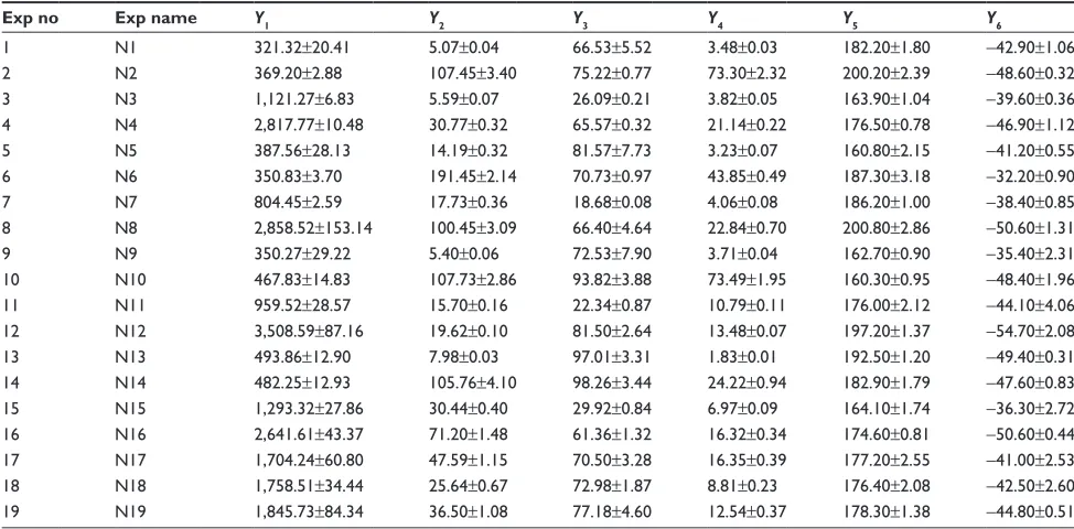
According to the experimental design, 19 experiments were carried out, and the results obtained for the evaluated responses are given in Table 3.
QbD approach
CQAs of liposomal CUR and DOX identified
by risk assessment
In the QbD approach, in order to achieve a desired QTPP, the CQAs of the product have to be established first. A CQA according to ICH Q8(R2) is “a physical, chemical, biological, or microbiological property or characteristic that should be within an appropriate limit, range, or distribution to ensure the desired product quality”.33 According to the current
scientific knowledge and our previous formulation studies, the size, size distribution, zeta potential, drug loading and encapsulation efficiency are the most important attributes of nanoparticulate systems, including liposomes.
The potential risk factors affecting the CQAs of the liposomes co-loaded with CUR and DOX were identified based on the literature and preliminary formulation studies. The factors were divided into two categories: formulation factors and process parameters. The formulation factors
Drug Design, Development and Therapy downloaded from https://www.dovepress.com/ by 118.70.13.36 on 21-Aug-2020
Dovepress
Tefas et al
chosen for our study were the phospholipid concentration, phospholipid:cholesterol molar ratio, CUR concentration and DOX concentration. The ratio between the main lipid (DPPC) and the PEGylated lipid (PEG-DSPE) was kept constant throughout the experimental plan (5% PEGylated lipid of the total phospholipid concentration) and was not considered a formulation factor. The reason behind PEGyla-tion is to improve the stability and biological performance of liposomes in vivo by extending their circulation time in the bloodstream.38 Since the proposed liposomal system was
not yet assessed in vivo, it was not considered necessary to study the influence of the degree of PEGylation on the char-acteristics of the liposomes. Also, usually, the proportion of PEGylated phospholipids which confers stealth properties is quite low, approximately 5–7 mol%, but enough to pre-vent fast elimination of liposomes from the bloodstream.39
Among the process parameters, the temperature and the pH of the buffer were evaluated. The temperature maintained during the extrusion and hydration steps was above the transition temperature of the phospholipids. Also, the pH of the phosphate buffer was set such that the stability of the drugs was ensured.
The size and polydispersity are critical physicochemi-cal properties which determine the in vivo uptake and fate of the liposomes.40 These parameters can be modulated in
order to specifically direct the distribution of liposomes in vivo. Generally, nanoparticulate systems of approximately
100 nm have longer circulation half-lives. Also, nanoparticles with a size of 100–200 nm have been shown to be able to extravasate through the fenestrations of the endothelial lin-ing of blood vessels and leak into tumors to a greater extent than in normal tissues due to the enhanced permeation and retention effect.41 However, by ensuring a small size for
the liposomes, often the necessary dose of active substance to be delivered is reduced. Thus, the most appropriate size for nanoparticles, including liposomes, which can deliver a corresponding dose of therapeutic agent, is usually in the range of 10–200 nm.42 A size within the previously
men-tioned range can be achieved by extruding the liposomes through membranes with a specific porosity at a tempera-ture above the phase transition temperatempera-ture of the lipids. According to the risk analysis carried out, the phospholipid concentration, the ratio of phospholipids to cholesterol and the extrusion temperature are the most influential parameters which affect the liposome size.
The zeta potential is an important parameter which can directly influence the stability of a liposomal dispersion. It is agreed that particles can aggregate due to insufficient electric repulsion between them when the zeta potential values range between -30 and +30 mV.43,44 Thus, the zeta
potential was chosen as a CQA of CUR-DOX-LCL. The most important factors influencing zeta potential, identified by the risk assessment, are the type and concentration of lipids and the physicochemical properties of the active substance.
Table 3 experimental data for the studied dependent variables
Exp no Exp name Y1 Y2 Y3 Y4 Y5 Y6
1 n1 321.32±20.41 5.07±0.04 66.53±5.52 3.48±0.03 182.20±1.80 -42.90±1.06
2 n2 369.20±2.88 107.45±3.40 75.22±0.77 73.30±2.32 200.20±2.39 -48.60±0.32
3 n3 1,121.27±6.83 5.59±0.07 26.09±0.21 3.82±0.05 163.90±1.04 -39.60±0.36
4 n4 2,817.77±10.48 30.77±0.32 65.57±0.32 21.14±0.22 176.50±0.78 -46.90±1.12
5 n5 387.56±28.13 14.19±0.32 81.57±7.73 3.23±0.07 160.80±2.15 -41.20±0.55
6 n6 350.83±3.70 191.45±2.14 70.73±0.97 43.85±0.49 187.30±3.18 -32.20±0.90
7 n7 804.45±2.59 17.73±0.36 18.68±0.08 4.06±0.08 186.20±1.00 -38.40±0.85
8 n8 2,858.52±153.14 100.45±3.09 66.40±4.64 22.84±0.70 200.80±2.86 -50.60±1.31
9 n9 350.27±29.22 5.40±0.06 72.53±7.90 3.71±0.04 162.70±0.90 -35.40±2.31
10 n10 467.83±14.83 107.73±2.86 93.82±3.88 73.49±1.95 160.30±0.95 -48.40±1.96
11 n11 959.52±28.57 15.70±0.16 22.34±0.87 10.79±0.11 176.00±2.12 -44.10±4.06
12 n12 3,508.59±87.16 19.62±0.10 81.50±2.64 13.48±0.07 197.20±1.37 -54.70±2.08
13 n13 493.86±12.90 7.98±0.03 97.01±3.31 1.83±0.01 192.50±1.20 -49.40±0.31
14 n14 482.25±12.93 105.76±4.10 98.26±3.44 24.22±0.94 182.90±1.79 -47.60±0.83
15 n15 1,293.32±27.86 30.44±0.40 29.92±0.84 6.97±0.09 164.10±1.74 -36.30±2.72
16 n16 2,641.61±43.37 71.20±1.48 61.36±1.32 16.32±0.34 174.60±0.81 -50.60±0.44
17 n17 1,704.24±60.80 47.59±1.15 70.50±3.28 16.35±0.39 177.20±2.55 -41.00±2.53
18 n18 1,758.51±34.44 25.64±0.67 72.98±1.87 8.81±0.23 176.40±2.08 -42.50±2.60
19 n19 1,845.73±84.34 36.50±1.08 77.18±4.60 12.54±0.37 178.30±1.38 -44.80±0.51
Notes: Y1, encapsulated curcumin concentration (μg/ml); Y2, encapsulated doxorubicin concentration (μg/ml); Y3, curcumin entrapment efficiency (%); Y4, doxorubicin entrapment efficiency (%); Y5, size (nm); Y6, zeta potential (mV). The data are presented as mean values ± standard deviation.
Abbreviation: exp, experiment.
Drug Design, Development and Therapy downloaded from https://www.dovepress.com/ by 118.70.13.36 on 21-Aug-2020
Dovepress antiproliferative liposomes co-encapsulating DOX and cUr
Liposome stability is reportedly related to the rigidity of the lipid bilayer, and thus, an important requirement is select-ing the appropriate lipids in terms of chain length, degree of unsaturation and phase transition temperature.45 We have
chosen DPPC and DSPE (PEGylated) as phospholipids for our study due to their suitable properties: long acyl chain (16C for DPPC and 18C for DSPE, respectively), saturated nature and high transition temperature. Also, cholesterol has been shown to have an impact on the rigidity of the lipid membrane. Studies investigating the role of cholesterol in liposome formulation have revealed that it increases the packing of phospholipids, tempers the particle aggregation and reduces the membrane permeability.45
Designing liposomes with optimum therapeutic payload is an essential prerequisite to ensure the delivery of an adequate dose of active substance to the site of action. The phospholipid concentration, content of cholesterol and drug concentrations have been previously shown to influence the drug loading and EE. Therefore, we have selected these two latter properties as CQAs of liposomal CUR and DOX.
After the risk analysis, our next focus was to investigate the impact of the risk factors, both formulation and process variables, on the CQAs of CUR-DOX-LCL, by an experi-mental design.
DOe
It is ideal to develop a pharmaceutical product having desirable quality characteristics by running a minimum number of experiments, in the shortest time possible. The traditional approach which involves changing one variable at a time, while keeping the others constant, requires a lot of time, energy and resources. As opposed to this classical technique, experimental design allows to simultaneously modulate several input variables.46 DOE is a powerful and
useful tool for evaluating the relation between a set of input variables and the investigated experimental responses.
The first step in conducting an experimental design is choosing the appropriate type of design, which depends on the number of investigated variables. In this study, six factors were evaluated, among which four, namely the phospholipid, CUR and DOX concentrations and the phospholipid:cholesterol molar ratio, were quantitative-type factors, while the remaining two factors, the temperature and the pH of the buffer, were qualitative-type variables. Another important aspect in planning an experimental design is to select an adequate level range for each formulation factor. All chosen independent variables were evaluated at two different levels, with the purpose of identifying the factors with a
significant influence on the output variables. Hence, for the reasons of saving time and resources, a two-level fractioned factorial design was proposed as the most suitable one.
Summary of fit, fitting the model to the experimental data and regression coefficient analysis
The most important statistical parameters to be calculated when fitting a regression model to the experimental data are the coefficient of determination or R2 and the coefficient
of prediction or Q2. R2 is known as “goodness of fit”, and
it shows the extent to which the data fit the model. On the other hand, Q2, called “goodness of prediction”, reflects the
model’s power of prediction. What is more, model validity indicates whether the appropriate model was chosen, while reproducibility denotes a summary of variability.47
According to Figure 1, the values of R2 and Q2 were
greater than 0.5 indicating that the chosen model fitted well to the set of experimental data and showing a sufficiently good predictive power. In addition, the model validity and the reproducibility were greater than 0.25 and 0.5, respectively, thus suggesting a reduced experimental error.
Also, the ANOVA test was performed to assess the sig-nificance of the regression model and to confirm the model accuracy (Table 4). The low probability values (P,0.05) obtained for the regression model indicated the significance of the model. The validity of the model was also supported
1
0.9 0.8 0.7 0.6 0.5 0.4
0.3 0.2 0.1 0
Y1 Y2 Y3 Y4 Y5 Y6
R2 Q2 Model validity Reproducibility
Figure 1 Summary of fit for the models.
Notes: The abscissa shows the statistical parameters R2 and Q2, the model
validity and the reproducibility for the evaluated responses (Y1, encapsulated cUr
concentration [μg/ml]; Y2, encapsulated DOX concentration [μg/ml]; Y3, cUr entrapment efficiency [%]; Y4, DOX entrapment efficiency [%]; Y5, size of the
liposomes [nm]; Y6, zeta potential of the liposomes [mV]). The ordinate indicates
the values for R2, Q2, the model validity and the reproducibility ranging from 0 to 1.
Abbreviations: cUr, curcumin; DOX, doxorubicin.
Drug Design, Development and Therapy downloaded from https://www.dovepress.com/ by 118.70.13.36 on 21-Aug-2020
Dovepress
Tefas et al
by the absence of lack of fit (P.0.05). Overall, the results confirm that the chosen model was adequate and reliable and had a good predictive power.
Model regression coefficients were estimated and are presented in Figure 2. The coefficient plots illustrate the influence of formulation and process variables on the studied response.48
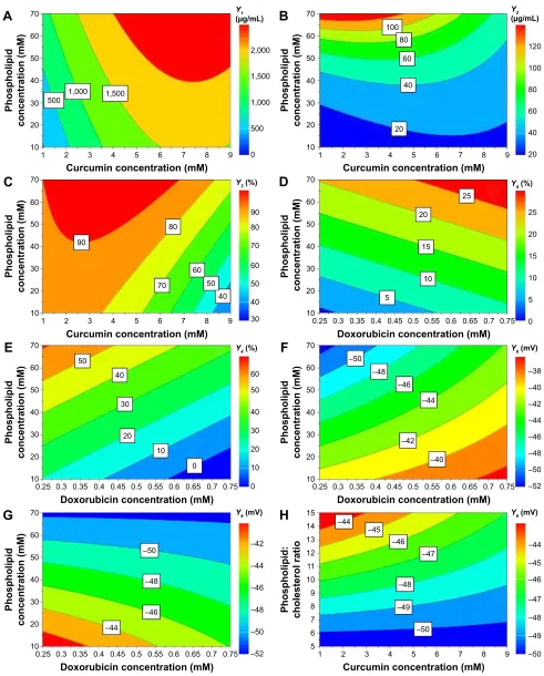
Contour plots were generated for a better understanding of the main effects and the interactions between the investi-gated variables and are illustrated in Figure 3.
Influence of variables on the encapsulated drug
concentration
The encapsulated CUR concentrations ranged between 321.32±20.41 and 3,508.59±87.16 μg/mL, while entrapped DOX concentrations varied from 5.07±0.04 to 191.45± 2.14 μg/mL. The results from the ANOVA test (Table 4) indicated a significant influence of the independent variables on the response (P=0.000) and absence of lack of fit (P=0.212 for CUR and P=0.449 for DOX).
According to Figure 2A and B, the concentrations of both encapsulated CUR and DOX were influenced by the phospholipid concentration. An increase in the amount of phospholipid led to an increase in the amount of CUR and DOX entrapped in the liposomes. The positive influence that the phospholipid concentration had on these responses could be explained by the fact that a larger amount of phospholipids could accommodate a larger amount of drug. Our results are in accordance with those published by Sailor et al.49
Both drugs positively influenced their own tions, but had no effect whatsoever on each other’s concentra-tion. By increasing the amount of drug during the preparation process, the concentration of entrapped drug increased, both for CUR and for DOX. This shows that the saturation concentration of liposomes was not reached, and more drug could be incorporated into the particles.50
No effect on the encapsulated drug concentration was observed when varying the phospholipid:cholesterol molar ratio, the temperature or the pH of the buffer solution.
The interactions between the variables for encapsulated drug concentration were also investigated and are depicted by the contour plots in Figure 3A. Results showed a synergistic effect of the phospholipid and CUR concentrations on the entrapped CUR concentration. It appears that entrapped CUR concentration increases when simultaneously increasing the phospholipid and CUR concentrations, while keeping the temperature at 55°C and the pH at 5. However, for phospholipid concentrations below 40 mM, the increase of CUR concen-tration over 6 mM does not result in a further increase of encapsulated CUR, so the saturation concentration is reached in these conditions. Regarding the entrapped DOX concentra-tion, the contour plot seen in Figure 3B reveals that on the one hand at low phospholipid concentration, the CUR concentration had a somewhat positive effect, but on the other hand at high concentrations of phospholipids, the entrapped DOX concen-tration decreased when increasing CUR concenconcen-tration.
Influence of variables on the encapsulation efficiency
The EE was found to be between 18.68%±0.08% and 98.26%±3.44% for CUR and from 1.83%±0.01% to 73.49%±1.95% for DOX. According to the results of the ANOVA test shown in Table 4, the investigated variables exhibited a significant effect on the entrapment efficiencies of CUR and DOX (P=0.000). Also, the chosen model did not show a significant lack of fit (P=0.118 for Y3 response and P=0.346 for Y4 response).
As shown in Figure 2C and D, increasing the phospho-lipid concentration significantly increased the percentage of encapsulated CUR and DOX, respectively.
It appears that both the drugs negatively influenced their own entrapment efficiencies, as indicated by the negative values of the regression coefficients. The EE is depen-dent on the initial amount of drug and the entrapped drug
Table 4 statistical parameters – one-way analysis of variance test for studied responses
Response SS df MS (variance) F P-value Lack of fit R2 Q2 Model validity
Y1 197,836a 11a 17,985.10a 125.64b 0.000b 0.212 0.986 0.962 0.611
Y2 0.323a 12a 0.027a 24.71b 0.000b 0.449 0.925 0.811 0.799
Y3 1,564.34a 12a 130.36a 21.23b 0.000b 0.118 0.914 0.780 0.465
Y4 315.39a 11a 28.67a 40.56b 0.000b 0.346 0.963 0.877 0.734
Y5 456.74a 12a 38.06a 11.61b 0.000b 0.659 0.853 0.607 0.895
Y6 28.94a 8a 3.62a 16.80b 0.000b 0.583 0.955 0.791 0.865
Notes: aValues correspond to residuals. bValues for regression. R2, regression coefficient; Q2, predictive power of the model; Y
1, encapsulated curcumin concentration
(μg/ml); Y2, encapsulated doxorubicin concentration (μg/ml); Y3, curcumin entrapment efficiency (%); Y4, doxorubicin entrapment efficiency (%); Y5, size (nm); Y6, zeta
potential (mV).
Abbreviations: ss, sum of squares; df, degrees of freedom; Ms, mean of square; F, Fisher’s ratio; P, probability.
Drug Design, Development and Therapy downloaded from https://www.dovepress.com/ by 118.70.13.36 on 21-Aug-2020
Dovepress antiproliferative liposomes co-encapsulating DOX and cUr
concentration, but it appears that it is a nonlinear relation, according to the EE equation. Therefore, it is possible for high initial amounts of drugs to translate to reduced EE values.
While DOX had no effect on the CUR EE, CUR led to a decrease in DOX EE. Due to the lipophilic nature of the mol-ecule, CUR is incorporated into the liposomes by insertion and entrapment in the lipid bilayer. On the other hand, DOX is readily soluble in water, and thus should be incorporated in the hydrophilic interior of the liposomes. However, it has been shown that due to its amphiphilic structure, DOX can
interact with the lipid membrane and possibly be incorporated in the bilayer of the liposomes.51 Our results suggested that
CUR, at higher concentrations, hindered the incorporation of DOX into the liposomes.
The EE was also affected by the working conditions so that CUR EE was influenced by temperature, and DOX EE was influenced by the pH of the buffer.
Concerning CUR, the EE was higher when working at 55°C and lower at 45°C. Only CUR EE was influenced by the temperature possibly because of its hydrophobic nature Figure 2 Regression coefficient plots showing the influence of formulation factors on the encapsulated CUR concentration (A), the encapsulated DOX concentration (B), the ee for cUr (C), the ee for DOX (D), the size of the liposomes (E) and the zeta potential of the liposomes (F).
Notes: Pho refers to X1, phospholipid concentration (mM); cur refers to X2, curcumin concentration (mM); Dox refers to X3, doxorubicin concentration (mM); Wor refers
to X4, working temperature (°c); ph refers to X5, buffer ph (units); Ph:ch refers to X6, phospholipid:cholesterol molar ratio.
Abbreviations: cUr, curcumin; DOX, doxorubicin.
&
'
$
±
± ± ± ±
±
± ±
± ±
± ± ± ± ± ±
± ± ± ±
3KR &XU 'R[
S+XQLWV S+XQLWV &XUî&XU
'R[îS+XQLWV'R[îS+XQLWV
3KR &XU 'R[
S+XQLWV S+XQLWV &XUî&XU 3KRî&XU
3KR 'R[
:RU& :RU&
S+XQLWV S+XQLWV S+&K
S+XQLWVîS+&KS+XQLWVîS+&K
3KR &XU 'R[
:RU&:RU& S+XQLWVS+XQLWV
S+&K 3KRî'R[
3KRî: RU&
3KRî: RU&
&XUî3K&K
'R[îS+XQLWV'R[îS+XQLWV ±
± ± ±
<
ȝJP/H[WHQGHG
<
H[WHQGHG
<
QPH[WHQGHG
<
H[WHQGHG
<
P9H[WHQGHG
<
ȝJP/H[WHQGHG
%
(
)
3KR &XU 'R[
:RU& :RU& &XUî&XU 3KRî&XU
3KR &XU 'R[
S+XQLWVS+XQLWV
3KRîS+XQLWV3KRîS+XQLWV&XUîS+XQLWV&XUîS+XQLWV'R[îS+XQLWV'R[îS+XQLWV
Drug Design, Development and Therapy downloaded from https://www.dovepress.com/ by 118.70.13.36 on 21-Aug-2020
Dovepress
Tefas et al
Figure 3 contour plots showing the interaction between variables on the responses: the effect of X1X2 on entrapped cUr concentration, at 55°c and ph 5 (A); the effect of X1X2
on entrapped DOX concentration, at 55°c and ph 5 (B); the effect of X1X2 on CUR entrapment efficiency, at 55°c and ph 5 (C); the effect of X1X3 on DOX entrapment efficiency,
at 55°c and ph 4.5 (D) and 5 (E); the effect of X1X3 on zeta potential, at 55°c and ph 4.5 (F) and 5 (G); and the effect of X2X6 on zeta potential, at 55°c and ph 5 (H).
Notes: X1, phospholipid concentration (mM); X2, curcumin concentration (mM); X3, doxorubicin concentration (mM); X6, phospholipid:cholesterol molar ratio.
Abbreviations: cUr, curcumin; DOX, doxorubicin.
&XUFXPLQFRQFHQWUDWLRQP0 3KRVSKROLSLG FKROHVWHUROUDWL R 3KRVSKROLSLG FRQFHQWUDWLRQP0 3KRVSKROLSLG FRQFHQWUDWLRQP0 3KRVSKROLSLG FRQFHQWUDWLRQP0 3KRVSKROLSLG FRQFHQWUDWLRQP0 3KRVSKROLSLG FRQFHQWUDWLRQP0 3KRVSKROLSLG FRQFHQWUDWLRQP0 3KRVSKROLSL G FRQFHQWUDWLRQP0 'R[RUXELFLQFRQFHQWUDWLRQP0 &XUFXPLQFRQFHQWUDWLRQP0 &XUFXPLQFRQFHQWUDWLRQP0 &XUFXPLQFRQFHQWUDWLRQP0 'R[RUXELFLQFRQFHQWUDWLRQP0 'R[RUXELFLQFRQFHQWUDWLRQP0 'R[RUXELFLQFRQFHQWUDWLRQP0 ± ± ± ± ± ± ± ± ± ± ± ± ± ± ± ± ± ± ± ± ± ± ± ± ± ± ± ± ± ± ± ± ± ± ± ± ± ±
$
%
&
'
(
)
*
+
<JP/ <JP/
< <
<P9
<P9 <P9
<
Drug Design, Development and Therapy downloaded from https://www.dovepress.com/ by 118.70.13.36 on 21-Aug-2020
Dovepress antiproliferative liposomes co-encapsulating DOX and cUr
and its ability to interact and mix with the melted lipids. On the other hand, DOX EE was increased when the pH of the buffer was 5, but decreased at pH 4.5.
Although we could not highlight the influence of the phospholipid:cholesterol molar ratio on drug encapsulation efficiency in this study, other reports state that drug loading was highly influenced by cholesterol concentrations. Gener-ally, a larger proportion of cholesterol results in increased EE. Due to greater rigidity, the permeability of the lipid bilayer is reduced and drug leakage is prevented, which translates into enhanced drug encapsulation.52
The contour plots in Figure 3C–E show a strong inter-action between the phospholipid concentration and the drug concentration on CUR encapsulation efficiency and DOX encapsulation efficiency. It can be deducted from the graphical representation that the phospholipid and CUR con-centrations had antagonistic effects on CUR encapsulation efficiency. As discussed earlier, increasing the phospholipid concentration led to higher CUR EE values, and the effect was more important at lower CUR concentrations. At CUR concentrations over 3 mM, the increase of EE with phos-pholipid concentration was not so important. Figure 3D and E illustrates the interaction between the phospholipid concentration and DOX concentration with regard to DOX EE, at different pH values of the phosphate buffer. At pH 4.5 (Figure 3D), the phospholipid and DOX concentrations showed a synergistic effect, while at pH 5 (Figure 3E), the two variables had an antagonistic effect on DOX EE. The DOX EE varied dramatically when increasing the phospho-lipid concentration, but the variation was rather moderate by modifying the DOX concentration, regardless of the pH. Hence, it can be concluded that the phospholipid concentra-tion had the most important impact on the interacconcentra-tion regard-ing the effect on DOX EE.
Influence of variables on the size of the liposomes
The mean size, polydispersity and zeta potential are important physicochemical parameters which can influence the release of the entrapped drug and the therapeutic outcome.43,53
The mean particle size varied from 160.30±0.95 to 200.80±2.86 nm. Regarding the particle size distribution, the polydispersity index of the liposomal formulations had values from 0.028±0.02 to 0.120±0.03 (data not shown). The statistical analysis of the data for particle size (Table 4) indicated the significant influence which the independent variables exhibited (P=0.000) and the absence of lack of fit (P=0.659).
Figure 2E reveals that the composition of the lipid bilayer influenced the size of the liposomes. Increasing the phos-pholipid concentration resulted in the formation of larger liposomes, since phospholipids are the main component of the liposomal lipid bilayer.
However, the phospholipid:cholesterol molar ratio had an opposite effect on the response. Cholesterol is frequently used in the preparation of liposomes and plays a key role in the formation and stability of these nanoparticulate systems. It has a direct influence on the fluidity and permeability of the liposomes.52 The decrease in size observed with an increase
in phospholipid:cholesterol molar ratio is the result of a higher flexibility of the lipid bilayer. A higher concentra-tion of cholesterol renders a more rigid structure of the lipid membrane, and thus a more stable formulation.54
Neither the concentration of drugs nor the working condi-tions influenced the size of the liposomes. The temperature was adequately selected for this study to be over the transi-tion temperature of DPPC (41°C).55 During melting, the
phospholipids undergo transformation from a gel to a fluid phase. According to some research groups, this phenomenon is accompanied by reduction in liposome size due to a more compact packing of the lipids.56 Although unobservable in
the present study, it is possible that the preparation at 45°C, which was closer to the transition temperature of the phos-pholipids, resulted in smaller-size liposomes. On the other hand, it was reported by Roy et al that above the transition temperature of the lipid, the size of the liposomes increased as a result of an increase in volume.56
Influence of variables on the zeta potential
All liposomal formulations showed a negative surface charge as zeta potential ranged from -54.70±2.08 to -32.20±0.90 mV. According to the ANOVA results from Table 4, the zeta potential response can be well described by the chosen model (P=0.000), and the validity of the model can be confirmed by the absence of lack of fit (P=0.583).
The formulation factors with the most impact on zeta potential were the phospholipid concentration and the phospholipid:cholesterol molar ratio, but the observed effects were opposite (Figure 2F). By increasing the phospholipid concentration, the surface charge of the particles decreased. The negative surface charge which the liposomes exhibited is due to the type of structural phospholipids and, according to the literature, is consistent with the liposomes prepared with phosphatidylcholine.43 These findings are in agreement
with those reported by others.55,57 DPPC is a zwitterionic-type
Drug Design, Development and Therapy downloaded from https://www.dovepress.com/ by 118.70.13.36 on 21-Aug-2020
Dovepress
Tefas et al
phospholipid which can change the orientation of its polar groups depending on the ionic strength of the medium. In low-ionic strength conditions, the net surface charge of the particles is negative, while the opposite can be observed with high ionic strength.55 Other research groups reported
that by adding a PEGylated lipid, the zeta potential would shift toward more negative values.52 The magnitude of the
zeta potential depends on the charge at the surface of the particles. The decrease in zeta potential with the increase in phospholipid concentration could be due to the presence of more phosphocholine moieties on the unit surface of the liposomes.
In contrast to these results, the zeta potential increased when increasing the phospholipid:cholesterol molar ratio. We observed that the higher the ratio, the lower the zeta potential absolute values. The bigger the concentration of phospho-lipids in the case of an increased phospholipid:cholesterol molar ratio, the higher the zeta potential value.
Although the drug concentrations had no influence on the response, the contrary was observed for the process parameters. We observed an increase in zeta potential values when the liposome preparation was carried out at 45°C, using phosphate buffer with pH 4.5, but the surface charge was lower when working at 55°C and pH 5. The shift in zeta potential could be related to the ionic strength of the buffer solution. The phosphate buffer solutions had different molari-ties, namely 0.05 M for the buffer with pH 4.5 and 0.02 M for the buffer with pH 5, and thus different ionic strengths. The underlying explanation would be that by increasing the ionic strength, a more compact layer of ions formed around the particles which consequently led to shielding the surface charge to a greater extent.58
Several interactions between variables were identified and showed significant effects on zeta potential (Figure 3F–H). As with the DOX EE discussed in the previous section, there was an interaction between phospholipid and DOX concentrations which influenced the zeta potential. It is clear from the contour plots that this interaction exhibited a different effect on zeta potential depending on the pH of the phosphate buffer. In both cases, an increase in phospholipid concentration resulted in decrease in zeta potential, but at different pH values, the DOX concentration had antagonistic effects on the zeta potential. At pH 4.5 (Figure 3F), by increasing the DOX concentration, the zeta potential absolute value decreased, while the opposite was observed at pH 5 (Figure 3G). Furthermore, the analysis showed a significant negative interaction between the phospholipid:cholesterol molar ratio and the CUR concentration on the zeta potential
of the liposomes, at 55°C and pH 5. zeta potential increased when increasing the phospholipid:cholesterol molar ratio, but for CUR concentrations over 5 mM, the increase was not so important.
establishment and evaluation of the design space
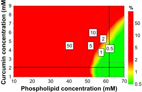
The design space is a region in the experimental domain in which any possible combination of formulation variables and process parameters assures obtaining formulations with desired characteristics.31 For CUR-DOX-LCL, the design
space is the green area of the plot shown in Figure 4, each point of this area being a possible formulation for which the prediction of the CQAs is made with a probability of failure of less than 1%. Moreover, the design space hypercube is shown in the same plot as the dotted frame inside, where factor values can vary independent from each other without influencing the obtaining of a product with the quality within specifications.
Based on the obtained results, four variables, namely the phospholipid concentration, the phospholipid:cholesterol molar ratio, the CUR concentration and DOX concentration, were selected as key parameters with the greatest impact on the CQAs of CUR-DOX-LCL. Therefore, the design space was constructed using these four variables, by set-ting the acceptance criteria for the investigated responses. In this sense, the size of the liposomes was minimized, while the entrapment efficiencies for both CUR and DOX were maximized. The zeta potential and the encapsulated drug concentrations were excluded from this evaluation. As long as the independent variables are kept within the limits of the hypercube (Table 5), the target quality of the liposomal formulations can be assured and controlled. The combina-tion of variables within the design space hypercube located at the intersection of the two perpendicular black lines
Figure 4 Design space for the formulation of cUr-DOX-lcl, represented as a function of cUr concentration and phospholipid concentration.
Abbreviations: cUr, curcumin; DOX, doxorubicin; lcl, long-circulating liposomes.
&XUFXPLQFRQFHQWUDWLRQP0
3KRVSKROLSLGFRQFHQWUDWLRQP0
Drug Design, Development and Therapy downloaded from https://www.dovepress.com/ by 118.70.13.36 on 21-Aug-2020
Dovepress antiproliferative liposomes co-encapsulating DOX and cUr
corresponds to the robust set point which has the lowest prediction error.
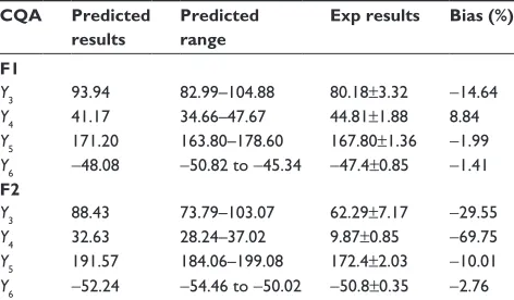
The validation was carried out by performing the prepa-ration and evaluation of one formulation within the defined design space hypercube (F1, corresponding to the robust set point) and one outside the established design space (F2). According to data, robustness of the manufacturing process was assured by using a higher phospholipid concentration (62 mM), a higher phospholipid:cholesterol molar ratio (13.67) and a higher DOX concentration (0.65 mM), but a lower CUR concentration (2.07 mM), and also by operating in conditions of 55°C and pH 5 of the phosphate buffer. Working in the previously described optimum conditions would ensure obtaining liposomal dispersions with desired CQAs (Table 6) and minimizing process variability. The formulation corresponding to the robust set point was pre-pared in triplicate, and the obtained experimental results were compared to the predicted ones. Except for the CUR EE, experimental values of CQAs were in the predicted range, and bias had low values, thus suggesting that the proposed model had a good ability of predicting the manufacturing process of liposomes. By operating in conditions outside the design space, namely 50 mM phospholipid concentra-tion, 5:1 phospholipid:cholesterol molar ratio, 5 mM CUR
concentration and 0.5 mM DOX concentration, at 55°C and using phosphate buffer of pH 5, we obtained CQAs outside the target range and high percentage biases, indicating a poor correlation between the experimental results and the predicted ones.
in vitro release study
In this paper, a dialysis bag method was employed to study the release of CUR and DOX from PEGylated liposomes. Prior to performing this assay, various release mediums were tested to find the best medium in which DOX and CUR show adequate solubility (data not shown). The most suitable release medium was found to be a mixture of phosphate buf-fer saline of pH 5 and ethanol (65:35, v/v). According to our previous solubility studies, 100 mL of this release medium provided sink conditions. The release test was carried out on the formulation corresponding to the robust set point. The release profile of CUR and DOX from liposomes is illus-trated in Figure 5. The results indicated that both CUR and DOX show a biphasic release from the liposomes, with rapid release in the first hours followed by sustained release up to 72 hours. The initial burst effect seen with both drugs could be attributed to the CUR and DOX incorporated at the sur-face of the liposomes.8 The further sustained-release pattern
might be due to the drugs encapsulated more deeply in the liposome structure. The accumulated drug release percent-age at 72 hours was 84.34% and 33.50% for DOX and CUR, respectively. The relatively low percentage of released CUR might be owed to the slow diffusion through the lipid bilayer. These findings are in agreement with those from other studies which reported that liposomes could provide a depot effect, especially in PEGylated formulations.59,60
in vitro cytotoxic effects of DOX and
cUr co-encapsulated in cUr-DOX-lcl
Several studies have demonstrated the ability of CUR to enhance the therapeutic potential of cytotoxic agents against
Table 5 Design space hypercube limits for independent variables, at 55°c and ph 5 of phosphate buffer
Independent variable Lower limit Upper limit
Phospholipid concentration (mM) 58 70
Phospholipid:cholesterol molar ratio 7.667 15
curcumin concentration (mM) 1 3.134
Doxorubicin concentration (mM) 0.617 0.75
Table 6 experimental and predicted results for the formulation within the design space and the formulation outside the design space
CQA Predicted
results
Predicted range
Exp results Bias (%)
F1
Y3 93.94 82.99–104.88 80.18±3.32 -14.64
Y4 41.17 34.66–47.67 44.81±1.88 8.84
Y5 171.20 163.80–178.60 167.80±1.36 -1.99
Y6 -48.08 -50.82 to -45.34 -47.4±0.85 -1.41 F2
Y3 88.43 73.79–103.07 62.29±7.17 -29.55
Y4 32.63 28.24–37.02 9.87±0.85 -69.75
Y5 191.57 184.06–199.08 172.4±2.03 -10.01
Y6 -52.24 -54.46 to -50.02 -50.8±0.35 -2.76
Notes: F1, formulation within the design space; F2, formulation outside the design space; Y3, curcumin entrapment efficiency (%); Y4, doxorubicin entrapment efficiency (%); Y5, size (nm); Y6, zeta potential (mV).
Abbreviations: cQa, critical quality attribute; exp, experimental.
Figure 5 In vitro release profile of CUR and DOX from liposomes.
Abbreviations: cUr, curcumin; DOX, doxorubicin.
Percentage released
100 90 80 70 60 50 40 30 20 10 0
0 10 20 30 40
Time (hours)
50 60 70 80
Doxorubicin Curcumin
Drug Design, Development and Therapy downloaded from https://www.dovepress.com/ by 118.70.13.36 on 21-Aug-2020
Dovepress
Tefas et al
various types of cancers including colon cancer.61,62
Par-ticularly, the coadministration of CUR has been ascribed to boost DOX, 5-fluorouracil, oxaliplatin, gemcitabine or camptothecin cytotoxic actions.25,63,64 In this experiment, we
aimed to evaluate the cytotoxic effects of LCL formulations based on the co-encapsulation of pharmacological agents, CUR and DOX, in C26 murine colon cancer cells. As the therapeutic potential of DOX is limited by the adverse side effects65 and in order to maximize its cytotoxic actions in
in vivo experiments, we encapsulated this agent in LCL and tested its cytotoxicity toward C26 cell proliferation. We reported herein that upon 48 hours of treatment of C26 murine colon cancer cells, liposomal DOX (DOX-LCL) was able to significantly reduce cell proliferation compared to its free form (DOX), at all tested concentrations (Figure 6A and B, P,0.05). Moreover, this effect was visible at con-centrations approximately two- to threefold lower than that of the free drug. This is in agreement with previous studies that demonstrated a remarkable enhancement of the cytotoxic actions of DOX when included in liposomes, nanoparticles or other lipid-based formulations.66–70 To enhance the
effi-cacy of DOX for further in vivo studies, we designed two liposomal formulations based on the co-encapsulation of DOX and CUR. The composition of these formulations was established using the Optimizer function in Modde 11 Pro software. Thus, the co-encapsulated liposomal formulations
were designed such as to get two different DOX:CUR molar ratios, that is, 1:9 and 1:167. The results related to the
cyto-toxic effects of CUR-DOX-LCL at DOX:CUR =1:9 molar
ratio are presented in Figure 6A and demonstrated a strong DOX concentration-dependent cytotoxic effect in murine C26 colon carcinoma cells (r=0.981, P=0.018 for free DOX;
r=0.954, P=0.046 for DOX-LCL; r=0.7954, P=0.204 for CUR-DOX-LCL). Irrespective of the concentration, the anti-proliferative effects of CUR-DOX-LCL (DOX:CUR =1:9) on C26 cells could not exceed that of DOX-LCL, at all concen-trations tested (P.0.05). On the other hand, CUR-DOX-LCL at DOX:CUR =1:167 molar ratio was able to inhibit cell pro-liferation with more than 75% compared to the propro-liferation of control cells at the highest concentrations tested (0.25 μM DOX +41.85 μM CUR) but to a similar extent to that noted after C26 cell incubation with 0.25 μM DOX administered as free as well as LCL form (Figure 6B). Interestingly, a synergistic cytotoxicity on cancer cells (about 75%) was assessed, when CUR-DOX-LCL was administered at the following concentrations of active agents: 0.10 μM DOX+16.74 μM CUR. The administration of 0.10 μM DOX as free and LCL form exerted only moderate antiprolifera-tive effects (29% and 45%, respecantiprolifera-tively) on C26 cells. Our results showed that the co-incorporation of DOX and CUR in LCL at a ratio of 1:9 did not improve the cytotoxicity of DOX-LCL. Moreover, by increasing the liposomal content of
Figure 6 effects of cUr-DOX-lcl on the proliferation of c26 murine colon carcinoma cells.
Notes: The results show cell proliferation at 48 hours after incubation of c26 cells with 0.05–0.25 μM DOX, as a free form, DOX-lcl, and lcl co-encapsulated with cUr and DOX at a molar ratio of (A) DOX:cUr =1:9 and (B) DOX:cUr =1:167. Data are presented as the mean ± standard deviation of triplicate measurements. The results are expressed as percentage of inhibition of c26 cell proliferation following DOX, DOX-lcl and cUr-DOX-lcl treatments, compared to the proliferation of control cells
(untreated cells). Significance was considered at values of P,0.05 (ns, P.0.05; *P,0.05; **P,0.01; ***P,0.001; ****P,0.0001). Abbreviations: CUR, curcumin; DOX, doxorubicin; LCL, long-circulating liposomes; ns, not significant.
DOX/DOX+CUR concentration (µM)
Inhibition of cell proliferation
(% of untreated cells
)
Inhibition of cell proliferation
(% of untreated cells
)
DOX/DOX+CUR concentration (µM)
0.05 0.05
0.05+0.45 0.100.100.10+0.90 0.150.150.15+1.3 5
0.250.25
0.25+2.25 0.050.050.05+8.37
0.100.10 0.10+16.74
0.150.15 0.15+25.13
0.25 0.25 0.25+41.85
125
100
75
50
25
0
125
100
75
50
25
0 ns
ns
ns
ns ns
ns ns ns
ns
*** *** *
*
** ***
***
*** ***
**** **** **
* *
***
A
B
DOX LCL-DOX LCL-DOX + CUR
Drug Design, Development and Therapy downloaded from https://www.dovepress.com/ by 118.70.13.36 on 21-Aug-2020
![Figure 1 Summary of fit for the models.entrapment efficiency [%]; liposomes [nm]; the values for Abbreviations:Notes: The abscissa shows the statistical parameters R2 and Q2, the model validity and the reproducibility for the evaluated responses (Y1, encap](https://thumb-us.123doks.com/thumbv2/123dok_us/8091812.1350579/7.612.325.557.429.645/entrapment-efficiency-liposomes-abbreviations-statistical-parameters-reproducibility-responses.webp)