715
© 2017 by the Serbian Biological Society How to cite this article: Stoyanova E, Oreshkova T, Mourdjeva M. Generation of human induced pluripotent stem cells from adipose-derived stromal/stem cells isolated from a 75-year-old patient. Arch Biol Sci. 2017;69(4):715-21.
Generation of human induced pluripotent stem cells from adipose-derived stromal/stem
cells isolated from a 75-year-old patient
Elena Stoyanova*, Tsvetelina Oreshkova and Milena Mourdjeva
Department of Molecular Immunology, Institute of Biology and Immunology of Reproduction, Bulgarian Academy of Sciences, Sofia, Bulgaria
*Corresponding author: [email protected]
Received: March 13, 2017; Revised: May 10, 2017; Accepted: May 17, 2017; Published online: June 6; 2017
Abstract: Human adipose-derived stromal/stem cells (hASCs) have been considered a valuable cell source for generating induced pluripotent stem cells (iPSCs). Adipose tissue is easy to obtain. Moreover, the isolated cells rapidly proliferate to reach the required number of cells. Some authors have already shown that iPSCs can be successfully obtained using adult hASCs. Nevertheless, little is known about the generation of iPSCs using hASCs isolated from the tissues of patients over the age of 70. In this study, we examined the generation of iPSCs from hASCs isolated from a 75-year-old man. We trans-duced hASCs with human transcription factors OCT4, SOX2, c-MYC and KLF4 and observed the formation of human embryonic stem cell (hESC)-like colonies. The efficiency of the reprogramming process was 0.08% at day 18 post-infection. Reprogrammed cells expressed pluripotent state-specific transcription factors OCT4, SOX2, NANOG and KLF4, and were able to differentiate into three germ layers in vitro.
Key words: adipose cells; stem cells; reprogramming; transcription factor
INTRODUCTION
The discovery that somatic cells can be induced into pluripotent cells provides an unique tool for study-ing disease pathogenesis, for toxicological teststudy-ing and for the identification of new therapeutic targets [1,2]. In principle, human induced pluripotent stem cells (iPSCs) can be obtained from various somatic cells and are capable of forming any cell type in the body. Different types of reprogrammed parental cells are not equivalent in their ability to convert into iPSCs.
The majority of iPSC studies use dermal fibro-blasts from adult skin biopsies as a parental cell source. However, the taking of punch biopsies is an invasive procedure and requires more than two weeks to obtain sufficient amounts of cells for reprogram-ming. Moreover, the reprogramming efficiency of adult human fibroblasts using OCT4, SOX2, c-MYC and KLF4 (‘‘Yamanaka’’ factors) is about 0.01% [3]. In comparison, the keratinocytes from a 4-year-old child and 28-35-year-old adult have been reprogrammed to a pluripotent state at least 100-fold more efficiently
and 2-fold faster when compared to the reprogram-ming of the human fibroblasts [4]. The main disad-vantage of using keratinocytes for this purpose is that they accumulate more mutations than cells inside the body due to higher UV exposure.
Sun et al. [8] have reported the efficient genera-tion of iPSCs using hASCs from 40- to 65-year-old patients by transduction with OCT4, SOX2, KLF4 and c-MYC. Recently, Kang et al. [10] described the reprogramming of stem cells and stromal cells derived from the adipose tissues of 27-53 year-old male or female patients. However, there are no reports on the generation of iPSCs using adipose-derived cells from patients aged over 70 years.
In this study, we focused on the investigation of reprogramming capability of hASCs derived from a 75-year-old man. Reprogramming of hASCs was ac-companied by changes in cell morphology, induction of endogenous pluripotency genes and the ability to differentiate into the three germ layers in vitro. Our results indicate that ASCs from elderly patients can be successfully reprogrammed into adult, individual-specific iPSCs.
MATERIALS AND METHODS
Cell culture
Primary hASCs were isolated from an adipose tissue sample of a 75-year-old man donor. Informed consent was obtained from the donor (Project 4/2006 NSF).
The tissue was washed with phosphate buffered sa-line (PBS), and digested with type II collagenase (Sigma-Aldrich) at 37oC for 2 h. Primary cultures of mouse
em-bryonic fibroblasts (MEFs, feeder cells) were prepared from 13-14-day old embryos from imprinting control region (ICR) mice after digestion with 0.05%/0.02% trypsin/EDTA (Sigma-Aldrich) at 37oC for 20 min. The
cells were then washed with PBS and cultured in Dul-becco’s modified Eagle’s medium (DMEM) low glucose (Sigma-Aldrich) supplemented with 10% fetal calf se-rum (FCS; Sigma-Aldrich) and penicillin/streptomycin/ amphotericin B (PAN-Biotech). The culture medium was changed 48 h after cell plating to remove debris and nonadherent cells. After 2 passages, 90% confluent MEFs were mitotically inactivated by 5 mg/mL mito-mycin C (Sigma-Aldrich) at 37oC for 1h.
Embryonic stem cells from the BG01V line (ATCC® Number SCRC-2002TM), as well as the
re-programmed hASCs, were maintained on amitotic
MEFs in embryonic stem cell (ESC) media containing DMEM/F12 (Sigma-Aldrich), 15% FCS, 5% knockout serum replacement (KOSR, Gibco, Invitrogen), 1% MEM non-essential amino acids (Gibco, Invitrogen), 2.0 mM L-alanyl-L-glutamine (Gibco, Invitrogen), 0.1 mM β-mercaptoethanol (Sigma-Aldrich), 4 ng/ ml hFGF-2 (Genaxxon) and penicillin/streptomy-cin/amphotericin B. Cells cultured without a feeder layer were maintained in DMEM/F12 supplemented with 15% KOSR, 10% MEFs-conditioned medium, 1% MEM non-essential amino acids, 2 mM L-alanyl-L-glutamine, 0.1 mM β-mercaptoethanol, 4 ng/ml hFGF-2 and penicillin/streptomycin/amphotericin B. The cultures were passaged mechanically. The plurip-otent cells were cultured on mitomycin-C-inactivated MEFs. All cultures were carried out in a humidified atmosphere at 37°C in 5% CO2.
Preparation of MEF-conditioned medium
MEFs at 80% confluence were cultured in DMEM/ F12, supplemented with 15% KOSR, 1% MEM non-essential amino acids, 2.0 mM L-alanyl-L-glutamine, 0.1 mM β-mercaptoethanol, 4 ng/ml hFGF-2 and penicillin/streptomycin/amphotericin B. Conditioned medium were collected after 48-h incubation.
Flow cytometry
Flow cytometry was used for phenotypic character-ization of hASCs. The cells were trypsinized, washed, and a single cell suspension was stained with fluoro-chrome-conjugated anti-human CD29 PE (BD Bio-sciences, Clone MAR4), CD73 PE (BD BioBio-sciences, Clone AD2), CD90 FITC (BD biosciences, Clone 5E10), and CD45 CD45 FITC (BD Biosciences, Clone HI30). Data were collected by FACSCalibur cell ana-lyzer (BD Biosciences) and analyzed with Cell Quest software (BD Biosciences).
Cellular reprogramming
supernatants were harvested 48 h later and used for first infection that was performed at day 0. For second infection, fresh media was added to infected 293T cells and collected after another 48 h. The supernatants con-taining OCT4, SOX2, c-MYC and KLF4 virus particles were pooled and concentrated 600-fold using Amicon Ultra concentrators (Millipore) with a cut-off 50000 nominal molecular weight limit (NMWL). Virus con-centrates were diluted in culture media at a ratio 1:50 and polybrene (Sigma-Aldrich) and a final concentra-tion of 8 µg/µl was added. The mixture was used for infection of 3x10⁵ hASCs/50 cm2 and the cells were
in-cubated overnight. Next, the wells were supplemented with 50% v/v culture media (DMEM low glucose with 10% fetal calf serum and penicillin/streptomycin/am-photericin B). On day 7, the reprogrammed cells were transferred to amitotic MEFs in ESC media.
Calculation of reprogramming efficiency
The reprogramming efficiency was determined as the number of ESC-like colonies formed per number of infected seeded cells on day 18. The colonies with non-ESC-like morphology were not included in the calculation.
Positive selection of reprogrammed cells
A method for the selection of human iPSCs based on the expression of transgenous OCT4 and surface pluripotent marker TRA-1-60 was used [11]. Briefly, 8-11 days after formation of the first colonies (on the 25th day after first infection), the puromycin-resistant
transgenous OCT4-expressing cells were selected by culturing in ESC media with 1 mg/mL puromycin for 5 days. The medium was changed every second day. Next, the cells were passaged and expanded for an-other 7 days. Then, 5x10⁵ OCT4-positive cells were
sorted by a magnetic column according to expression of TRA-1-60 on the 37th day after first infection. The
positive OCT4, TRA-1-60 fraction was resuspended in ESC media and transferred onto amitotic MEFs.
Evaluation of alkaline phosphatase activity
OCT4 and TRA-1-60 positive hASCs cells were cul-tured on MEFs for a week and alkaline phosphatase
activity was evaluated 18 and 65 days after first infec-tion. The cells were washed with buffer (0.1 M Tris, 0.1 M NaCl, 0.005 M MgCl2, pH 9.5) and incubated with 0.15 mg 5-Br-4-Cl-3-indolyl phosphate and 0.3 mg nitro blue tetrazolium diluted in washing buffer for 20 min at room temperature (RT). After washing with PBS, images were taken using a Leica DMI3000B microscope (Leica Microsystems, Germany).
Quantitative real-time polymerase chain reaction
Total RNA was isolated using Tri Reagent (Sigma-Aldrich), followed by cDNA synthesis using Affinity Script QPCR cDNA Synthesis Kit (Stratagene) accord-ing to the manufacturer’s recommendation. Quan-titative PCR was performed using a Go Taq qPCR master Mix Kit (Promega) and primers to endog-enous transcripts as in [12]: OCT4 (forward primer 5´-GCACTGTACTCCTCGGTCCCTTTCCC-3´; reverse primer 5´-CTTCCCTCCAACCAGTT-GCCCCAAAC-3´); NANOG (forward primer 5´-AAAGAATCTTCACCTATGCC-3´; reverse primer 5´-GAAGGAAGAGGAGAGACAGT-3´) and reference gene β-actin (forward primer 5´TGAC-GGGGTCACCCACACACTGTGCCCATCTA-3´, re-verse primer 5´ CTAGAAGCATTTGCGGACGATG-GAGGG-3´).
For each sample, the threshold cycle (Ct) for the internal control (β-actin) amplification was subtracted from the threshold cycle of the corresponding tran-scription factor amplification (Ct, TF) to yield ∆Ct. The Pfaffl method was used to calculate the ratio of relative gene expression related to β-actin (internal control), and represented in the bar graphs [13]. All values were expressed as means±SD. Statistical analy-sis was performed by Student’s t test. A probability value of p<0.05 was considered statistically significant.
Embryoid body-based in vitro test of pluripotency
The colonies of reprogrammed cells were scraped and gravity sedimented for 10 min at 37°C. The cells were then seeded on a nonadherent surface, precoated with 0.8 mg/cm2 polyHEMA (Sigma-Aldrich). The
0.1 mM β-mercaptoethanol and penicillin/streptomy-cin/amphotericin B) was changed every second day.
Immunofluorescence staining of pluripotency-associated proteins
To detect pluripotency-specific or germ layer-specific marker expression, the reprogrammed cells were cultured on confluent amitotic MEF feeder cells on coverslips for 7-9 days until defined colonies were obtained. The embryoid bodies (EBs) were seeded onto gelatin-coated coverslips 30 days after formation and cultured for 5 more days to allow attachment and monolayer spreading of EB cells. The coverslips were collected, washed with PBS and the cells fixed with 4% paraformaldehyde for 20 min at RT. Next, the cells were blocked in PBS with 1% bovine serum albumin and permeabilized with 0.1% Triton X 100 for 1h.
Immunofluorescence staining was performed us-ing primary antibodies against pluripotency-specific markers: SOX2 ((R&D Systems, MAB2018, 1:50OC-T4A (R&D Systems, MAB17591, 1:50), NANOG (R&D Systems, AF1997, 1:20), KLF4 (R&D Systems, AF3640, 1:20), and germ layer-specific markers: VIMENTIN (eBioscience, 14-9897-82, 1:500; NES-TIN (R&D Systems, MAB1259, 1:100), α-ACNES-TININ (Abcam, ab78505, 1:500), DESMIN (R&D Systems, AF3844, 1:20), AFP (Sigma-Aldrich, A8452, 1:500), and GATA4 (Abcam, ab61767, 1:500). The cells were incubated with primary antibodies overnight. They were then washed and incubated for 1 h at RT with secondary antibodies conjugated with Alexa Fluor 488 (Invitrogen, A21202, 1:1000) or Alexa Fluor 594 (Invitrogen, A21203, 1:1000). Cell nuclei were stained with l μg/ml Hoechst 33258 (Sigma-Aldrich) for 5 min at RT. Images were taken using a confocal microscope Leica TCS SPE (Leica Microsystems).
RESULTS
Reprogramming of hASCs
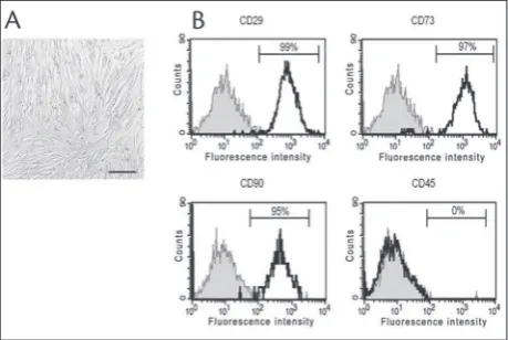
hASCs were isolated from a 75-year-old man. When the hASCs were cultured in DMEM with 10% FCS, they grew attached to the culture surface and exhib-ited spindle-shaped fibroblast-like morphology (Fig. 1A). At passage four, 95% of the cells were positive
for mesenchymal surface markers CD29, CD73 and CD90 and negative for leukocyte common antigen CD45 (Fig. 1B).
A schematic diagram of the iPSC generation pro-tocol is shown in Fig. 2A. The cells were infected with a mix of lentiviruses carrying one of the four genes (OCT4, SOX2, c-MYC and KLF4) produced in 293T cells [11]. On day 10, we detected the first non-ESC-like colonies (Fig. 2B) that had a morphology simi-lar to the ‘‘early colonies’’ or ‘‘background colonies’’ described previously [8,14,15]. Four days later, the treated hASCs were evaluated for the appearance of tightly-packed colonies, resembling to hESCs (Fig. 2C). The colonies were composed of small,
fast-grow-Fig. 1. Characteristics of hASCs. A – monolayer of ASCs (adher-ent fibroblast-like cells). Scale bar 200 μm. B – flow cytometry analysis showed that more than 95% of hASCs are positive for the mesenchymal surface markers CD29, CD73 and CD90, and all cells are negative for leukocyte common antigen CD45.
ing cells and were positive for alkaline phosphatase (AP) activity (Fig. 2D) on day 18. Forty-one ESC-like colonies, representing 0.08% mean reprogramming efficiency, were obtained.
In our previous study, we reported a method for the selection of human iPSCs based on the expres-sion of transgenous OCT4 (the OCT4 vector has a puromycin resistance gene) and the surface plu-ripotent marker TRA-1-60 [11]. Here, we used the same protocol to pick out reprogrammed cells. We selected 1x10⁵ cells positive for OCT4- and
TRA-1-60-reprogrammed hASCs and expanded them on MEFs. hASC-derived ESC-like colonies were mor-phologically indistinguishable from hESCs. We manu-ally picked six individual colonies. A further three iPSCs clones, iP-hASC-1, iP-hASC-2 and iP-hASC-3, were successfully established from this reprogram-ming experiment and characterized with regard to pluripotency-associated properties.
Characterization of human iPSCs
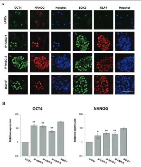
Two human iPSC clones (iP-hASC-1, iP-hASC-2) were continuously cultured for approximately 3 months and characterized to demonstrate their fully reprogrammed character. The expression of pluri-potency-specific markers OCT4, SOX2, KLF4 and NANOG, was examined in clones hASC-1 and iP-hASC-2 (Fig. 3A). The analyzed clones were positive for these markers. Although ASC were not negative for the tested markers, the observed expression was definitely much lower than in the iP-hASC clones. The expression intensities for the four markers in the iP-hASC clones were comparable with those detected in the embryonic stem cell line BG01V.
Reprogramming of ASCs was induced by virus de-livery of transgenous oct4, sox2, c-myc and klf4, which activated the transcription of corresponding endo-gens. The activation of endogenous oct4 and nanog
transcription is a key step in the reprogramming of somatic cells into induced pluripotent stem cells. A statistically significant increase in mRNA expression levels of endogenous oct4 and nanog in reprogrammed hASCs compared to parental cells was determined. In reprogrammed cells, the endogenous oct4 were from 6- to 15-fold higher than in parental cells. As regards en-dogenous oct4, its expression was increased from 3- to
4-fold. These genes were found to be from 2- to 3-fold lower in reprogrammed cells than in hESCs (Fig. 3B).
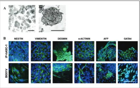
Finally, the ability to differentiate into nearly all tissue types as a hallmark of pluripotent stem cells was analyzed employing embryoid body (EB)-based differentiation in vitro. The reprogrammed cells were cultured in differentiation medium and by prevent-ing cell adhesion to the bottom of the culture dishes. Under these conditions, the cells formed spherical aggregates (EBs) shown in Fig. 4A. For evaluation of the ability for in vitro differentiation of reprogrammed cells, we investigated the expression of nestin (neural stem cells, pancreatic islet progenitors), vimentin (glial cells, pancreatic precursor cells), α-actinin (muscle
cells), desmin (muscle cells), α-fetoprotein (AFP) (visceral endodermal cells) and GATA4 (primitive endodermal cells) in 35-day-old EBs. Vimentin and nestin were expressed in the periphery of the adherent EBs, suggesting their expression in migrating cells. Muscle markers α-actinin and desmin and endoder-mal markers GATA4 and AFP were found inside the EBs as well as at the periphery (Fig. 4B). the EBs of BG01V cells showed similar expression of examined differentiation markers.
DISCUSSION
At present, it is possible to reprogram cells derived from patients over 60 years to obtain more youth-ful features, resetting telomere lengths and gene ex-pression profiles similar to those observed in hESCs [16,17]. Several groups have succeeded in the estab-lishment of iPSCs from healthy elderly donors or diseased aged persons. Ohmine et al. [18] generated iPSC lines from epidermal keratinocytes derived from elderly type 2 diabetes patients of different age, using the four Yamanaka factors. Although reprogramming efficiency is not high – 2 to 10 expandable clones per 105 (~0.002-0.01%), iPSC lines could be derived and
these lines expressed a range of pluripotency markers and exhibited the ability to differentiate into the three germ layers. Also, Sun et al. [8] reported the genera-tion of iPSCs from patients’ hASCs (40- to 65-year-old) by transduction with OCT4, SOX2, KLF4 and
c-MYC, with an efficiency of 0.01-0.03%. Neverthe-less, there are no reports on the reprogramming of adipose-derived iPSCs from patients aged over 70 years.
The present work contributes to the development of knowledge for the reprogramming of hASCs. Here we report the successful establishment of iPSCs from hASCs of a 75-year-old man. hASC-iPSCs with hESC-like morphology were generated with an efficiency of 0.08%, on day 18. However, the efficiency is a higher than reported for iPSCs generated from the fibroblasts of a 74-year-old donor, with a mean reprogramming efficiency of 0.06% [16]. We suggest that using hASCs as parental cells might resolve the low efficiency of transduction. Because adipose tissue often accumu-lates in the human body, hASCs might represent an abundant cell source for reprogramming.
The reprogrammed cells were characterized by alkaline phosphatase expression which, along with expression of normal pluripotent stem cell markers OCT4, NANOG, KLF4 and SOX2, indicated un-differentiated cells with the potential to self-renew. Moreover, all iPSC clones activated the transcription of the endogenous homeodomain transcription fac-tors, OCT4 and NANOG, which have been identi-fied as important to both early embryo development and pluripotency maintenance in hESCs [19,20]. We found a significant increase in mRNA levels for en-dogenous OCT4 and NANOG at more than 3 months post-infection, providing proof of a successful repro-gramming procedure.
The differentiation potential of the in vitro -formed iPSCs was investigated using the protocol for differentiation by subjecting the cells to EB forma-tion. EBs were formed by a spontaneous aggregation of cells [20,21]. This structure facilitates multicellular interactions comprising ectodermal, mesodermal, and endodermal tissues and leading to cell differentiation during early mammalian embryogenesis [18,19]. In the tested clones, we demonstrated the expression of the molecular markers nestin, vimentin, α-actinin, desmin, AFP and GATA4. Together, these results indi-cate that reprogrammed elderly hASCs have pluripo-tent properties, since only pluripopluripo-tent cells are able to differentiate towards the three embryonic germ tissues (ectoderm, mesoderm and endoderm).
In conclusion, herein we report the efficient gen-eration of iPSCs from hASCs obtained from an elderly patient. iPSCs generated from patients of different age should provide a valuable experimental model for in-vestigating the cellular reprogramming process as well as practical alternatives for the generation of patient-specific cells.
Acknowledgments: This work was partly supported by the grants ReProForceFP-7- REGPOT-2009-1 аnd №BG-051PO001-3.3.06-0059.
Authors’ contribution: ES, the main author, provided the design and implementation of the experiment, data collection, data analy-sis and manuscript compilation. TO contributed to the analyanaly-sis of data. MM coordinated the research.
Conflict of interest disclosure: The authors declare no conflict of interest in the study.
REFERENCES
1. Han W, Zhao Y, Fu X. Induced pluripotent stem cells: the dragon awakens. BioScience. 2010;60(4):278-85.
2. Lee Y-H, Mottillo EP, Granneman JG. Adipose tissue plastic-ity from WAT to BAT and in between. Biochim Biophys Acta. 2014;1842(3):358-69.
3. Takahashi K, Tanabe K, Ohnuki M, Narita M, Ichisaka T, Tomoda K, Yamanaka S. Induction of pluripotent stem cells from adult human fibroblasts by defined factors. Cell. 2007;131(5):861-72.
4. Aasen T, Raya A, Barrero MJ, Garreta E, Consiglio A, Gon-zalez F, Vassena R, Bilic J, Pekarik V, Tiscornia G, Edel M, Boue S, Izpisua Belmonte JC. Efficient and rapid generation of induced pluripotent stem cells from human keratinocytes. Nat Biotech. 2008;26(11):1276-84.
5. Kim JB, Sebastiano V, Wu G, Araúzo-Bravo MJ, Sasse P, Gen-tile L, Ko K, Ruau D, Ehrich M, van den Boom D, Meyer J, Hubner K, Bernemann C, Ortmeier C, Zenke M, Fleis-chmann BK, Zaehres H, Scholer HR.Oct4-Induced pluripo-tency in adult neural stem cells. Cell. 2009;136(3):411-9. 6. Zuk PA, Zhu M, Ashjian P, De Ugarte DA, Huang JI, Mizuno
H, Huang J, Futrell JW, Katz AJ, Benhaim P, Lorenz HP, Hed-rick MH. Human adipose tissue is a source of multipotent stem cells. Mol Biol Cell. 2002;13(12):4279-95.
7. Dicker A, Le Blanc K, Aström G, van Harmelen V, Göther-ström C, Blomqvist L, Arnera P, Rydén M.. Functional studies of mesenchymal stem cells derived from adult human adipose tissue. Exp Cell Res. 2005;308(2):283-90.
8. Sun N, Panetta NJ, Gupta DM, Wilson KD, Lee A, Jia F, Hu S, Cherry AM, Robbins RC, Longaker MT, Wu JC.
Feeder-free derivation of induced pluripotent stem cells from adult human adipose stem cells. Proc Natl Acad Sci U S A. 2009;106(37):15720-5.
9. González-Cruz RD, Fonseca VC, Darling EM. Cellular mechanical properties reflect the differentiation potential of adipose-derived mesenchymal stem cells. Proc Natl Acad Sci U S A. 2012;109(24):E1523-E1529.
10. Kang S-J, Park Y-I, Kwon M-J, Yang Y-H, Bang S-I, Sohn S-H, Park YH, So BJ, Kang H-GE.. Adipose stromal cells are a more efficient source than adipose stem cells in retrovirus-mediated iPS induction. Cell Mol Bioeng. 2015;8(1):224-35. 11. Stoyanova E, Mourdjeva M, Kyurkchiev S. Early selection
of human fibroblast-derived induced pluripotent stem cells. Biotechnol Biotechnol Equip 2015:29(5):942-8.
12. Kim D, Kim CH, Moon JI, Chung YG, Chang MY, Han BS, Ko S, Yang E, Cha KY, Lanza R, Kim KS, Generation of human induced pluripotent stem cells by direct delivery of repro-gramming proteins. Cell stem cell. 2009;4(6):472-6. 13. Pfaffl MW. A new mathematical model for relative
quantifica-tion in real-time RT–PCR. Nucleic Acids Res 2001;29(9):e45. 14. Takahashi K. Induction of pluripotent stem cells from adult human fibroblasts by defined factors. Cell. 2007;131:861-72. 15. Lowry WE, Richter L, Yachechko R, Pyle AD, Tchieu J, Srid-haran R, Clark AT, Plath K.. Generation of human induced pluripotent stem cells from dermal fibroblasts Proc Natl Acad Sci U S A. 2008;105(8):2883-8.
16. Lapasset L, Milhavet O, Prieur A, Besnard E, Babled A, Aït-Hamou N, Leschik J, Pellestor F, Ramirez J-M, Vos JD, Lehm-ann S, Lemaitre J-M Rejuvenating senescent and centenarian human cells by reprogramming through the pluripotent state. Genes Dev. 2011;25(21):2248-53.
17. Prigione A, Hossini AM, Lichtner B, Serin A, Fauler B, Megges M, Lurz R, Lehrach H, Makrantonaki E, Zouboulis CC, Adjaye J. Mitochondrial-associated cell death mecha-nisms are reset to an embryonic-like state in aged donor-derived iPS cells harboring chromosomal aberrations. PLoS ONE. 2011;6(11):e27352.
18. Ohmine S, Squillace KA, Hartjes KA, Deeds MC, Armstrong AS, Thatava T, Sakuma T, Terzic A, Kudva Y, Ikeda Y. Repro-grammed keratinocytes from elderly type 2 diabetes patients suppress senescence genes to acquire induced pluripotency. Aging (Albany NY). 2012;4(1):60-73.
19. Mitsui K, Tokuzawa Y, Itoh H, Segawa K, Murakami M, Taka-hashi K, Maruyama M, Maeda M, Yamanaka S. The homeo-protein Nanog is required for maintenance of pluripotency in mouse epiblast and ES cells. Cell. 2003;113(5):631-42. 20. Nichols J, Zevnik B, Anastassiadis K, Niwa H,
Klewe-Nebe-nius D, Chambers I, Schöler H, Smith A. Formation of plu-ripotent stem cells in the mammalian embryo depends on the POU transcription factor Oct4. Cell. 1998;95(3):379-91. 21. Banito A, Rashid ST, Acosta JC, Li S, Pereira CF, Geti I, Pinho