INTRODUCTION
The severe acute respiratory syndrome coronavirus 2 (SARS-CoV-2) is responsible for an ongoing pandemic that has already killed over 320,000 people and paralyzed the global economy (1). Currently, the main method for labora-tory diagnosis of SARS-CoV-2 is PCR testing of nasopharyn-geal swabs. There is an urgent need for highly specific and sensitive antibody detection assays to answer fundamental questions about the epidemiology and pathogenesis of SARS-CoV-2 and to implement and evaluate population-level con-trol programs (2). Efforts to understand the pathogenesis and define risk factors for severe SARS-CoV-2 disease have been
hampered by our inability to identify all infected individuals, irrespective of clinical symptoms. To contain the pandemic, many countries resorted to the widespread quarantine of cit-ies and regions. By deploying reliable antibody assays for population-level testing, it will be possible to obtain the high-resolution spatial data needed to implement policies for con-taining the epidemic and informing strategies for re-opening communities and cities.
Studies with SARS-CoV-2 and other human CoVs demon-strate that people rarely develop specific antibodies within the first 7 days after onset of symptoms (3–7). By 10-11 days after onset of symptoms, greater than 90% of SARS-CoV-2
The receptor binding domain of the viral spike protein is
an immunodominant and highly specific target of
antibodies in SARS-CoV-2 patients
Lakshmanane Premkumar1*, Bruno Segovia-Chumbez1, Ramesh Jadi1, David R. Martinez2, Rajendra Raut1, Alena Markmann3, Caleb Cornaby5, Luther Bartelt3, Susan Weiss3, Yara Park3, Caitlin E. Edwards2, Eric Weimer4, Erin M. Scherer8, Nadine Rouphael8, Srilatha Edupuganti8, Daniela Weiskopf6, Longping V. Tse2, Yixuan J. Hou2, David Margolis1,2,3, Alessandro Sette6,7, Matthew H. Collins8, John Schmitz4, Ralph S. Baric1,2, Aravinda M. de Silva1*
1Department of Microbiology and Immunology, University of North Carolina School of Medicine, Chapel Hill NC 27599, USA. 2Department of Epidemiology, UNC Chapel Hill
School of Public Health, University of North Carolina at Chapel Hill, Chapel Hill, North Carolina, USA. 3Departments of Medicine, University of North Carolina School of
Medicine, Chapel Hill NC 27599, USA. 4Department of Pathology & Laboratory Medicine, University of North Carolina School of Medicine, Chapel Hill NC 27599, USA. 5Immunology/Histocompatibility and Immunogenetics Laboratories, University of North Carolina School of Medicine, Chapel Hill NC 27599, USA. 6 Center for Infectious
Disease and Vaccine Research, La Jolla Institute for Immunology (LJI), La Jolla, CA 92037, USA. 7Department of Medicine, Division of Infectious Diseases and Global Public
Health, University of California, San Diego (UCSD), La Jolla, CA 92037, USA. 8Hope Clinic of the Emory Vaccine Center, Division of Infectious Diseases, Department of
Medicine, School of Medicine, Emory University, Decatur, Georgia, USA.
*Corresponding author. Email: Lakshmanane Premkumar ([email protected]) or Aravinda M. de Silva ([email protected])
patients develop specific IgG and IgM (3–6). For SARS-CoV-1 and the more distantly related MERS-CoV, IgG antibodies have been observed to persist for at least one year after infec-tion (8, 9). These observations strongly support the feasibility of using antibody assays for identifying recent and remote SARS-CoV-2 infections and for conducting population-level surveillance.
SARS-CoV-2 is a β-coronavirus, a subgroup that includes the closely related SARS-CoV-1 and the more distantly related MERS-CoV and the common-cold human CoVs (HCoV-OC43 and HCoV-HKU1) (10). Many companies have quickly devel-oped tests for SARS-CoV-2 antibody detection. These assays utilize the inactivated whole virion, viral nucleocapsid pro-tein or viral spike propro-tein as antigens in ELISA, lateral flow or other testing platforms. While the performance of these assays has not been fully evaluated, some assays appear quite sensitive when used 10 days or more after the onset of symp-toms (6, 11). The specificity of SARS-CoV-2 antibody assays has not been adequately addressed. Humans are frequently infected with HCoV-OC43 and HCoV-HKU1 and most adults have antibodies to these viruses (10). Any antibody cross-activity between common HCoVs and SARS-CoV-2 would re-sult in false-positive rere-sults interfering with antibody-based testing and surveillance for SARS-CoV-2.
SARS-CoV-1 and HCoV OC43 elicit antibodies that cross-react against related CoVs (12, 13). Following the SARS-CoV-1 outbreak in 2003, the overall specificity of serological assays utilizing the nucleocapsid protein of SARS-CoV-1 was poor, whereas assays based on the spike protein were more specific (14–16). In recent studies, the receptor binding domain (RBD) of the spike protein of SARS-CoV-2 has shown promise as an antigen for specific antibody detection (4, 17, 18). Here we re-port the production of properly folded recombinant receptor binding domains (RBDs) from the spike proteins of SARS and common-cold HCoVs in mammalian cells. We use these re-combinant antigens and a large diverse panel of human and animal sera to evaluate the RBD as an antigen for 2 serology. We demonstrate that the recombinant SARS-CoV-2 RBD antigen is highly sensitive and specific for detection of antibodies induced by SARS-CoVs. We also observed a strong correlation between the levels of RBD-binding antibodies and levels of SARS-CoV-2 neutralizing antibodies in patients. Our results support the use of RBD-based antibody assays for se-rology and as a correlate of neutralizing antibody levels in symptomatic people who have recovered from SARS-CoV-2 infections.
RESULTS
Expression and characterization of recombinant RBD antigens from pathogenic coronaviruses
The S1 and S2 subunits of the spike (S) protein of Corona-viruses are required for viral entry. The surface accessible
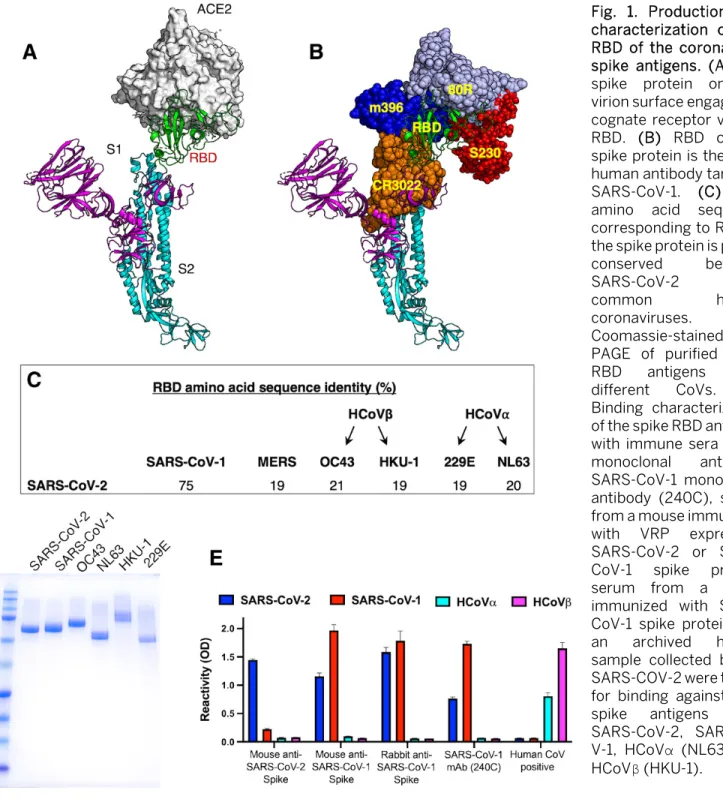
receptor binding domain (RBD) on the S1 subunit binds to receptors on target cells, whereas the exposure of the fusion loop in the S2 subunit induces fusion of the viral envelope to the host cellular membranes (19). The RBDs of SARS-CoVs, which bind to angiotensin-converting enzyme 2 (ACE2) re-ceptor on the host cells, are also a major target of human an-tibodies (Fig. 1A and B). As the RBD is a common target of human antibodies and poorly conserved between SARS-CoVs and other pathogenic human coronaviruses (Fig. 1C), this do-main is a promising candidate for use in antibody-based di-agnostic assays. We expressed the RBD of 2003 and 2019 SARS-Co-Vs and four common human coronaviruses (HCoV-HKU-1, -OC43, -NL63 and -229E) as fusion proteins that were secreted from human cells. The recombinant RBDs were pu-rified from the cell culture medium by affinity chromatog-raphy and purity was confirmed by SDS-PAGE (Fig. 1D). We used sera and monoclonal antibodies from animals immun-ized with SARS-CoV-1 or -2 spike proteins to assess the struc-tural integrity of the purified recombinant RBD antigens. Pooled serum from mice immunized with SARS-CoV-2 spike protein had antibodies that bound well to the RBD of SARS-CoV-2 and poorly to the RBDs of SARS-CoV-1 and other com-mon HCoVs (Fig. 1E). Sera from mice or rabbits immunized with SARS-CoV-1 or cross-reactive monoclonal antibody 240C reacted with the RBDs of SARS CoV-1 and -2 but not common human CoVs (Fig. 1E). Human serum collected before SARS-CoV-2 emerged contained antibodies to common α- and β -HCoVs (NL63 and HKU-1) but not to SARS-CoV RBD antigens (Fig. 1E). These results suggest that the purified recombinant RBD antigens retain native structures required for specific antibody binding.
Evaluating the specificity of SARS-CoV-2 RBD for serol-ogy
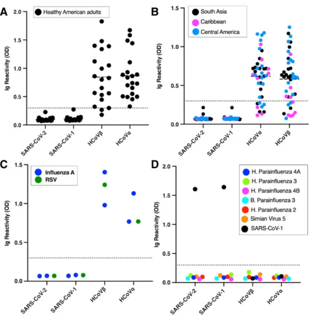
confirmed influenza A and respiratory syncytial virus infec-tions and sera from guinea pigs immunized with a panel of different human respiratory viruses (Fig. 2 C and D). Except guinea pigs immunized with SARS-CoV-1, none of the sera had detectable levels of antibodies to the recombinant RBD of SARS-CoVs.
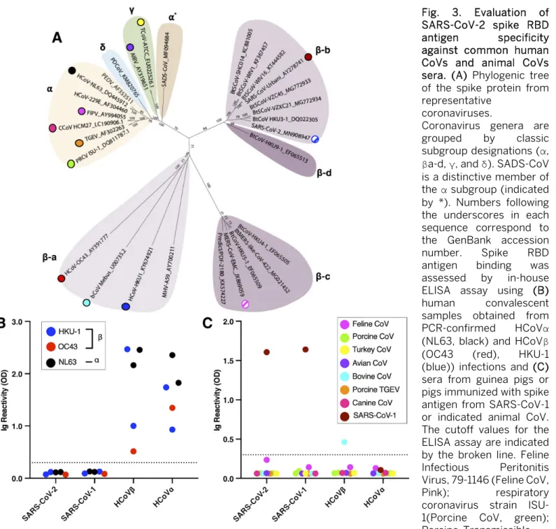
The known pathogenic human CoVs are members of the α-coronavirus and β-coronavirus genera (Fig. 3A). HCoV-NL63 and 229E are two α-coronaviruses that frequently in-fect and cause a mild common-cold-like illness in most
peo-ple. HCoV-OC43 and HKU-1 are two group 2A β
-coronaviruses that also commonly infect people and cause mild disease. Most adults (>90%) have antibodies to these common-cold HCoVs. SARS-CoV-1 and -2 and MERS-CoV are group 2B and 2C zoonotic β-coronaviruses that have recently crossed into humans and caused severe illness. The α- and β -coronavirus genera also contain a large number of zoonotic viruses that infect different animal hosts, which have not been implicated in human disease to date. To further assess the specificity of SARS-CoV-2 RBD for serology, we obtained and tested sera from people who had recently recovered from a laboratory-confirmed common-cold HCoV infection and sera from guinea pigs immunized with different animal CoVs (Fig. 3 B and C). None of the immune sera from people ex-posed to recent HCoV infections cross-reacted with the re-combinant RBD of SARS-CoVs. None of the guinea pigs vaccinated with different zoonotic CoVs had antibodies that cross-reacted with the recombinant SARS-CoV RBDs (Fig. 3B and C). These results establish that most individuals, includ-ing people who have been recently exposed to acute common HCoV infections, do not have detectable levels of cross-reac-tive antibodies to the recombinant RBD of SARS-CoVs. Evaluating the sensitivity of SARS-CoV-2 RBD for serol-ogy
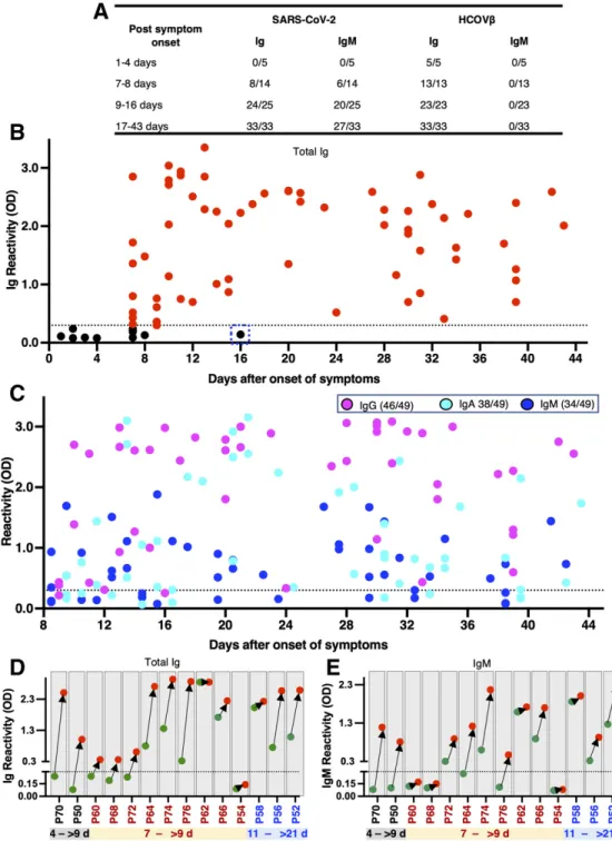
To evaluate the sensitivity of the RBD of SARS-CoV-2 for identifying infected individuals, we obtained a total of 77 se-rum samples from 63 patients with laboratory-confirmed (i.e., PCR positive) SARS-CoV-2 infections collected at differ-ent times after the onset of symptoms. All the samples were tested for binding of total immunoglobulin (Ig) and IgM an-tibodies to recombinant RBD antigens from SARS-CoVs and common-cold HCoVs. The sensitivity of the assay was high (98% and 81% respectively for Ig and IgM) for specimens col-lected 9 days or more after onset of symptoms (Fig. 4A). As expected, overall sensitivity was lower (57% and 43% respec-tively for Ig and IgM) for specimens collected between 7 and 8 days after onset of symptoms (Fig. 4A). With samples col-lected 9 days or more after onset of symptoms, we observed some Ig and IgM antibody cross reactivity with the RBD of SARS-CoV-1 (67% and 30% respectively for Ig and IgM), which was anticipated as these viruses are closely related group 2B β-coronaviruses (20, 21). When the specimens were
further analyzed to estimate the timing of seroconversion, we observed a marked transition from seronegative to seroposi-tive for both Ig and IgM about 9 days after the onset of symp-toms (Fig. 4A and B). By day 9 after onset of sympsymp-toms, most patients had high end-point titers in the RBD Ig ELISA (Fig. S1). To analyze the kinetics of all three of the major isotypes of serum antibodies within the first 6 weeks after the onset of symptoms, we separately measured IgG, IgA, and IgM in 49 serum samples obtained from SARS-CoV-2 infected pa-tients at >9 days after onset of symptoms. Most individuals (46/49) developed IgG responses (Fig. 4C). IgA and IgM re-sponses were observed less frequently (IgA = 38/49, IgM =34/49) than IgG (Fig. 4C). For 14 individuals with labora-tory-confirmed SARS-CoV-2 infection, we had two specimens collected at different times early in the infection (Fig. 4D). Two subjects (P70 and P50) were seronegative within the first 4 days and seropositive for both Ig and IgM 9 or more days after onset (Fig. 4D). For three subjects (P58, P56, P52) the acute samples were collected after 9 days and the convales-cent samples were collected 21 days or more after onset. In these individuals both acute and convalescent samples were positive, and we observed an increase in Ig and IgM levels in the second specimen. For the remaining 9 subjects, the acute specimen was collected on day 7 after onset and the conva-lescent specimen was collected >9 days after onset. Six out of the 9 subjects already had specific Ig, IgM or both in the acute specimen collected on day 7. All the subjects except one (P54) seroconverted or had elevated levels of antibody in the con-valescent sample collected >9 days after onset of symptoms. These results indicate that most people seroconvert between days 7 and 9 after onset of symptoms. Subject P54 was an outlier and did not develop specific Ig or IgM antibodies. All the individuals with documented SARS-CoV-2 had Ig but not IgM antibodies that bound to the RBD of common HCoVs, which is consistent with their high prevalence in humans (Fig. 4A). These results demonstrate that the RBD of SARS-CoV-2 is a highly sensitive antigen for antibody detection in patients 9 days or more after onset of symptoms.
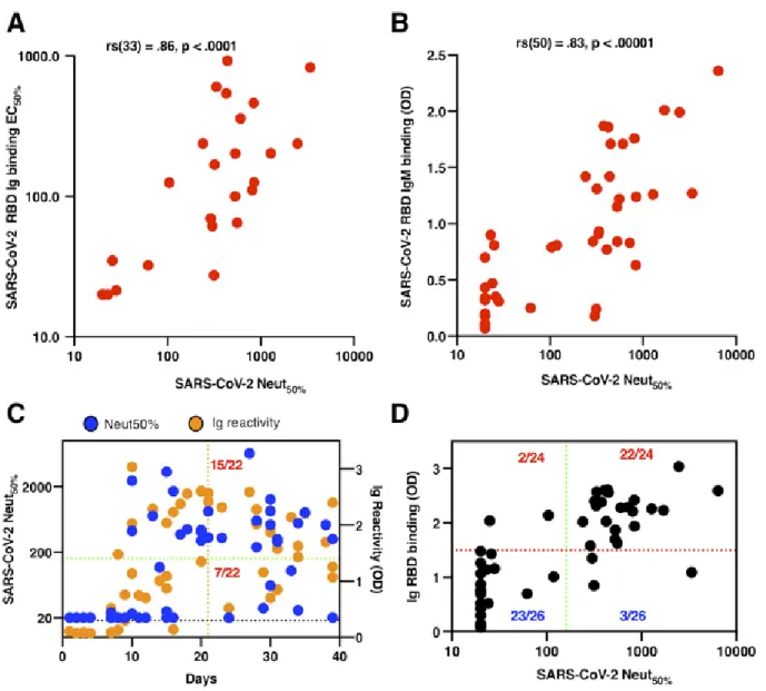
Antibodies to the RBD of SARS-CoV-2 as a correlate of neutralizing antibody response
the relationship between the RBD-binding activity and the neutralizing antibody response, we tested 50 PCR-confirmed SARS-COV-2 patient immune sera in a SARS-CoV-2 luciferase neutralization assay (Fig. 5). As judged by the Spearman test (ρ = 0.86, P < 0.0001), we observed that the magnitude of the total RBD-binding Ig antibody strongly correlated with the levels of neutralizing antibodies in SARS-CoV-2 patients (Fig. 5A). Moreover, the patient samples with high levels of IgM antibodies were strongly associated with the highest neutral-izing antibody titers in early convalescence (Spearman ρ = 0.83, P < 0.0001; Fig. 5B, <6 weeks after onset of symptoms). The neutralizing antibody kinetics in patients mirrored the kinetics of RBD antibody development (Fig. 5C and Fig. S2). None of the patients with confirmed SARS-CoV-2 infection (0/8) had any detectable levels of neutralizing antibodies within the first eight days after the onset of symptoms. While low levels of neutralizing antibody titers were detectable in 91% of patients (20/22) 21 days after the onset of symptoms, only 73% of patients (16/22) had a neutralization titer of at least 1:80.
Currently, patients who have had a documented SARS-CoV-2 infection identified by RT-PCR or a serologic test, and who are clear of symptoms for at least 14 days, are recruited for convalescent plasma donation. We evaluated the neutral-izing potency in patient samples collected between 1 and 40 days with a titer of at least 1:160 (Fig. 5D). We observed that 32% of patients (7/22) developed weak to no neutralizing an-tibodies even 21 days after onset of symptoms, suggesting that days after the start of symptoms is a poor determinant of the levels of SARS-CoV-2 neutralizing antibodies in the pa-tients included in our study, particularly within the early con-valescent phase (<6 weeks). To evaluate whether a simple RBD ELISA can be used as a surrogate for neutralizing po-tency in SARS-COV-2 patients, we analyzed the relationship between the level of total Ig antibody to RBD and a neutral-izing antibody titer of at least 1:160. We observed that 22/24 people who had a substantial total Ig binding antibody to RBD (>1.5 OD) also developed a robust neutralizing antibody titer (Fig. 5E). Notably, only 3/26 people who developed a rel-atively weak RBD-binding antibody had a neutralizing anti-body titer higher than 1:160. One subject (P54) neither seroconverted for RBD antigen nor developed neutralizing antibodies to SARS-CoV-2 (Fig. 4D and E, and Fig. S2).
DISCUSSION
Serology is critical to understanding the transmission, pathogenesis, mortality rate and epidemiology of emerging viruses. In the few months after the discovery of SARS-CoV-2 as a human pathogen, scientists have developed a large num-ber of antibody assays and many commercial tests are now available. Although none of the assays have been fully vali-dated yet, the FDA has granted emergency use authorization
(EUA) for multiple tests, while stressing the need for further validation. Investigators have already encountered problems with the specificity and sensitivity of commercial assays rushed to market (4, 22). Widespread use of inaccurate anti-body assays could lead to policies that exacerbate the current SARS-CoV-2 pandemic instead of containing it.
We designed the assay for separate detection of RBD-specific total Ig and IgM. As the pandemic is ongoing and most infections are likely to have occurred within the past few months, infected individuals have variable levels of anti-gen-specific IgG, IgM and IgA (Fig. 4C). To maximize assay sensitivity and to prevent different antibody isotypes compet-ing for bindcompet-ing sites and reduccompet-ing assay signal, we measured total Ig. We did not observe any decrease in assay specificity by designing the assay to monitor levels of total Ig instead of IgG binding to the RBD even at high serum concentration or with hyperimmune sera. Our study showed that IgM and IgA antibodies can also be detected using RBD-based serological assays. Both IgA and IgM antibodies are relatively short lived and indicative of a recent exposure. When conducting large scale population level surveillance for SARS-CoV-2 antibod-ies, it will be possible to distinguish recent from remote in-fections by measuring both total Ig and IgM (or IgA) binding to the RBD.
Antibody assays that correlate with protective immune re-sponses in individuals who have recovered from SARS-CoV-2 infection and also reflect herd immunity at a population level are urgently needed to define each individual’s risk of disease and to identify communities at high risk for new waves of infection. In animal studies with SARS-CoV-1, virus-neutral-izing antibodies were strongly correlated with protective im-mune responses (19). We observed a striking correlation between the levels of RBD antibodies in patients and the abil-ity of patient sera to neutralize SARS-CoV-2 virus. Other groups have recently reported finding a strong correlation be-tween spike/RBD antibodies and SARS-CoV-2 neutralization in patients infected with SARS-CoV-2 (4, 17, 18). Our results point out that roughly one-third of patients develop very low or no neutralizing antibodies to SARS-CoV-2 and that Ig and IgM antibodies are useful predictors of neutralizing antibody levels in patients in the early convalescent phase (<6 weeks). As people developing a high level of RBD-binding antibodies (>1.5 OD) also have a robust neutralizing response, a simple RBD-based ELISA can be a useful tool to identify blood plasma donors. While further studies are needed to fully eval-uate RBD antibodies as correlate of protective immunity, the results to date indicate that RBD antibodies are a promising correlate of protection in the early convalescent phase. A sim-ple antibody detection assay that also predicts individual-level risk of disease will be a major advance for vaccine de-velopment and immunogenicity of vaccines because SARS-CoV-2 neutralization assays are time-consuming and require BSL-3 containment.
One SARS-CoV-2 patient (P54) who tested positive for vi-ral RNA and required hospitalization did not develop RBD-specific Ig, IgM or neutralizing antibodies, even at 16 days after the onset of symptoms. This was the only person among the 68 PCR positive subjects who did not seroconvert by 9
days after onset of symptoms in the RBD-based assay. While we cannot rule out the possibility of a false positive PCR test result, others have also reported rare instances where people infected with SARS-CoVs have atypical, dampened immune responses (23). Further studies are needed to establish the frequency and significance of atypical antibody responses in SARS-CoV-2 patients and characterize the serological reper-toire and epitopes targeted by the antibodies in convalescent sera.
As SARS-CoV-2 infections in the southeastern U.S. have started to increase relatively recently, all convalescent sam-ples used in this study were collected within 90 days follow-ing onset of symptoms. In most patients, the convalescent sera had high end-point titers (>1:1000) in the RBD Ig ELISA supporting the utility of this assay even as antibody levels start to wane over time. We need to prioritize studies to pro-spectively monitor SARS-CoV-2 patients to determine the long-term kinetics of antibody levels and the performance of antibody detection assays over time.
All the SARS-CoV-2 human immune sera used for this study were collected from symptomatic patients that in-cluded many with serious illness requiring hospitalization. The research community currently does not know if individ-uals experiencing mild/inapparent symptoms after SARS-CoV-2 infection have similar kinetics and levels of RBD-binding antibodies as those experiencing symptomatic infec-tions. Studies must be done with individuals experiencing mild/inapparent SARS-CoV-2 infections to define the kinetics and levels of RBD antibodies before implementing large pop-ulation-level antibody testing.
MATERIALS AND METHODS Study design
The goal of the study was to evaluate the performance of RBD-based spike antigen for reliable detection of SARS-CoV-2-specific antibodies. We produced properly folded RBD from the spike proteins of SARS and common-cold HCoVs in mam-malian cells and used this antigen to evaluate a large panel of human sera from documented SARS-CoV-2 patients and control subjects, and hyperimmune sera from animals ex-posed to zoonotic CoVs. We also used a SARS-CoV-2 lucifer-ase neutralization assay to assess the dynamics of the neutralizing antibody response and its association with the RBD-binding activity.
Structural analysis
structural alignments and analysis were as follows: SARS-CoV-2 spike (6VSB), SARS-CoV-1 spike (6CRV), SARS-CoV-1 spike/S230 (6NB6), SARS-Co-V1 spike RBD/80R (2GHW), SARS-CoV-1 spike RBD/ m396 (2DD8), SARS-CoV-1 spike RBD/F26G19 (3BGF), SARS-CoV-2 spike RBD/CR3022 (6W41).
Protein expression and purification
We used the following structure coordinates of the coro-navirus spike proteins from the PDB to define the boundaries for the design of RBD expression constructs: SARS-CoV-2 (6VSB), SARS-CoV-1 (6CRV), HKU-1 (5I08), OC43 (6NZK), 229E (6U7H) NL63 (6SZS). Accordingly, a codon-optimized gene encoding for S1-RBD [SARS-CoV-1 (318 – 514 aa, P59594), SARS-CoV-2 (331 – 528 aa, QIS60558.1), OC43 (329 – 613 aa, P36334.1), HKU-1 (310 – 611 aa, Q0ZME7.1), 229E (295 – 433 aa, P15423.1) and NL63 (480 – 617 aa, Q6Q1S2.1)] containing human serum albumin secretion signal sequence, three purification tags (6xHistidine tag, Halo tag, and Twin-Strep tag) and two TEV protease cleavage sites was cloned into the mammalian expression vector pαH. S1 RBDs were expressed in Expi293 cells (ThermoFisher) and purified from the culture supernatant by nickel-nitrilotriacetic acid agarose (Qiagen).
Generation of SARS-CoV-2 Spike VRP and immunized mouse sera
To generate virus replicon particles (VRPs), the SARS-CoV-2 S gene was inserted into pVR21 3526 as previously de-scribed (24). In summary, the SARS-CoV-2 S gene was ligated into pVR21 following digestion by restriction endonuclease sites, PacI and ApaI. T7 RNA transcripts were generated us-ing the SARS-CoV-2-S-pVR21 construct in conjunction with plasmids containing the Venezuelan equine encephalitis vi-rus envelope glycoproteins and capsid protein. The RNA tran-scripts were then electroporated into baby hamster kidney fibroblasts and monitored for cytopathic effect. VRP were harvested 48 hours after electroporation and purified via high-speed ultra-centrifugation. To generate serum samples against SARS-CoV-2, 10-week-old BALB/c mice (Jackson Labs) were inoculated via footpad injection with the VRP and boosted with the same dose one time three weeks later. Se-rum samples were then collected from individual animals at 2 weeks post-boost and pooled for use in assays.
Human specimens
All human specimens used in these studies were obtained after informed consent under good clinical research practices (GCP) and compliant with oversight by the relevant institu-tional review boards (IRBs). A list of the SARS-CoV-2 patient samples included in the study with basic demographic and clinical information can be found in Table S1.
UNC Hospital Specimens: Sera for this study were rem-nants from samples submitted to the UNC Hospital McLen-don Clinical Laboratories or Blood Bank. SARS-CoV-2 patient
samples were obtained from patients with positive RT-PCR test result (in-house assay developed and validated by UNC Hospital McLendon Clinical Laboratory) for SARS-CoV-2. SARS-CoV-2 negative samples were obtained from patients with other diagnoses or from samples collected prior to De-cember 2019 and cryopreserved at -80°C.
Emory University School of Medicine Specimens: Speci-mens were obtained from patients with symptomatic illness and clinical testing confirming SARS-CoV-2 by PCR (CDC SARS-CoV-2 test). De-identified specimens were shared with researchers at UNC consistent with local IRB protocols (Emory IRB# 00110683 and 00022371).
Blood plasma donor study: Convalescent sera was ob-tained from donors who volunteered for plasma collections at the UNC Donation Center. Fresh sera collected as part of the standard plasmapheresis procedure were saved for re-search from donors who signed informed consent. UNC IRB 20-1141 is conducted under good clinical research practices (GCP) and is compliant with institutional IRB oversight. All donors had confirmed SARS-CoV-2 infection by nasopharyn-geal swab indicating the presence of SARS-CoV-2 RNA as per-formed by EUA approved qRT-PCR in a US laboratory with a Clinical Laboratory Improvement Amendments (CLIA) certi-fication. All donors had recovered from their SARS-CoV-2 ill-ness and were at least 14 days post last symptoms. Donors who presented for plasma collection prior to 28 days from their last symptoms had a confirmed negative nasopharyn-geal RT-PCR test done within 72 hours prior to donation.
Healthy Unexposed Donors: Samples from healthy U.S. adult donors were obtained by the La Jolla Institute for Im-munology (LJI) Clinical Core or provided by a commercial vendor (Carter Blood Care) for prior, unrelated studies be-tween early 2015 and early 2018, at least one year before the emergence of SARS-CoV-2. The LJI Institutional Review Board approved the collection of these samples (LJI; VD-112). Samples from the Caribbean, Central America and South Asia were obtained from archived samples at UNC collected before December 2019 for other studies.
Infectious Bronchitis Virus, Massachusetts obtained from guinea pig (NR-2515), Anti-Turkey Coronavirus, Indiana ob-tained from guinea pig (NR-9465), Anti-Canine Coronavirus, UCD1 obtained from guinea pig (NR-2727), Anti-Human Parainfluenza Virus 2 obtained from guinea pig (NR-3231), Anti-Simian Virus 5 obtained from guinea pig (NR-3232), Anti-Human Parainfluenza Virus 3 obtained from guinea pig (NR-3235), Anti-Bovine Parainfluenza Virus 3 obtained from guinea pig (NR-3236), Anti-Human Parainfluenza Virus 4A obtained from guinea pig (NR-3239), Anti-Human Parainflu-enza Virus 4B obtained from guinea pig (NR-3240), Human Convalescent Serum 001 to 2009 H1N1 Influenza A Virus (NR-18964), Human Convalescent Serum 002 to 2009 H1N1 Influenza A Virus (NR-18965), and Human Reference Antise-rum to Respiratory Syncytial Virus (NR-4020). For some ani-mal CoV anti-serum samples, the certificate of analysis provided by the BEI Resources confirmed the presence of neutralizing and binding antibodies (see Table S1).
In-house RBD Ig and IgM ELISA
All serum specimens tested by ELISA assay were heat-in-activated at 56°C for 30 min to reduce risk from any possible residual virus in serum. Briefly, 50 μl of spike RBD antigen at 4 μg/ml in Tris Buffered Saline (TBS) pH 7.4 was coated in the 96-well high-binding microtiter plate (Greiner Bio-One cat # 655061) for 1 hour at 37°C. Then the plate was washed three times with 200 μl of wash buffer (TBS containing 0.2% Tween 20) and blocked with 100 μl of blocking solution (3% milk in TBS containing 0.05% Tween 20) for 1 hour at 37°C. The blocking solution was removed, and 50 μl of serum sam-ple at 1:20 or indicated dilutions in blocking buffer was added for 1 hour at 37°C. The plate was washed in the wash buffer, 50 μl of alkaline phosphatase-conjugated secondary goat anti-human secondary antibody at 1:2500 dilution was added for 1 hour at 37°C. For measuring total Ig, a mixture of anti-IgG (Sigma Cat # A9544), anti-IgA (Abcam Cat # AB97212), and anti-IgM (Sigma Cat # A3437] were added together. For measuring specific antibody isotype, only secondary goat anti-human IgG or IgA or IgM was used. The plate was
washed, and 50 μl p-Nitrophenyl phosphate substrate
(SIGMA FAST, Cat No N2770) was added to the plate and ab-sorbance measured at 405nm using a plate reader (Biotek Epoh, Model # 3296573). For testing animal sera, the second-ary antibody was matched to the species as follows: goat anti-mouse IgG (Sigma, A3688), goat anti-rabbit IgG (Abcam, ab6722), goat pig IgG (Abcam, ab6916), and goat anti-guinea pig IgG (Abcam, ab7140).
SARS-CoV-2-Washington neutralization assays
Full-length viruses expressing luciferase were designed and recovered via reverse genetics and described previously (25, 26). Viruses were tittered in Vero E6 USAMRID cells to obtain a relative light units (RLU) signal of at least 20X the cell only control background. Vero E6 USAMRID cells were
plated at 20,000 cells per well the day prior in clear bottom black-walled 96-well plates (Corning 3904). Neutralizing an-tibody serum samples were tested at a starting dilution of 1:20, and were serially diluted 4-fold up to eight dilution spots. Antibody-virus complexes were incubated at 37°C with 5% CO2 for 1 hour. Following incubation, growth media was removed and virus-antibody dilution complexes were added to the cells in duplicate. Virus-only controls and cell-only con-trols were included in each neutralization assay plate. Fol-lowing infection, plates were incubated at 37°C with 5% CO2 for 48 hours. After the 48 hour incubation, cells were lysed and luciferase activity was measured via Nano-Glo Luciferase Assay System (Promega) according to the manufacturer’s specifications. SARS-CoV-2 neutralization titers were defined as the sample dilution at which a 50% reduction in RLU was observed relative to the average of the virus control wells. Statistical analysis
Each data points in Fig. 1E, Fig. 2, Fig. 3B and 3C, Fig. 4 and 5 are presented as means of technical duplicates. The cor-relation of RBD binding and neutralization titers shown in Fig. 5A and Fig. 5B was evaluated using a Spearman correla-tion coefficient (rs) and the associated two-tailed p-value (GraphPad Prism, version 8). Receiver operating characteris-tic (ROC) analyses were performed to establish cutoff values for SARS-CoV-2 seropositivity using SPSS software. Statistical analyses were performed using SPSS software ver. 26.0 (IBM, Armonk, NY, USA).
SUPPLEMENTARY MATERIALS
immunology.sciencemag.org/cgi/content/full/5/48/eabc8413/DC1 Fig. S1. Titration curves of sera from SARS-CoV-2 positive patients. Fig. S2. Seroconversion of SARS-CoV-2 neutralizing antibodies. Fig. S3. Estimation of RBD ELISA assay cutoff.
Table S1. Summary of samples tested and associated characteristics (Excel spread-sheet).
Table S2. Raw data file (Excel spreadsheet).
REFERENCES AND NOTES
1. Y. Jin, H. Yang, W. Ji, W. Wu, S. Chen, W. Zhang, G. Duan, Virology, Epidemiology, Pathogenesis, and Control of COVID-19. Viruses 12, 372 (2020).
doi:10.3390/v12040372Medline
2. A. K. Winter, S. T. Hegde, The important role of serology for COVID-19 control.
Lancet Infect. Dis. S1473-3099(20)30322-4 (2020). doi:10.1016/S1473-3099(20)30322-4Medline
3. L. Guo, L. Ren, S. Yang, M. Xiao, D. Chang, F. Yang, C. S. Dela Cruz, Y. Wang, C. Wu, Y. Xiao, L. Zhang, L. Han, S. Dang, Y. Xu, Q. Yang, S. Xu, H. Zhu, Y. Xu, Q. Jin, L. Sharma, L. Wang, J. Wang, Profiling Early Humoral Response to Diagnose Novel Coronavirus Disease (COVID-19). Clin. Infect. Dis. ciaa310 (2020).
doi:10.1093/cid/ciaa310Medline
Medline
5. K. K. To, O. T.-Y. Tsang, W.-S. Leung, A. R. Tam, T.-C. Wu, D. C. Lung, C. C.-Y. Yip, J.-P. Cai, J. M.-C. Chan, T. S.-H. Chik, D. P.-L. Lau, C. Y.-C. Choi, L.-L. Chen, W.-M. Chan, K.-H. Chan, J. D. Ip, A. C.-K. Ng, R. W.-S. Poon, C.-T. Luo, V. C.-C. Cheng, J. F.-W. Chan, I. F.-N. Hung, Z. Chen, H. Chen, K.-Y. Yuen, Temporal profiles of viral load in posterior oropharyngeal saliva samples and serum antibody responses during infection by SARS-CoV-2: An observational cohort study. Lancet Infect. Dis. 20, 565–574 (2020). doi:10.1016/S1473-3099(20)30196-1Medline
6. J. Zhao, Q. Yuan, H. Wang, W. Liu, X. Liao, Y. Su, X. Wang, J. Yuan, T. Li, J. Li, S. Qian, C. Hong, F. Wang, Y. Liu, Z. Wang, Q. He, Z. Li, B. He, T. Zhang, Y. Fu, S. Ge, L. Liu, J. Zhang, N. Xia, Z. Zhang, Antibody responses to SARS-CoV-2 in patients of novel coronavirus disease 2019. Clin. Infect. Dis. ciaa344 (2020).
doi:10.1093/cid/ciaa344Medline
7. P. R. Hsueh, L. M. Huang, P. J. Chen, C. L. Kao, P. C. Yang, Chronological evolution of IgM, IgA, IgG and neutralisation antibodies after infection with SARS-associated coronavirus. Clin. Microbiol. Infect. 10, 1062–1066 (2004).
doi:10.1111/j.1469-0691.2004.01009.xMedline
8. P. G. Choe, R. A. P. M. Perera, W. B. Park, K.-H. Song, J. H. Bang, E. S. Kim, H. B. Kim, L. W. R. Ko, S. W. Park, N.-J. Kim, E. H. Y. Lau, L. L. M. Poon, M. Peiris, M. D. Oh, MERS-CoV Antibody Responses 1 Year after Symptom Onset, South Korea, 2015. Emerg. Infect. Dis. 23, 1079–1084 (2017). doi:10.3201/eid2307.170310 Medline
9. W. Liu, A. Fontanet, P.-H. Zhang, L. Zhan, Z.-T. Xin, L. Baril, F. Tang, H. Lv, W.-C. Cao, Two-year prospective study of the humoral immune response of patients with severe acute respiratory syndrome. J. Infect. Dis. 193, 792–795 (2006).
doi:10.1086/500469Medline
10. J. Cui, F. Li, Z. L. Shi, Origin and evolution of pathogenic coronaviruses. Nat. Rev. Microbiol. 17, 181–192 (2019). doi:10.1038/s41579-018-0118-9Medline
11. Z. Li, Y. Yi, X. Luo, N. Xiong, Y. Liu, S. Li, R. Sun, Y. Wang, B. Hu, W. Chen, Y. Zhang, J. Wang, B. Huang, Y. Lin, J. Yang, W. Cai, X. Wang, J. Cheng, Z. Chen, K. Sun, W. Pan, Z. Zhan, L. Chen, F. Ye, Development and clinical application of a rapid IgM-IgG combined antibody test for SARS-CoV-2 infection diagnosis. J. Med. Virol.
jmv.25727 (2020). doi:10.1002/jmv.25727Medline
12. K. H. Chan, J. F.-W. Chan, H. Tse, H. Chen, C. C.-Y. Lau, J.-P. Cai, A. K.-L. Tsang, X. Xiao, K. K.-W. To, S. K.-P. Lau, P. C.-Y. Woo, B.-J. Zheng, M. Wang, K.-Y. Yuen, Cross-reactive antibodies in convalescent SARS patients’ sera against the emerging novel human coronavirus EMC (2012) by both immunofluorescent and neutralizing antibody tests. J. Infect. 67, 130–140 (2013).
doi:10.1016/j.jinf.2013.03.015Medline
13. D. M. Patrick, M. Petric, D. M. Skowronski, R. Guasparini, T. F. Booth, M. Krajden, P. McGeer, N. Bastien, L. Gustafson, J. Dubord, D. Macdonald, S. T. David, L. F. Srour, R. Parker, A. Andonov, J. Isaac-Renton, N. Loewen, G. McNabb, A. McNabb, S.-H. Goh, S. Henwick, C. Astell, J. P. Guo, M. Drebot, R. Tellier, F. Plummer, R. C. Brunham, An Outbreak of Human Coronavirus OC43 Infection and Serological Cross-reactivity with SARS Coronavirus. Can. J. Infect. Dis. Med. Microbiol. 17, 330–336 (2006). doi:10.1155/2006/152612Medline
14. M. Maache, F. Komurian-Pradel, A. Rajoharison, M. Perret, J.-L. Berland, S. Pouzol, A. Bagnaud, B. Duverger, J. Xu, A. Osuna, G. Paranhos-Baccalà, False-positive results in a recombinant severe acute respiratory syndrome-associated coronavirus (SARS-CoV) nucleocapsid-based western blot assay were rectified by the use of two subunits (S1 and S2) of spike for detection of antibody to SARS-CoV. Clin. Vaccine Immunol. 13, 409–414 (2006).
doi:10.1128/CVI.13.3.409-414.2006Medline
15. X. Y. Che, L. W. Qiu, Z. Y. Liao, Y. D. Wang, K. Wen, Y. X. Pan, W. Hao, Y. B. Mei, V. C. C. Cheng, K. Y. Yuen, Antigenic cross-reactivity between severe acute respiratory syndrome-associated coronavirus and human coronaviruses 229E and OC43. J. Infect. Dis. 191, 2033–2037 (2005). doi:10.1086/430355Medline
16. B. Meyer, C. Drosten, M. A. Müller, Serological assays for emerging
coronaviruses: Challenges and pitfalls. Virus Res. 194, 175–183 (2014).
doi:10.1016/j.virusres.2014.03.018Medline
17. F. Amanat, D. Stadlbauer, S. Strohmeier, T. H. O. Nguyen, V. Chromikova, M. McMahon, K. Jiang, G. A. Arunkumar, D. Jurczyszak, J. Polanco, M. Bermudez-Gonzalez, G. Kleiner, T. Aydillo, L. Miorin, D. S. Fierer, L. A. Lugo, E. M. Kojic, J. Stoever, S. T. H. Liu, C. Cunningham-Rundles, P. L. Felgner, T. Moran, A. García-Sastre, D. Caplivski, A. C. Cheng, K. Kedzierska, O. Vapalahti, J. M. Hepojoki, V. Simon, F. Krammer, A serological assay to detect SARS-CoV-2 seroconversion in humans. Nat. Med. (2020). doi:10.1038/s41591-020-0913-5Medline
18. R. A. Perera, C. K. P. Mok, O. T. Y. Tsang, H. Lv, R. L. W. Ko, N. C. Wu, M. Yuan, W. S. Leung, J. M. C. Chan, T. S. H. Chik, C. Y. C. Choi, K. Leung, K. H. Chan, K. C. K. Chan, K.-C. Li, J. T. Wu, I. A. Wilson, A. S. Monto, L. L. M. Poon, M. Peiris, Serological assays for severe acute respiratory syndrome coronavirus 2 (SARS-CoV-2), March 2020. Euro Surveill. 25 (2020).
doi:10.2807/1560-7917.ES.2020.25.16.2000421Medline
19. L. Du, Y. He, Y. Zhou, S. Liu, B.-J. Zheng, S. Jiang, The spike protein of SARS-CoV—A target for vaccine and therapeutic development. Nat. Rev. Microbiol. 7, 226–236 (2009). doi:10.1038/nrmicro2090Medline
20. H. Lv, N. C. Wu, O. T.-Y. Tsang, M. Yuan, R. A. P. M. Perera, W. S. Leung, R. T. Y. So, J. M. C. Chan, G. K. Yip, T. S. H. Chik, Y. Wang, C. Y. C. Choi, Y. Lin, W. W. Ng, J. Zhao, L. L. M. Poon, J. S. M. Peiris, I. A. Wilson, C. K. P. Mok, Cross-reactive antibody response between SARS-CoV-2 and SARS-CoV infections. bioRxiv, 10.1101/2020.03.15.993097 (2020);
www.biorxiv.org/content/10.1101/2020.03.15.993097v1.
21. X. Tian, C. Li, A. Huang, S. Xia, S. Lu, Z. Shi, L. Lu, S. Jiang, Z. Yang, Y. Wu, T. Ying, Potent binding of 2019 novel coronavirus spike protein by a SARS coronavirus-specific human monoclonal antibody. Emerg. Microbes Infect. 9, 382–385 (2020). doi:10.1080/22221751.2020.1729069Medline
22. M. Döhla, C. Boesecke, B. Schulte, C. Diegmann, E. Sib, E. Richter, M. Eschbach-Bludau, S. Aldabbagh, B. Marx, A.-M. Eis-Hübinger, R. M. Schmithausen, H. Streeck, Rapid point-of-care testing for SARS-CoV-2 in a community screening setting shows low sensitivity. Public Health 182, 170–172 (2020).
doi:10.1016/j.puhe.2020.04.009Medline
23. F. Wu, A. Wang, M. Liu, Q. Wang, J. Chen, S. Xia, Y. Ling, Y. Zhang, J. Xun, L. Lu, S. Jiang, H. Lu, Y. Wen, J. Huang, Neutralizing antibody responses to SARS-CoV-2 in a COVID-19 recovered patient cohort and their implications. medRxiv, 10.1101/2020.03.30.20047365 (2020);
www.medrxiv.org/content/10.1101/2020.03.30.20047365v2.
24. S. Agnihothram, V. D. Menachery, B. L. Yount Jr., L. C. Lindesmith, T. Scobey, A. Whitmore, A. Schäfer, M. T. Heise, R. S. Baric, Development of a Broadly Accessible Venezuelan Equine Encephalitis Virus Replicon Particle Vaccine Platform. J. Virol. 92, e00027-18 (2018). doi:10.1128/JVI.00027-18Medline
25. T. Scobey, B. L. Yount, A. C. Sims, E. F. Donaldson, S. S. Agnihothram, V. D. Menachery, R. L. Graham, J. Swanstrom, P. F. Bove, J. D. Kim, S. Grego, S. H. Randell, R. S. Baric, Reverse genetics with a full-length infectious cDNA of the Middle East respiratory syndrome coronavirus. Proc. Natl. Acad. Sci. U.S.A. 110, 16157–16162 (2013). doi:10.1073/pnas.1311542110Medline
26. B. Yount, K. M. Curtis, E. A. Fritz, L. E. Hensley, P. B. Jahrling, E. Prentice, M. R. Denison, T. W. Geisbert, R. S. Baric, Reverse genetics with a full-length infectious cDNA of severe acute respiratory syndrome coronavirus. Proc. Natl. Acad. Sci. U.S.A. 100, 12995–13000 (2003). doi:10.1073/pnas.1735582100Medline
Acknowledgments: We gratefully acknowledge BEI Resources
Burroughs Wellcome Fund Postdoctoral Enrichment Program Award (D.M.). Author contributions: Conceptualization: L.P., A.M.d S. Investigation: L.P., B.S., R.J., D.R.M., R.R., C.E.E. Resources: A.M, C.C, L.B., S.W., Y.P., E.W., E.M.S., N.R., S.E., D.W., L.V.T., Y.J.H., D.M., A.S., M.H.C., J.S., R.S.B. Writing: L.P., A.M.d S. Supervision: L.P, R.S.B, A.M.d S. Competing interests: The authors declare that they have no competing interests. Data and Materials Availability: The recombinant RBD antigens from the spike proteins used in this study are available under a standard MTA with the University of North Carolina. Please contact Lakshmanane Premkumar ([email protected]) or Aravinda M. de Silva ([email protected]). All data needed to evaluate the conclusions in the paper are present in the paper or the Supplementary Materials. This work is licensed under a Creative Commons Attribution 4.0 International (CC BY 4.0) license, which permits unrestricted use, distribution, and reproduction in any medium, provided the original work is properly cited. To view a copy of this license, visit https://creativecommons.org/licenses/by/4.0/. This license does not apply to figures/photos/artwork or other content included in the article that is credited to a third party; obtain authorization from the rights holder before using such material.
Submitted 16 May 2020 Accepted 9 June 2020
Fig. 1. Production and characterization of the RBD of the coronavirus spike antigens. (A) The spike protein on the virion surface engages its cognate receptor via the RBD. (B) RBD of the spike protein is the main human antibody target in SARS-CoV-1. (C) The amino acid sequence corresponding to RBD of the spike protein is poorly conserved between
SARS-CoV-2 and
common human
coronaviruses. (D)
Fig. 3. Evaluation of SARS-CoV-2 spike RBD antigen specificity against common human CoVs and animal CoVs sera. (A) Phylogenic tree of the spike protein from representative
coronaviruses.
Coronavirus genera are grouped by classic subgroup designations (α,
βa-d, γ, and δ). SADS-CoV is a distinctive member of the α subgroup (indicated by *). Numbers following the underscores in each sequence correspond to the GenBank accession number. Spike RBD antigen binding was assessed by in-house ELISA assay using (B) human convalescent samples obtained from
PCR-confirmed HCoVα
(NL63, black) and HCoVβ
(OC43 (red), HKU-1 (blue)) infections and (C) sera from guinea pigs or pigs immunized with spike antigen from SARS-CoV-1 or indicated animal CoV. The cutoff values for the ELISA assay are indicated by the broken line. Feline Infectious Peritonitis Virus, 79-1146 (Feline CoV,
Pink); respiratory coronavirus strain