MOLECULAR MECHANISM OF THE INTERACTION BETWEEN LGL/TOMOSYN HOMOLOG, SRO7, AND THE RAB GTPASE SEC4 IN POLARIZED EXOCYTOSIS
Kelly Ann Watson
A dissertation submitted to the faculty of the University of North Carolina at Chapel Hill in partial fulfillment of the requirements for the degree of Doctor of Philosophy in the Department
of Cell Biology and Physiology in the School of Medicine
Chapel Hill 2015
Approved by: Patrick Brennwald Jean Cook
iii
ABSTRACT
Kelly Ann Watson: Molecular mechanism of the interaction between Lgl/Tomosyn homolog, Sro7, and the Rab GTPase Sec4 in Polarized Exocytosis
(Under the direction of Patrick Brennwald)
Polarized exocytosis requires the proper localized delivery, docking and fusion of secretory vesicles with sites of active growth on the plasma membrane. Members of the Tomosyn/Lgl/Sro7 family play important roles in vesicle trafficking and cell polarity in
eukaryotic cells. The yeast homolog, Sro7, is thought to act as a downstream effector of the Sec4 Rab GTPase to promote SNARE assembly during Golgi to cell surface vesicle transport. Here we report the identification of a Sec4 binding site on the surface of Sro7 that is contained within a cleft created by the junction of two adjacent β-propellers which form the core structure of Sro7. We combined in vitro results with in vivo suppression studies and in silico modeling to validate the Sro7-Sec4 docking interaction interface. Close examination of this docking model suggests a structural basis for the high substrate and nucleotide selectivity in effector binding by Sro7. Analysis of the surface variation within the homologous interaction site on Tomosyn-1 and Lgl-1 structural models suggests a possible conserved Rab GTPase effector function in Tomosyn vertebrate homologs. Additionally, overexpression of either Sro7 or the Exocyst complex
component Rab effector, Sec15, results in the formation of a cluster of post-Golgi vesicles within the cell. We describe a novel assay that recapitulates post-Golgi vesicle clustering in vitro
Sro7-iv
v
vi
ACKNOWLEDGEMENTS
I would first like to thank my advisor, Dr. Patrick Brennwald, for his guidance these past six years. He has trained me how to think critically and ask exciting, but fruitful questions. He has challenged me to improve both my speaking and writing abilities. Most importantly, he inspires in us excitement and curiosity to attack the many unsolved scientific questions of cell biology.
I was very fortunate to be in a lab environment with extremely engaging, helpful and supportive scientists throughout my time in graduate school. Dr. Guendalina Rossi, thank you for being a mentor and dear friend. The impact you have had on my growth as both a scientist and person is incalculable. Dr. Leah Watson, the advice you have given these past 6 years is priceless. Thank you for all of the laughter the three of us shared during my time at UNC.
I would like to thank my committee, Dr. Brenda Temple, Dr. Doug Cyr, Dr. Bob Duronio, Dr. Stephanie Gupton and Dr. Jean Cook, for asking constructive questions and encouraging me during my study.
I am thankful for my family. My parents, Joseph and Cathleen Watson, brother, Ryan Watson, and partner, Richard Strakosch, have always been supportive of me in every aspect of my life. Thank you for your advice, motivation and belief in me.
vii
PREFACE
viii
TABLE OF CONTENTS
LIST OF TABLES……….….….….………..……….x
LIST OF FIGURES………..….……….xi
LIST OF ABBREVIATIONS .……….……….…..xiii
CHAPTER 1: INTRODUCTION ...1
1.1 Polarized Exocytosis ... 1
1.2 Yeast Exocytic Machinery ... 2
1.3 Lethal Giant Larvae Family and Trafficking ... 7
1.4 Thesis Contributions ... 12
1.5 Figures ... 13
CHAPTER 2: STRUCTURAL BASIS FOR RECOGNITION OF THE SEC4 RAB GTPASE BY ITS EFFECTOR, THE LGL/TOMOSYN HOMOLOG, SRO7 ...16
2.1 Overview ... 16
2.2 Introduction ... 17
2.3 Results ... 18
2.4 Discussion ... 30
2.5 Materials and Methods ... 33
2.6 Tables and Figures ... 37
CHAPTER 3: IN VITRO RECONSTITUTION OF RAB GTPASE-DEPENDENT VESICLE CLUSTERING BY THE YEAST LETHAL GIANT LARVAE/TOMOSYN HOMOLOG, SRO7 ...53
ix
3.2 Introduction ... 54
3.3 Results ... 55
3.4 Discussion ... 63
3.5 Materials and Methods ... 66
3.6 Figures ... 72
CHAPTER 4: CONCLUDING REMARKS ...85
x
LIST OF TABLES
Table 2.1 - Complementation of sro7Δ,sro77Δ by SRO7 mutants used in this
xi
LIST OF FIGURES
Figure 1.1 - Schematic of S. cerevisiae post-Golgi secretory machinery. ...13 Figure 1.2 - Structural conservation within the lethal giant larvae family. ...14 Figure 1.3 - Phylogenetic tree of Lgl/Tomosyn family of proteins. ...15 Figure 2.1 - The interaction between Sro7 and the yeast Rab GTPase Sec4 is
specific and GTP-dependent. ...37 Figure 2.2 - Biochemical screen identifies two Sro7 mutants deficient in binding
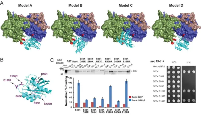
to Sec4-GTP ...38 Figure 2.3 - Computational docking studies extracted interacting elements from
the best scoring complexes of Sro7 and Sec4-GTP to produce four models ...40 Figure 2.4 - Novel mutations in Sec4 were designed to discriminate between
predicted in silico docking models ...41 Figure 2.5 - Mutations in Sec4 predict a precise model for the docking of Sec4
onto the binding cleft of Sro7 ...42 Figure 2.6 - Sec4 effector specificity for Sro7 interaction is attributed to the Sec4
N-terminal half of the Switch I domain ...44 Figure 2.7 - Conservation of the Sro7-Sec4 binding interface within the Lgl
family of proteins ...45 Figure 2.8 - SRO7 alleles defective in binding to Sec4-GTP complement the salt
sensitivity of sro7Δ,sro77Δ as the sole source of SRO7 in the cell ...47 Figure 3.1 - In vitro system for clustering post-Golgi vesicles in the presence of
Sro7, MgCl2 and GTPγS. ...72 Figure 3.2 - Post-Golgi vesicles do not require functional SNARE proteins,
Snc1/2 or Sso1/2, to cluster in vitro in the presence of Sro7, MgCl2, and GTPγS. ...74 Figure 3.3 - Purified post-Golgi vesicles cluster in vitro in the presence of Sro7,
MgCl2, and GTPγS. ...75 Figure 3.4 - In vitro post-Golgi vesicle clustering depends on the presence of
GTP-Sec4 on vesicles. ...77 Figure 3.5 - Novel mutant Sro7, sro7-R189D,R222D fails to induce clustering and
xii
Figure 3.6 - Biochemical characterization of Sro7-D189,D222 shows that
although the novel mutant cannot cluster vesicles in the in vitro clustering assay,
it can still bind to Sec4-GTP, Myo2, and Exo84 in GST pulldown assays ...80 Figure 3.7 - N914K,S942F behaves genetically and biochemically like
Sro7-R189D,R222D. ...81 Figure 3.8 - Sro7 mutants that favor an open conformation inhibit Sro7-mediated
xiii
LIST OF ABBREVIATIONS
aPKC atypical protein kinase C
ER endoplasmic reticulum
GDI GDP dissociation inhibitor
GDP guanosine 5’-diphosphate GST glutathione S-transferase GTPases guanosine triphosphatases
GTPγS guanosine 5’-3-O-(thio)triphosphate
HSP high speed pellet
Lgl Lethal giant larvae
SNARE soluble N-ethylmaleimide-sensitive factor adaptor protein receptor
t-SNARE target membrane SNARE
1
CHAPTER 1
Introduction
1.1 Polarized Exocytosis
The establishment of cell polarity is essential for all eukaryotic cells to perform numerous diverse cellular processes, such as cell movement, axonal outgrowth, secretion of hormones and cell differentiation. Maintaining cell polarity requires the proper transport of lipids, proteins and other materials to the correct cellular compartment. Much of this trafficking process is mediated by small, fluid-filled organelles enclosed by lipid bilayers, or secretory vesicles, that bud from the Golgi apparatus and fuse with the plasma membrane. This membrane trafficking pathway is called polarized exocytosis and requires the precise localized delivery, docking and fusion of secretory vesicles with specific sites of active growth on the plasma membrane. In the last three decades, researchers have made significant progress exploring the details of this elegant
pathway, however the precise mechanisms that regulate the spatial and temporal transport of secretory vesicles and their cargo to the correct cellular destinations remain to be fully elucidated.
One of the most pivotal moments in the trafficking field came in 1980, when Randy Schekman and his student, Peter Novick, conducted a density-based screen for mutants that were conditionally defective for secretion in the budding yeast, Saccharomyces cerevisiae, identifying 23 essential genes involved at various stages of the secretory pathway [1]. Developing
2
Golgi to plasma membrane [2]. In 2013, Schekman, along with Thomas Südhof and James Rothman, won the Nobel Prize for their work in budding yeast, neurons and in vitro systems, respectively, that was collectively responsible for discovering the intracellular exocytic trafficking machinery which regulates protein and membrane transport within all eukaryotic cells [3].
The identification of a near complete collection of genes critical for vesicle transport in budding yeast and mammalian cells has reinforced the belief that secretion is a conserved process, as almost every yeast member has a clear ortholog present in mammalian cells
performing analogous functions. Therefore, what is understood in one system is likely to have implications in the other. Additionally, because of its well-defined genetic and biochemical tools available, budding yeast is a particularly good model system for studying vesicle transport and exocytosis.
1.2 Yeast Exocytic Machinery
Polarized exocytosis can be organized into three basic steps. In yeast, the first step involves budding of vesicles from the Golgi, followed by transport of vesicles along actin cables by the type V myosin motor, Myo2, [4] and delivery to the correct plasma membrane site. Next, secretory vesicles are tethered to the target membrane, which is coordinated by the interaction between the Rab GTPase protein, Sec4, on vesicles and the Exocyst tethering complex on the plasma membrane [5-7], (Figure 1.1). Following tethering, a trans-SNARE complex forms, which drives vesicle fusion with the plasma membrane.
3
1.2.1 Rab GTPases
Rab proteins are a large family of small GTPases that direct and regulate vesicular trafficking events in the cell at all steps in the secretory pathway. The Rab family is part of the Ras superfamily of small GTPases. There are at least 66 Rab proteins present in mammalian cells and 11 yeast Rab proteins. Rab GTPases function like molecular switches, cycling from an inactive, GDP-bound state, to an active, GTP-bound state [8]. Several upstream regulators catalyze Rab cycling between active and inactive nucleotide states: Guanine nucleotide
Exchange Factors (GEFs) initiate the GDP- to-GTP activating switch, while GTPase Activating Proteins (GAPs) stimulate GTP-hydrolysis and the return to an inactive, GDP-bound state. Activated Rab proteins bind to numerous effector proteins that regulate many different membrane transport pathways and processes.
The founding member of the Rab family is the yeast Rab GTPase, Sec4 [1]. Sec4 is required for transport of post-Golgi vesicles to the plasma membrane [9], and therefore localizes to post-Golgi vesicles and sites of polarized growth on the plasma membrane [9]. The nucleotide state of Sec4 is regulated by its GEF, Sec2. Mutants of SEC2 show an accumulation of vesicles with random distribution throughout the cell, confirming that the activation of Sec4 by Sec2 directs polarized transport of secretory vesicles [10-11]. Sec4 is thought to mediate vesicle tethering at least in part through interaction with its effector, the Exocyst tethering complex subunit, Sec15 [12].
1.2.2 SNAREs
membrane-4
proximal helical SNARE motifs [14]. Biochemical reconstitution studies via in vitro transport systems pioneered by James Rothman and colleagues led to the SNARE hypothesis, suggesting that SNARE proteins on the surface of vesicles, or v-SNAREs, bind specifically with SNARE proteins on the target membrane, or t-SNAREs, to form an active trans-SNARE complex which cinches opposing membranes close together [15].
Alternatively, SNARE proteins are referred to as Q-SNAREs or R-SNAREs based on the presence of highly conserved glutamine or arginine residues in the polar “zero layer” of the SNARE motif [16]. The coiled-coil interactions in helical SNARE motifs drive pairing of 3 Q-SNARE domains with 1 R-Q-SNARE domain into a tight bundle [17-20]. The energy that is released during the formation of this stable 4-helical SNARE bundle is the major driving force behind vesicle fusion [21]. In general, there is correlation of R-SNAREs with v-SNAREs and of Q-SNAREs with t-SNAREs, however, the 3Q:1R SNARE motif ratio is not required within the yeast exocytic SNARE complex, as complexes containing four glutamine residues are fully functional [22].
5
While different sets of SNARE proteins function at distinct steps throughout trafficking pathways, aiding in vesicle transport specificity, individual SNARE proteins localize to unspecific areas on their respective membranes. For example, in yeast, Sso1 / Sso2 distributes across the entire plasma membrane, yet secretory vesicles find a way to specifically dock and fuse with sites of active growth [26]. Biochemical and genetic studies have identified several proteins that function in membrane transport after vesicle formation, but prior to vesicle fusion. This intermediate step, where an initial connection is formed between a vesicle and its target membrane, is known as vesicle tethering and may be responsible for the earliest stages of fusion specificity [1-2].
1.2.3 Tethering Factors
Tethering factors are thought to mediate the initial attachment between a vesicle and its membrane target. There are two general classes of tethering factors: large homodimeric coiled-coil tethering proteins and multi-subunit tethering complexes (MTCs). Coiled-coiled-coil tethers are mostly long, rod-like proteins, while MTCs are hetero-oligomers containing 3-10 protein subunits. This section will focus primarily on MTCs.
6
CATCHR family, the largest class of tethering proteins [28-30]. Tethering factors both physically and functionally interact with a multitude of trafficking proteins—namely Rab GTPases and SNAREs—to orchestrate the poorly understood linkage between vesicle docking and vesicle fusion.
Three of the four CATCHR family tethering proteins have been shown to act as direct effectors for Rab GTPases. In yeast, the Exocyst subunit, Sec15, is a downstream effector of the Rab GTPase Sec4 [7]. This Rab effector role is conserved for the Exocyst in higher eukaryotes where, for example, Sec15 is a downstream effector of Rab11, the Rab GTPase that mediates vesicle transport between endosomes and the plasma membrane [30-31]. The COG complex interacts with activated Ypt1 and Ypt6 during retrograde intra-Golgi trafficking in yeast,
essential for proper glycosylation of secretory proteins [32-36]. Furthermore, of the 66 identified mammalian Rabs, 12 of them have been shown to interact with different COG subunits [32, 36-38]. The GARP complex facilitates fusion of endosome-derived vesicles to the trans-Golgi network [39-40]. GARP interacts with GTP-bound Ypt6 in yeast (ortholog to mammalian Rab6) [41]. While diverse Rab GTPases mediate vesicle docking and tethering in the multitude of membrane trafficking pathways, it is their interactions with distinct tethering effectors, however, that imparts specificity to vesicle targeting.
7
release of Sec9 by Sec6, triggered by Exocyst assembly, promotes SNARE complex assembly at the plasma membrane. Of the remaining CATCHR family complexes, the Dsl1 complex interacts with the endoplasmic reticulum-localized STX18 SNARE complex [44-45], the COG complex has been shown to interact with numerous intra-Golgi and trans-Golgi SNARE complexes [36, 46] and the GARP complex interacts with the trans-Golgi STX16 SNARE complex [40-41].
1.3 Lethal Giant Larvae Family and Trafficking
Genetic screens in yeast to detect additional exocytic machinery associated with the SNARE complex identified a novel Sec9 (t-SNARE)-binding protein, Sro7 [47]. Sro7 is a member of the Lethal Giant Larvae (Lgl) / Tomosyn family of proteins that function in
maintenance of cell polarity. Lgl proteins are highly structurally conserved, containing multiple WD40 domain repeats and sequence similarity within many elements of the domain interface [48] (Figure 1.2). Lgl homologs are present in vertebrates (Lgl1 and Lgl2—also known as Hugl-1
and Hugl-2—and neuronal Tomosyn-1 and Tomosyn-2), in Drosophila melanogaster (Lgl and
Tomosyn), in Caenorhabditis elegans (Lgl and Tomosyn), and in budding yeast (SRO7 and
SRO77) (Figure 1.3).
The mechanisms of Lgl function in cell polarity remain controversial. Two different, yet not mutually exclusive hypotheses have been suggested to explain the function of the Lgl family in cell polarity: regulation of polarization of the actomyosin cytoskeleton and regulation of polarized exocytosis.
1.3.1 Lgl
8
resulting from a loss of cell polarity and die before entering metamorphosis [50]. Additionally,
lgl mutant flies share many of the properties of human tumors, such as loss of tissue architecture, cell shape and failure of cells to differentiate [51-56]. There is a strong correlation between loss of human Lgl1 and pancreatic carcinoma, malignant melanoma and colorectal cancer [57]. Lgl proteins play a crucial role in regulating cell polarity. Lgl, along with dlg and scrib, comprise a class of functionally related neoplastic tumor suppressors termed the Scribble
complex, where mutations in any of these genes cause overproliferating cells to lose the ability to organize an epithelial monolayer and differentiate [56, 58-62]. Lgl localizes to the basolateral membrane and regulates apical-basal polarity in epithelial cells through interactions with the Par6-atypical protein kinase C (aPKC) polarity complex [56, 61-62]. Par6 binds to mislocalized Lgl, where aPKC-mediated phosphorylation of Lgl at a conserved phosphorylation site prevents it from associating with the apical membrane [63-66]. Lgl also inhibits aPKC from localizing to the baso-lateral membrane [61, 67-68]. This antagonistic interaction maintains the identities of the apical and basolateral membranes.
Numerous studies suggest that the role of Lgl in cell polarity is as a regulator of the actin cytoskeleton. Experiments in Drosophila demonstrate that lgl physically associates with the nonmuscle myosin II [69-70]. This interaction, regulated by phosphorylation of Lgl by aPKC, localizes myosin II to the apical cortex of metaphase neuroblasts to modify the actin cytoskeleton [70]. Additionally, myosin II is mislocalized in the neural progenitor cells of Lgl1-/- mice [71]. The cell polarity defects in the Lgl1-/- brain are very similar to the brain phenotype of myosin II-B mutant mice [72].
9
(MDCK) epithelial cells, Lgl associates with the basolateral specific t-SNARE, Syntaxin4, as well as in complex with Syntaxin4/Snap-23 [63]. The interaction with t-SNAREs is a structurally conserved and functionally important feature of the Lgl family, as chimeric Lgl proteins in which large regions of yeast Sro7 and mammalian Lgl were exchanged could functionally rescue growth and secretion defects associated with loss of Sro7 when chimeras retained the ability to bind the yeast t-SNARE, Sec9 [74]. In neurons, Lgl was also shown to interact with the Rab GTPase, Rab10 at a common state of axonal membrane protrusion [75]. Additionally,
Drosophila lgl is required for signaling and secretion of Decapentaplegic (DPP)—a member of the transforming growth factor beta (TGFbeta) family [73].
1.3.2 Tomosyn
10
Tomosyn-1 has been suggested to act as a negative regulator of exocytosis based on studies in PC12 and chromaffin cells, and in several neurosecretory cells and neurons [80, 82-85]. In neuronal-like PC12 cells, overexpression of tomosyn-1 leads to a reduction in calcium-dependent human growth hormone secretion [76, 85]. When overexpressed in 3T3-L1
adipocytes, tomosyn interacts with the adipocyte t-SNARE complex, syntaxin-4 and SNAP-23, inhibiting VAMP-2 association and blocking insulin-stimulated fusion of GLUT4 vesicles [86]. In mouse pancreatic β-cells, overexpression of m-tomosyn significantly decreases insulin secretion and siRNA knockdown of tomosyn expression was associated with an increase in growth hormone exocytosis [87]. Taken together, these results combined with numerous other studies suggest a function for tomosyn in polarized exocytosis and regulation of neurotransmitter release by affecting the formation of trans-SNARE complexes.
1.3.3 Sro7
Whereas both Lgl and Tomosyn demonstrate the ability to bind SNARE proteins, the yeast Lgl family member, Sro7, was the first family member to suggest an in vivo functional role for the Lgl family in polarized exocytosis. SRO7 and its redundant protein homolog, SRO77, were isolated from a multicopy screen for suppressors of Rho3, a small GTPase critical for maintenance of cell polarity[88]. The following year, our lab identified Sro7 in a screen for binding partners of the SNAP25-like yeast t-SNARE, Sec9 (Figure 1.1) [47]. This interaction is regulated by the C-terminal autoinhibitory tail of Sro7 in that when it binds to Sro7, the SNARE domain of Sec9 is precluded from binding [48]. This conformational switch between an “open” form, with an unbound tail, and “closed” form with the tail bound, likely plays a role in
11
Upon deletion of SRO7 and SRO77, secretory vesicles accumulate in the cell, specifically in the polarized bud, while the actin cytoskeleton and its polarized distribution remains
unperturbed [47]. This phenotype is similar to that of late secretory mutants and suggests that the Lgl family functions in polarized exocytosis rather than in cytoskeletal regulation. Consistent with the exocytic defect being due to a role in SNARE assembly, Sro7 was also found associated with ternary SNARE complexes of Sec9/Sso/Snc.
Sro7 is the only Lgl / Tomosyn family member whose crystal structure has been determined [48] (Figure 1.2A), however structural sequence alignment of Sro7 with Lgl and Tomosyn suggests significant structural conservation (Figure 1.2B). This structure consists of two, 7-bladed WD40 beta-propeller domains followed by a 60-residue-long C-terminal tail that binds back to the protein. Structural conservation among the Lgl family members is especially clear within the 14 WD40 repeats and in many elements of the domain interface (Figure 1.2B). The beta-propeller domains are arranged in a way that resembles an open clamshell structure. Both Lgl and Tomosyn have insertions in the 10D-11A loop containing conserved sites for phosphorylation by aPKC and cAMP-dependent protein kinase (PKA), respectively.
Interestingly, many residues in the C-terminal regulatory tail of Sro7 are conserved in Tomosyn, but not in Lgl.
Recent evidence suggests that Sro7 could also act as a direct effector of the Rab GTPase, Sec4 [89] (Figure 1.1). Sro7 was shown to bind preferentially to Sec4 in vitro in the presence of GTP and has genetic properties consistent with it functioning downstream of Sec4. More
GTP-12
dependent interaction and in this case Lgl has been suggested to function upstream of Rab10 to activate it, instead of act as its effector.
1.4 Thesis Contributions
The goal of my thesis was to understand the molecular details and functional significance of the interaction between Sro7 and the Rab GTPase Sec4. Chapter 2 details studies directed at identifying the interaction interface between Sro7 and the Rab GTPase, Sec4. Biochemical results combined with in vivo suppression studies and computational modeling pinpointed the Sro7-Sec4 docking interface. Specifically, we found that mutations in Sro7 which disrupt the Sro7-Sec4 interaction also demonstrate a clear deficiency in vivo to overcome defects in Exocyst complex function. Chapter 3 describes the development of a novel in vitro assay that
13
1.5 Figures
Figure 1.1 - Schematic of S. cerevisiae post-Golgi secretory machinery.
14
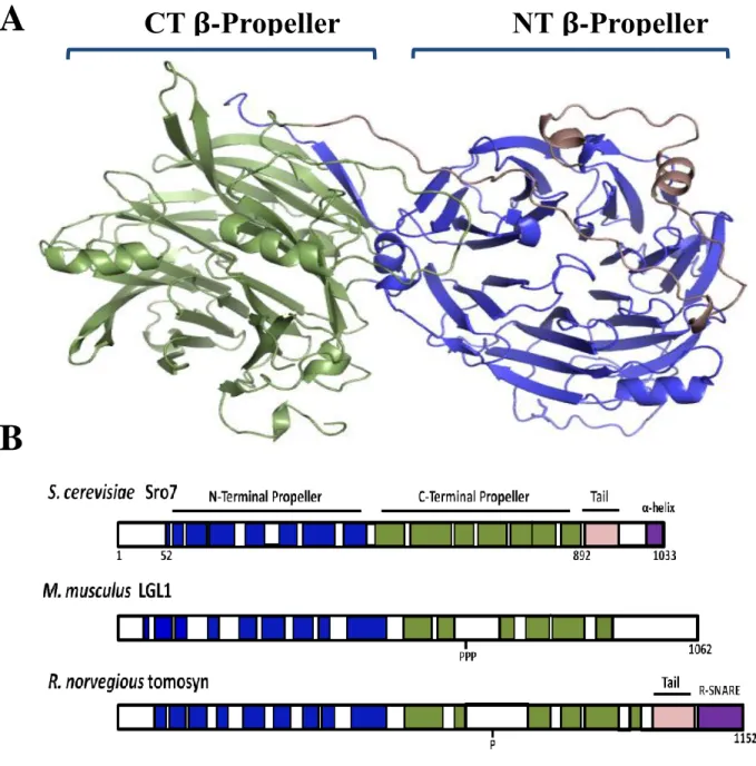
Figure 1.2 – Structural conservation within the lethal giant larvae family.
A, Crystal structure of Sro7 showing the N-terminal propeller in blue, the C-terminal propeller in green and the autoinhibitory tail in light pink. B, Schematic illustrating the structural alignments between Sro7 and the Lgl and Tomosyn family members. Phosphorylation sites are indicated on Lgl1 and tomosyn.
CT
β
-Propeller NT
β
-Propeller
A
15
Figure 1.3 – Phylogenetic tree of Lgl/Tomosyn family of proteins.
Lgl/Tomosyn homologs are present in humans, Mus musculus mice, and Rattus norvegicus rats (Lgl1 and Lgl2—also known as Hugl-1 and Hugl-2—and neuronal Tomosyn-1 and Tomosyn-2), in Drosophila melanogaster fuit flies (Lgl and Tomosyn), in Caenorhabditis elegans worms (Lgl
16
CHAPTER 2
Structural Basis for Recognition of the Sec4 Rab GTPase by its Effector, the Lgl/Tomosyn Homolog, Sro71
2.1 Overview
Members of the Tomosyn/Lgl/Sro7 family play important roles in vesicle trafficking and cell polarity in eukaryotic cells. The yeast homolog, Sro7, is thought to act as a downstream effector of the Sec4 Rab GTPase to promote SNARE assembly during Golgi to cell surface vesicle transport. In this paper, we describe the identification of a Sec4 binding site on the surface of Sro7 that is contained within a cleft created by the junction of two adjacent
β-propellers which form the core structure of Sro7. Computational docking experiments suggested four models for interaction of GTP-Sec4 with the Sro7 binding cleft. Further mutational and biochemical analyses confirmed that only one of the four docking arrangements is perfectly consistent with our genetic and biochemical interaction data. Close examination of this docking model suggests a structural basis for the high substrate and nucleotide selectivity in effector binding by Sro7. Finally, analysis of the surface variation within the homologous interaction site on Tomosyn-1 and Lgl-1 structural models suggests a possible conserved Rab GTPase effector function in Tomosyn vertebrate homologs.
1
17
2.2 Introduction
Polarized exocytosis requires the proper localized delivery, docking and fusion of secretory vesicles with sites of active growth on the plasma membrane. In the budding yeast,
Saccharomyces cerevisiae, secretory vesicles are delivered to specific plasma membrane sites where the Rab GTPase Sec4 mediates vesicle tethering through its interaction with the Exocyst complex subunit, Sec15 [7, 10]. Following vesicle tethering, a trans-SNARE complex forms between the vesicle SNARE proteins Snc1/2 and the plasma membrane SNARE proteins Sec9 and Sso1/2, which drives vesicle fusion [23-25].
18
in the emerging bud while the actin cytoskeleton is unperturbed [47]. This is phenotype is similar to that of late secretory mutants and implicates that the Lgl / Tomosyn family functions in
polarized exocytosis rather than cytoskeletal regulation.
Recent evidence suggests that Sro7 could also act as a direct effector of Sec4 [89]. Sro7 was shown to bind to Sec4 in the presence of GTP and has genetic properties consistent with it functioning downstream of Sec4. However, there has not been rigorous testing that proves the physical interaction with Sec4 is required for Sro7 function in vivo, so the mechanism of this interaction and if it plays a role in exocytosis still remains unknown.
Sro7 is the only Lgl / Tomosyn family member whose X-ray structure has been determined [48]. In this study, we utilized the crystal structure of Sro7 to identify charged surface patches on Sro7 and screened for their involvement in the Sro7-Sec4 interaction. We combined these in vitro results with in vivo suppression studies and in silico modeling to validate the Sro7-Sec4 docking interaction interface. We found that disruption of the Sro7-Sec4
interaction results in a reduction of Sro7 function in vivo. Moreover, bioinformatic analysis suggests the possibility that the Sro7-Sec4 Rab-binding interface may be conserved in vertebrate Tomosyn-1.
2.3 Results
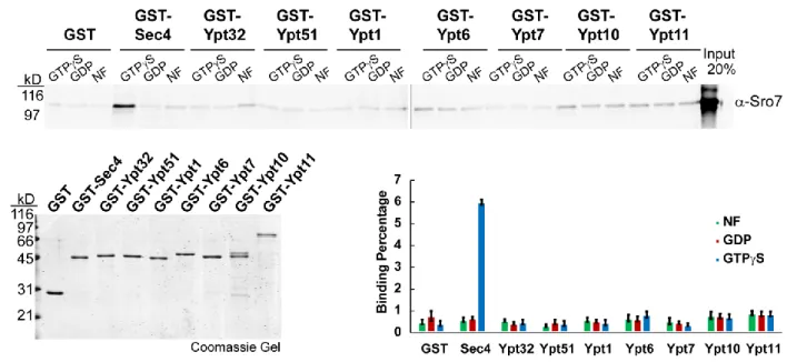
2.3.1 The interaction between Sro7 and the yeast Rab GTPase Sec4 is specific and
GTP-dependent.
19
interaction suggests that Lgl and Rab10 interact in a GDP- rather than GTP-dependent manner. This further suggests that Lgl1 may act as a GDI displacement factor (GDF) facilitating the activation of Rab10, instead of as a Rab GTPase effector transducing the GTP-Rab function.
This prompted us to further examine nucleotide and Rab-binding specificity of Sro7 with the yeast Rab GTPases. We therefore examined binding properties of Sro7 with representatives of each subgroup of the well-characterized yeast Rab GTPase family [92-94]. Representatives of each of the 8 yeast Rab subfamilies—Sec4, Ypt1, Ypt32, Ypt51, Ypt6, Ypt7, Ypt10 and
Ypt11—were purified from E.coli as N-terminally-tagged GST fusions, immobilized on
glutathione sepharose beads and exchanged with GTPγS, GDP or no nucleotide (Figure 2.1). As seen in Figure 2.1, purified full-length Sro7 binds specifically to GTPγS-Sec4 and fails to show significant binding to any of the other 7 Rab proteins tested (Figure 2.1). Importantly, Sro7 binding is completely dependent on Sec4 being in a GTP-bound, activated state and no detectable binding was seen to GDP or nucleotide-free forms of any of the 8 Rab GTPases in yeast. Taken together, these results indicate that the interaction between Sro7 and the yeast Rabome is highly specific to Sec4 in its active or GTP-bound state.
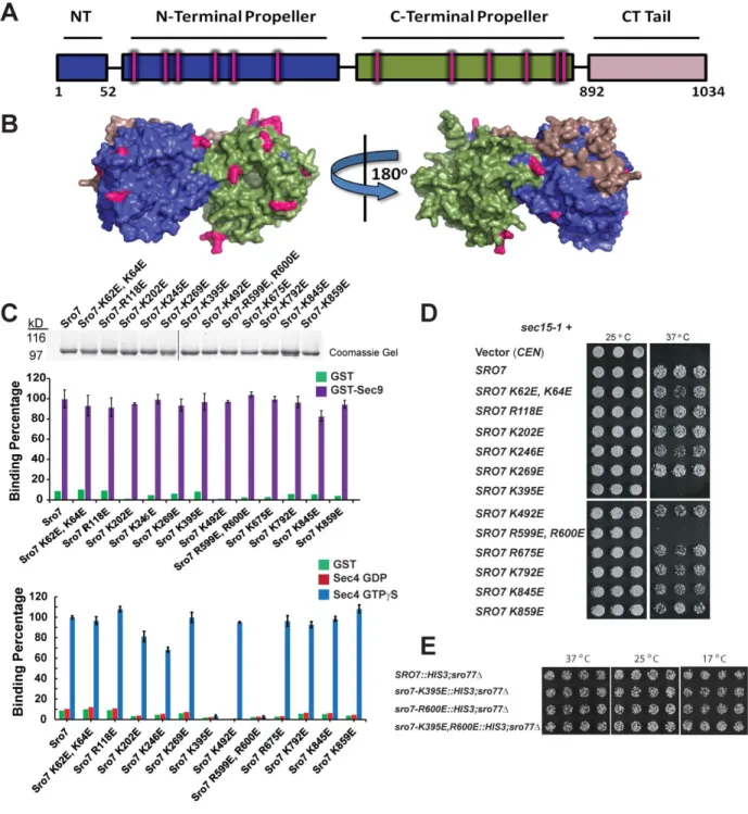
2.3.2 Biochemical screen identifies two Sro7 mutants deficient in binding to Sec4-GTP.
20
patch on charged residues by creating charge-reversal mutations because of their high likelihood to disrupt protein-protein interactions. We therefore generated a collection of 12 different
charge-reversal mutations that span both β-propeller domains of Sro7 and exclude known sites of interaction with the Sec9 N-terminus and with the regulatory tail of Sro7 (Figure 2.2A, B) [48]. Wildtype Sro7 and the Sro7 mutants were purified to homogeneity from yeast using a multi-step procedure we have previously described [95] and all proteins were subjected to SDS-PAGE and Coomassie staining to assess both purity and quantity of each preparation (Figure 2.2C).
As a first examination of overall protein integrity, we compared wildtype Sro7 and the Sro7 mutant proteins for binding to a known Sro7 ligand, the t-SNARE Sec9[47]. All twelve charge-reversal mutants bind GST-Sec9 comparably to wildtype Sro7 and statistical analysis of the binding data demonstrates that differences are not significant (Figure 2.2C).
To determine if any of the 12 surface patches on Sro7 were involved in mediating specific binding to Sec4-GTP, we subjected each of the purified mutant proteins to binding assays with Sec4 that had been exchanged with GTPγS or GDP. Of the 12 Sro7 mutant proteins, two—Sro7-K395E and Sro7-R599E, R600E—no longer bind to Sec4-GTP, while the remaining 10 bind Sec4-GTP at levels statistically similar (p >0.05) to wildtype Sro7 (Figure 2.2C). None of the mutations have any detectable effect on the nucleotide-specificity of the interaction with Sec4.
21
[47, 96]. As the sole source of Sro7, all twelve of the charge-reversal mutants complement the cold and salt sensitivity of sro7∆, sro77∆ like wildtype SRO7 (Table 2.1). To determine if the lack of any detectable growth phenotype for the two Sec4-binding deficient mutants was related to the presence of the mutants on an extra-chromosomal plasmid, we integrated each defective allele (sro7-K395E and sro7-R600E), as well as an allele with both mutations (
sro7-K395E,R600E) into the native SRO7 locus by gene replacement (see Figure 2.8 for details on the integration). The results of this analysis, shown in Figure 2.2E and Figure 2.8, demonstrate that each of the single mutants are able to fully restore growth as the sole source of SRO7 at all temperatures and media conditions examined, including 17 oC and 0.7M NaCl. Furthermore, the absence of a growth phenotype is unlikely due to residual binding present in each mutant as the phenotype is identical to wildtype even when both mutations are combined in a single allele (sro7-K395E,R600E).
The second genetic assay makes use of our previous observation that Sro7 plays a role as an effector of Sec4 that is parallel to the Exocyst complex, as overexpression of Sro7 strongly suppresses growth defects associated with deletions or temperature-sensitive mutations in subunits of the Exocyst complex[47, 87, 89]. Temperature-sensitive alleles of the Sec15
component of the Exocyst (sec15-1) are particularly sensitive to small increases in SRO7 dosage, as just a single additional copy (CEN plasmid) is sufficient to suppress temperature-sensitivity of
sec15-1 at 37oC (Figure 2.2D). When the sec15-1 suppression analysis was extended to the collection of surface patch mutants, we found that only the sro7-K395E and sro7-R599E, R600E
alleles demonstrated loss of suppression. The other 10 surface patch alleles suppress sec15-1
22
data in Figure 2.2C, these results identify a potential surface(s) on Sro7 involved in the interaction with Sec4 both in vitro and in vivo.
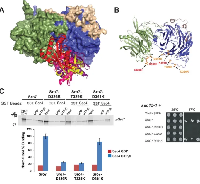
2.3.3 Computational docking studies extracted interacting elements from the best scoring
complexes of Sro7 and Sec4-GTP to produce four models.
To examine the structural implications of the in vitro and in vivo effects of the Rab binding mutants described above, we mapped the sites of the Sro7-K395 and Sro7-R599, R600 residues on the crystal structure of Sro7. Interestingly, these sites suggest that both the N-terminal and C-N-terminal β-propeller domains contribute to Sec4 binding and implicate a cleft formed by the intersection of the two propeller domains of Sro7 (Figure 2.3B). The
23
Ras GTPases that change in response to the nucleotide state and are therefore strongly predicted to be involved in the interaction with a Sec4 effector protein such as Sro7.
All four Sro7-Sec4 docking models included the same Sro7 interface for Sec4 binding (Figure 2.3A). This region is in a pocket on the opposing side of the protein from where the Sro7 regulatory tail and the t-SNARE, Sec9 bind. To confirm that this Sro7 interface is involved in binding to Sec4, as predicted by these four docking models, and to further delineate the site of Sec4 binding on Sro7, we created a second set of Sro7 mutants at this interface (shown in
orange, Figure 2.3B). We characterized the Sro7 mutants both in vitro by binding to Sec4-GTP and in vivo by analysis of their ability to suppress the growth defect of sec15-1 at 37oC. The results, shown in Figure 2.3C, demonstrate that two of the three mutant proteins—Sro7-D326R and Sro7-S327A, T329E—are deficient in binding to GTP, while Sro7-D361K binds Sec4-GTP comparably to wildtype Sro7. Likewise, when we examined the Sro7 mutants in vivo by testing their ability to suppress the temperature sensitivity of sec15-1 we found that the same two mutants that are deficient in binding to Sec4-GTP—sro7-D326R and sro7-S327A, T329E—are
also unable to suppress growth at the restrictive temperature (Figure 2.3C). Also consistent with the binding data, we see that sro7-D361K suppresses sec15-1 at 37oC similarly to wild-type
24
Sec4-GTP also demonstrate a clear defect in vivo to overcome the loss in Exocyst complex function present in sec15-1.
2.3.4 Novel mutations in Sec4 were designed to discriminate between predicted in silico
docking models.
While the same binding interface of Sro7 is involved in all four docking models, the orientation of Sec4 with respect to Sro7 is substantially different in each model (Figure 2.4A). We therefore generated a second set of mutations in surface-exposed, charged Sec4 residues with the aim of distinguishing between the four models. To accomplish this, we chose six residues to mutate with high predictive value in distinguishing between the four models based on differences in their predicted distance from Sro7 in the four models. Residues were scored for high
interaction potential when the distance was less than 4Å and low interaction potential when the distance was greater than 4Å (Figure 2.5A). For example, the Sec4-D56R mutant is predicted to be involved in Sec4-Sro7 docking in Models A, C and D, but not in Model B. Therefore, this particular mutation will discriminate Model B from the other models. The locations of the novel set of Sec4 mutations tabulated in Figure 2.5A are shown by a ribbon diagram in Figure 2.4B.
Wildtype Sec4 and the discriminatory Sec4 mutants were purified as GST fusion proteins, exchanged with either GTPγS or GDP and tested for binding to wildtype Sro7. The binding data shown in Figure 2.4C demonstrates that 4 of the 6 mutant proteins tested exhibit a significant defect in binding to Sro7, while 2 of the mutant Sec4 proteins bind to Sro7 at levels similar to wildtype Sec4. In order to analyze the in vivo consequences of Sec4 mutations
resulting in a loss of binding to Sro7, we utilized the fact that, like SRO7, one additional copy of
25
binding to Sro7 in vitro also have completely lost the ability to suppress the sec15-1 mutant temperature-sensitivity. Likewise, the two mutant SEC4 alleles which encode proteins that bind to Sro7 at levels similar to that of wildtype Sec4, also demonstrate suppression of sec15-1
temperature-sensitivity in a manner indistinguishable from wildtype SEC4. The strong
correlation between the biochemical and genetic analyses strongly supports the notion that the interaction between Sec4 and Sro7 observed in vitro is also important for the function of both proteins within the cell.
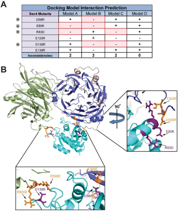
26
affected the Sro7-Sec4 interaction both in vitro and in vivo. Therefore, it is unlikely that Models A and B are the correct docking arrangement between Sro7 and Sec4 and this is scored as an inconsistency (in red) in Figure 5A. As can be seen, Models A, B and C all contain several inconsistencies when comparing the predicted effects of the mutations with the effects observed
in vitro and in vivo. In contrast, Model D (shown in Figure 2.5B) is the sole model with perfect correlation between its interaction predictions and the actual in vitro and in vivo data (Figure 2.5A). Additionally, 4 of the 5 mutated Sec4 residues in Model D are predicted to be within 4 Å of the Sro7-Sec4 binding interface and demonstrate a strong effect on both binding and
suppression.
Three distinct patches of mutations on both proteins affecting the Sro7-Sec4 interaction correspond nicely between Sro7 (orange residues) and Sec4 (purple residues)—Sro7-R599E, Sro7-R600E and Sec4-D136R on the C-terminal β-propeller front side of the binding cleft (Figure 2.5, Inset Below), Sro7-K395E and Sec4-D56R on the N-terminal β-propeller front side of the binding cleft (Figure 2.5, Inset Below), and Sro7-D326R, Sro7-T329K, Sec4-E80K and Sec4-R83D on the N-terminal β-propeller back side of the binding cleft (Figure 2.5, Inset Right). Based on this extensive analysis, we invalidated 3 of the 4 docking models and have substantial evidence that Model D is the native docking arrangement for interaction between Sro7 and Sec4-GTP.
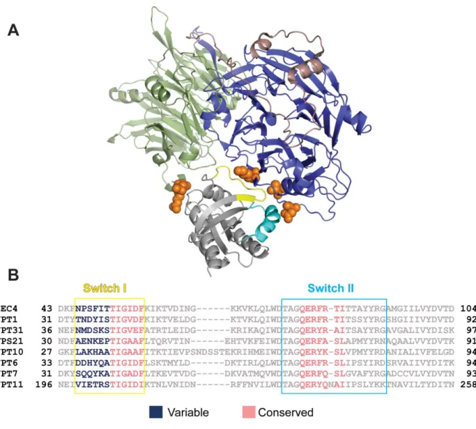
2.3.6 How does Sro7 selectively bind GTP-bound Sec4?
27
model for Sro7-Sec4 docking allows us to identify elements of the binding interaction that are likely responsible for these two effector specificity aspects of Sro7 with Sec4. Close
examination of the model illustrates four regions within Sec4 that are in most intimate contact with Sro7 (Figure 2.6). Two of the regions in close contact with Sro7 are the switch I and switch II domains of Sec4—the two regions that undergo the most conformational change when
comparing GDP and GTP-bound structures and are therefore critical to nucleotide-specific recognition of small GTPases by effectors and accessory proteins [99]. The four Sro7 contact regions within Sec4 are: residues 46-58 (the entire switch I domain), residues 79-84 (within the switch II domain), Sec4-135-140 and Sec4-162-167. The first two regions make contact with the Sro7 N-terminal propeller domain, while the latter two interact with the Sro7 C-terminal β-propeller domain.
28
switch I domain demonstrates the kind of variability that one would expect of a site responsible for the Sro7 substrate specificity. Taken together, we conclude that while Sro7 contact with both the switch I and switch II domains is likely responsible for recognition of the GTP-bound state of Sec4, it is the specific interaction with the N-terminal segment of the switch I domain that
provides the high degree of Rab specificity for recognition of Sec4-GTP by the Sro7 effector protein.
2.3.7 Conservation of the Sro7-Sec4 binding interface within the Lgl family of proteins.
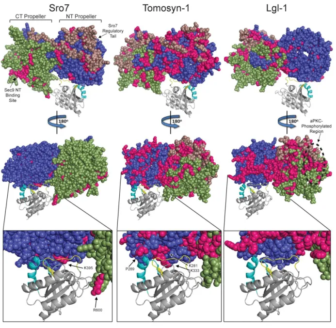
Structural and evolutionary examination of surface residue variation has demonstrated that protein-protein interfaces are significantly more constrained in their variability when compared to non-interaction surfaces [100-101]. Structural alignments of Sro7 with its closest vertebrate homologs Tomosyn and Lgl demonstrate that the overall dual β-propeller domain structure of Sro7 is likely shared between all three members of this family [48]. Since the
SNARE regulatory function is shared between yeast and vertebrate homologs [102], it is possible that the Rab effector function is also shared with one or more of the vertebrate homologs. If so, one might expect to see a reduced surface variability within the region predicted to form the homologous Sro7-Sec4 binding cleft. We therefore used a combined structural and phylogenetic approach to examine the surface variability of vertebrate members of Tomosyn and Lgl,
29
residues were also identified from an alignment of 47 Tomosyn-1 vertebrate (fish, frog, bird, and mammal) homologs (Figure 2.7 Middle Panels) and from 34 Lgl-1 vertebrate homologs (Figure 2.7 Right Panels). Conserved invariant residues are indicated in pink on the Sro7 crystal structure and the Tomosyn and Lgl structural models, respectively (Figure 2.7). While rates of surface change are overall much greater in the yeasts compared to vertebrates (presumably due to both functional redundancy with the Exocyst complex and much shorter generation time), specific sites of low variability are apparent in all three family members.
There are three areas on the surface of Sro7 with decreased variability within the yeast family—regions homologous to where the Sro7 regulatory tail binds back to the N-terminal β-propeller, the binding site for the N-terminus of the t-SNARE Sec9 [48], and residues within the Sro7-Sec4 binding pocket (Figure 2.7). When we focus specifically on the Sec4 binding cleft on Sro7, the variability among the yeast members reveals two conserved sites in the Sro7-Sec4 binding pocket—one on the C-terminal β-propeller including the Sro7-R600 residue and one on the N-terminal β-propeller including the Sro7-K395 residue (Sro7 Inset, Figure 2.7). Residues Sro7-R600 and Sro7-K395 were previously shown to be directly involved in the Sro7-Sec4 interaction (Figure 2.2). The conserved Sro7-K395 area in the docked Sro7-Sec4 structure interacts with the Sec4 switch I domain (yellow), the region of Sec4 responsible for effector specificity, consistent with the correlation between decreased protein surface variability and functional importance (Sro7 Inset, Figure 2.7).
30
conserved regions spanning both faces of its dual propeller structure, likely attributed to increased surface fixation from acquired functionality (Figure 2.7). Like Tomosyn, Lgl
vertebrates also developed greater surface residue conservation, however, the invariant residues are located primarily on one protein face—the same face as where the aPKC phosphorylation sites reside (Figure 2.7). A focused examination of the region of Tomosyn that is homologous to the Sro7-Sec4 binding pocket reveals that vertebrate Tomosyn-1 family members maintain significant conservation within the interaction interface. Notably, there is a cluster of invariant residues in Tomosyn-1 that correspond with the part of the binding cleft in Sro7 containing the critical K395 residue (Tomosyn Inset, Figure 2.7).
Interestingly, the surface conservation in vertebrate Lgl-1 members is quite distinct from the conservation observed in vertebrate Tomosyn-1 proteins. While there is a significant increase in overall Lgl surface residue conservation when compared to Sro7 and Tomosyn-1, the region corresponding to the Sec4 binding pocket in Sro7 is significantly more variable in Lgl-1 (Lgl Inset, Figure 2.7). This suggests that of the two vertebrate branches of the Sro7/Lgl/Tomosyn family, Tomosyn is the most likely to have a conserved role as an effector for a vertebrate Rab GTPase.
2.4 Discussion
C-31
terminal propellers [48]. Here we map the binding site utilized by the Sec4 GTPase in its
interaction with Sro7 to a cleft formed at the intersection of these two propellers—an interaction which is highly specific to the yeast Rab family member Sec4 in its activated, GTP-bound form. Our bioinformatic analysis of the sequence variation found in vertebrate members of the Tomosyn1/2 family suggests that this cleft may also be important for the interaction between Tomosyn and a related small GTPase—perhaps as part of an ancestral function for this family of proteins that predates the divergence of the family members [104-105]. Interestingly, there is significantly less conservation in the homologous Sro7-Sec4 binding interface for members of the Lgl1/2 family when compared to Tomosyn1/2. We can only speculate about the precise significance of this difference, but it could be attributed to divergence or loss of the ancestral Rab effector function as the Lgl family evolved distinct functions from Tomosyn in metazoans. This functional separation between family members could have occurred in parallel to the loss of the C-terminal R-SNARE motif in members of the Lgl family in metazoans [104]. Interestingly, Wang et al. [75] have reported a direct interaction between Lgl1 and the Rab10 GTPase. However, unlike the GTP-dependent Sro7-Sec4 interaction, Lgl1 appears to interact specifically with the GDP- rather than GTP-bound form of Rab10.
32
33
2.5 Materials and Methods
2.5.1 Media and Reagents
Yeast growth media used in this study includes: YPD (1% yeast extract, 2% bacto-peptone, 2% dextrose [Difco, Sparks, MD]), S minimal (0.67% yeast nitrogen base without amino acids and 2% dextrose [Difco, Sparks, MD]), agar [Fisher Scientific, Pittsburgh, PA], and dropout media (0.67% yeast nitrogen base without amino acids, synthetic complete amino acid supplement minus appropriate amino acid(s) and 2% dextrose [US Biological, Swampscott, MA]).
Bacteria growth media used in this study includes: Terrific Broth (TB; 4.7% bacto-TB, 1% glycerol [Fisher Scientific, Pittsburgh, PA]), Super Optimal Broth (SOB; 2% tryptone
[Difco, Sparks, MD], 0.5% bacto-yeast extract, 2.5mM KCl [Sigma Aldrich, St. Louis, MO], 1M NaCl, 10mM MgCl2, 10mM MgSO4 [Fisher Scientific, Pittsburgh, PA]), Super Optimal Broth with Catabolite Repression (SOC; SOB + 2.5% glucose), and Lysogeny Broth (LB; 1% bacto-tryptone, 0.5% bacto-yeast extract, and 1% NaCl).
Reagents used in this study: GTPγS, Triton X-100, GDP, sodium azide, sodium fluoride, dithiothreitol (DTT), β-mercaptoethanol, Pepstatin A and LAA components (Leupeptin 1
34
MA]. IgG Sepharose 6 Fast Flow beads were obtained from Amersham Biosciences [Piscataway, NJ].
2.5.2 Yeast Strains and Genetic Analysis
The yeast strains that were constructed and used for this study are listed in Table 2.2. Yeast transformations were performed using the lithium acetate method [107]. For genetic analysis, at least three different spores were analyzed per experiment.
2.5.3 Plasmids and Molecular Biology
The plasmids that were constructed and used for this study are listed in Table 2.3. Sro7 charge reversal mutants were generated by site-directed mutagenesis on pB2129 (Sro7, CEN,
HIS3 plasmid). Protein A-tagged Sro7 constructs were generated as BamHI/HindIII subclones in pB966 (Protein A, 2 µ plasmid) as previously described [95]. Sro7 charge-reversal mutants were generated by site-directed mutagenesis on pB1931 (Sec4, CEN, LEU2 plasmid). GST-tagged Sec4 constructs were generated as BamHI-SalI fragments in pB 2173 (pGEX-6P1 plasmid). GST-tagged Rab protein constructs were generated using genomic DNA as BamHI-SalI fragments (Ypt32, Ypt51, Ypt1, Ypt6, Ypt7, Ypt10) or BglII-SalI (Ypt11) fragments and subcloned into pB 2173 (pGEX-6P1 plasmid). Constructs were confirmed by sequencing.
2.5.4 Protein Purification
Wildtype Sro7 and Sro7 charge reversal mutant proteins were purified as previously described [95]. GST fusion proteins (Sec4, Sec4 charge reversal mutants, Ypt32, Ypt51, Ypt1, Ypt6, Ypt7, Ypt10, and Ypt11) were transformed into BL21 Escherichia coli and expressed as previously described [89]. Sec9 with a C-terminal His6 tag was purified from E. coli as
35
2.5.5 In Vitro Binding Assays
In vitro binding of wildtype Sro7 and Sro7 charge reversal mutants to GST fusions of Sec9, Sec4, Sec4 mutants and the 7 Rab family proteins were performed using previously described conditions [89] with the following modifications: During nucleotide exchange, beads were incubated with 100 µM GTPγS, GDP or nucleotide free for 30 minutes at 25 degrees Celsius. Following this incubation, MgCl2 was added to a final concentration of 30 mM and incubated for 1 hour at 25 degrees Celsius. Binding buffer consisted of 20 mM Tris-HCl, pH 7.4, 140 mM NaCl, 10 mM McCl2, 1mM DTT and 0.5% Triton X-100. In vitro binding of wildtype Sro7 and Sro7 charge reversal mutants to GST-Sec9 (Full length) was performed using
previously described conditions [74]. Binding percentages for all in vitro binding experiments were expressed as percent binding relative to wildtype Sro7 binding. P values were determined using Student's t test from three separate binding experiments for each protein.
2.5.6 Protein-Protein Docking Analysis
36
scoring functions included 9 poses which involved both the Sec4 and Sro7 residues of interest. These 9 poses could be separated into 4 unique poses, as some poses were identified as large clusters for more than 1 scoring function. A contact analysis for residues in Sec4 within 4Å of Sro7 interface residues was calculated using PyMOL tools.
2.5.7 Tomosyn-1 and Lgl-1 Model Building
The sequences for human Tomosyn-1 and Lgl-1 were submitted to the HHpred fold recognition server (http://toolkit.tuebingen.mpg.de/hhpred). The 14 WD-repeat structure of yeast Sro7 was identified as a top hit. The models of Tomosyn-1 and Lgl-1 were built using MODELLER [109] based on the yeast Sro7 (PDB ID 2OAJ) template.
2.5.8 Sequence Analysis
Sequences for vertebrate Tomosyn-1 and for Tomosyn homologs in the Hemichordate acorn worm (Saccoglossus kowalevskii) and the Echinoderm purple sea urchin
37
2.6 Tables and Figures
Figure 2.1 - The interaction between Sro7 and the yeast Rab GTPase Sec4 is specific and GTP-dependent.
38
Figure 2.2 -Biochemical screen identifies two Sro7 mutants deficient in binding to Sec4-GTP.
39
wildtype Sro7 binding set to 100%. Quantitation in each graph was based on 4 independent experiments. (D) The mutant strain sec15-1 was transformed with a plasmid (CEN) expressing
40
Figure 2.3 - Computational docking studies extracted interacting elements from the best scoring complexes of Sro7 and Sec4-GTP to produce four models.
41
Figure 2.4 - Novel mutations in Sec4 were designed to discriminate between predicted in silico docking models.
(A) In the surface-filling models (A-D) of Sro7, the N-terminal propeller is shown in blue, the C-terminal propeller in green, and the C-C-terminal tail in light pink. The Sro7 mutations defective in binding Sec4-GTP (cyan) are shown in red. (B) Ribbon diagram of Sec4-GTP shown with
discriminatory mutations in purple. (C) Wildtype Sec4 or Sec4 mutants were purified as GST fusion proteins and bound to Sro7 as previously described. Western blot of binding and
quantitation from 4 independent experiments is shown. Sec15-1 was transformed with a plasmid (CEN) expressing SEC4, the discriminatory mutants or vector only. Three independent
42
Figure 2.5 - Mutations in Sec4 predict a precise model for the docking of Sec4 onto the binding cleft of Sro7.
43
44
Figure 2.6 - Sec4 effector specificity for Sro7 interaction is attributed to the Sec4 N-terminal half of the Switch I domain.
45
Figure 2.7 - Conservation of the Sro7-Sec4 binding interface within the Lgl family of proteins.
Crystal structure of Sro7 (left) and structural models of Tomosyn-1 (middle) and Lgl-1 (right) docked with Sec4-GTP (grey). Structural models were built with MODELLER using the crystal structure of Sro7 as a template. The Tomosyn-1 model is similar to that shown by Williams et al.
46
47
Figure 2.8 - SRO7 alleles defective in binding to Sec4-GTP complement the salt sensitivity of sro7Δ,sro77Δas the sole source of SRO7 in the cell
48
YPD YPD+0.7M NaCl ______________ _____
37O
C 14O
C 25O
C
Vector (CEN) + - -
SRO7 + + +
SRO7 K62E,K64E + + +
SRO7 R118E + + +
SRO7 K202E + + +
SRO7 K246E + + +
SRO7 K269E + + +
SRO7 K395E + + +
SRO7 K492E + + +
SRO7 R599E,R600E + + +
SRO7 R675E + + +
SRO7 K792E + + +
SRO7 K845E + + +
SRO7 K859E + + +
SRO7 D326R + + +
SRO7 S327A,T329K + + +
SRO7 D361K + + +
Table 2.1 - Complementation of sro7Δ,sro77Δ by SRO7 mutants used in this study
The sro7Δ,sro77Δ plasmid shuffle strain containing wildtype SRO7 (CEN, URA3) was transformed with a plasmid (CEN, HIS3) expressing SRO7, SRO7 mutants orvector only. Transformants were transferred and grown on 5-FOA media at the permissivetemperature (37OC) to select against the SRO7 (CEN, URA3) plasmid. Three independenttransformants were then picked into microtiter wells and transferred to YPD or selective mediaat various
49
Strain Genotype Source
BY706 sro7∆::LEU2; his3-∆200; leu2-3,112; ura3-52 P. B. collection
BY1612 sec15-1; his3-∆200; ura3-52 P. B. collection
BY2885 sro7∆::HIS3; pep4∆::NATR; his3-∆200; ura3-52
P. B. collection
BY2906 sro7∆::HIS3; pep4∆::NATR; his3-∆200; ura3-52; SRO7
in pB966 (ProtA, 2µ, URA3) P. B. collection
BY2971 sro7∆::HIS3; pep4∆::NATR; his3-∆200; ura3-52
; SRO7-R118E in pB966 (ProtA, 2µ, URA3) This Study BY2972 sro7∆::HIS3; pep4∆::NATR; his3-∆200; ura3-52; SRO7-K202E in pB966 (ProtA, 2µ, URA3) This Study BY2973 sro7∆::HIS3; pep4∆::NATR; his3-∆200; ura3-52; SRO7-K246E in pB966 (ProtA, 2µ, URA3) This Study BY2974 sro7∆::HIS3; pep4∆::NATR; his3-∆200; ura3-52; SRO7-R675E in pB966 (ProtA, 2µ, URA3) This Study
BY2998 sro7∆::HIS3; pep4∆::NATR; his3-∆200; ura3-52; SRO7-K62E,K64E in pB966 (ProtA, 2µ, URA3) This Study
BY2999 sro7∆::HIS3; pep4∆::NATR; his3-∆200; ura3-52; SRO7-K395E in pB966 (ProtA, 2µ, URA3) This Study
BY3000 sro7∆::HIS3; pep4∆::NATR; his3-∆200; ura3-52
; SRO7-K859E in pB966 (ProtA, 2µ, URA3) This Study BY3001 sro7∆::HIS3; pep4∆::NATR; his3-∆200; ura3-52
; SRO7-K792E in pB966 (ProtA, 2µ, URA3) This Study BY3012 sro7∆::HIS3; pep4∆::NATR; his3-∆200; ura3-52
; SRO7-K269E in pB966 (ProtA, 2µ, URA3) This Study BY3016 sro7∆::HIS3; pep4∆::NATR; his3-∆200; ura3-52; SRO7-K492E in pB966 (ProtA, 2µ, URA3) This Study BY3018 sro7∆::HIS3; pep4∆::NATR; his3-∆200; ura3-52; SRO7-K845E in pB966 (ProtA, 2µ, URA3) This Study BY3019 sro7∆::HIS3; pep4∆::NATR; his3-∆200; ura3-52; SRO7-R599E,R600E in pB966 (ProtA, 2µ, URA3) This Study
BY3061 sec15-1; leu2-3,112; his3-∆200; ura3-52; SRO7 in pRS316 (CEN, URA3) This Study
BY3073 sro7∆::LEU2; his3-∆200; leu2-3,112; ura3-52 This Study
BY3090 sro7∆::LEU2; his3-∆200; leu2-3,112; ura3-52;sec15-1; SRO7 in pRS316 (CEN, URA3) This Study BY3105 sro7∆::LEU2; sro77∆::KANR; his3-∆200; leu2-3,112; ura3-52
; SRO7 in pRS316 (CEN, URA3) This Study
BY3109 sec4∆::KANR ;his3-∆200; ura3-52; leu2-3,112; SEC4
in pRS316 (CEN, URA3) This Study
BY3112 sec4∆::KANR; his3-∆200; ura3-52; leu2-3,112; pRS315 (CEN, LEU2) This Study
BY3113 sec4∆::KANR; his3-∆200; ura3-52; leu2-3,112; SEC4 in pRS315 (CEN, LEU2) This Study
BY3114 sec4∆::KANR ;his3-∆200; ura3-52; leu2-3,112; SEC4-D56R in pRS315 (CEN, LEU2) This Study
BY3115 sec4∆::KANR; his3-∆200; ura3-52; leu2-3,112; SEC4-E80K in pRS315 (CEN, LEU2) This Study
BY3116 sec4∆::KANR ;his3-∆200; ura3-52; leu2-3,112; SEC4-E120R in pRS315 (CEN, LEU2) This Study
BY3117 sec4∆::KANR ;his3-∆200; ura3-52; leu2-3,112
; SEC4-D136R in pRS315 (CEN, LEU2) This Study BY3124 sro7∆::HIS3; pep4∆::NATR; ura3-52; his3-∆200
; SRO7-R600E in pB966 (ProtA, 2µ, URA3) This Study BY3136 sro7∆::HIS3; pep4∆::NATR; ura3-52; his3-∆200
50
BY3139 sro7∆::HIS3; pep4∆::NATR; ura3-52; his3-∆200; SRO7-D361K in pB966 (ProtA, 2µ, URA3) This Study BY3140 sro7∆::HIS3; pep4∆::NATR; ura3-52; his3-∆200; SRO7-D326R in pB966 (ProtA, 2µ, URA3) This Study
BY3166 sro7∆::LEU2; sro77∆::NATR; sec15-1; ura3-52; leu2-3,112; his3-∆200; SRO7 (CEN, URA3) This Study
BY3167 sro7∆::LEU2; sro77∆::NATR
; sec15-1; ura3-52; leu2-3,112; his3-∆200; SRO7 (CEN, HIS3) This Study BY3168 sro7∆::LEU2; sro77∆::NATR
; sec15-1; ura3-52; leu2-3,112; his3-∆200; SRO7-K395E (CEN, HIS3) This Study BY3169 sro7∆::LEU2; sro77∆::NATR
; sec15-1; ura3-52; leu2-3,112; his3-∆200; SRO7-R600E (CEN, HIS3) This Study
BY3178 SRO7:: HIS3; sro77∆:: KANR; ura3-52; his3-∆200 This Study
BY3179 sro7-K395E:: HIS3; sro77∆:: KANR; ura3-52; his3-∆200 This Study
BY3180 sro7-R600E:: HIS3; sro77∆:: KANR; ura3-52; his3-∆200 This Study
BY3181 sro7-K395E,R600E:: HIS3; sro77∆:: KANR; ura3-52; his3-∆200 This Study
51
Plasmid Host Description Source
pB38 DH5α GAL1 Promoter in pRS313 (CEN, HIS3) P.B. collection
pB39 DH5α GAL1 Promoter with ADH1 Terminator in pRS313 P.B. collection
pB331 DH5α pRS316 (CEN, URA3) P.B. collection
pB844 DH5α pRS315 (CEN, LEU2) P.B. collection
pB966 DH5α ADH1 Promoter with ProtA-TEV tag in pRS426 (2µ, URA3) P.B. collection
pB1331 DH5α pRS313 (CEN, HIS3) P.B. collection
pB1931 DH5α SEC4 in pB844 (CEN, LEU2) P.B. collection
pB2042 DH5α YPT32 in pGEX6P-1 This Study
pB2043 DH5α YPT51 in pGEX6P-1 This Study
pB2044 DH5α YPT1 in pGEX6P-1 This Study
pB2045 BL21 YPT32 in pGEX6P-1 This Study
pB2046 BL21 YPT51 in pGEX6P-1 This Study
pB2047 BL21 YPT1 in pGEX6P-1 This Study
pB2104 DH5α SRO7-K492E in pB1331 (CEN, HIS3) This Study
pB2105 DH5α SRO7-K845E in pB1331 (CEN, HIS3) This Study
pB2107 DH5α SRO7-R599E,R600E in pB1331 (CEN, HIS3) This Study
pB2108 DH5α SRO7-R118E in pB1331 (CEN, HIS3) This Study
pB2109 DH5α SRO7-R202E in pB1331 (CEN, HIS3) This Study
pB2110 DH5α SRO7-K246E in pB1331 (CEN, HIS3) This Study
pB2111 DH5α SRO7-R675E in pB1331 (CEN, HIS3) This Study
pB2112 DH5α SRO7-K62E,K64E in pB1331 (CEN, HIS3) This Study
pB2113 DH5α SRO7-K395E in pB1331 (CEN, HIS3) This Study
pB2114 DH5α SRO7-K801E in pB1331 (CEN, HIS3) This Study
pB2115 DH5α SRO7-K859E in pB1331 (CEN, HIS3) This Study
pB2116 DH5α SRO7-K792E in pB1331 (CEN, HIS3) This Study
pB2117 DH5α SRO7-K492E in pB966 This Study
pB2118 DH5α SRO7-K845E in pB966 This Study
pB2120 DH5α SRO7-R599E,R600E in pB966 This Study
pB2121 DH5α SRO7-R118E in pB966 This Study
52
pB2123 DH5α SRO7-K246E in pB966 This Study
pB2124 DH5α SRO7-R675E in pB966 This Study
pB2125 DH5α SRO7-K62E,K64E in pB966 This Study
pB2126 DH5α SRO7-K859E in pB966 This Study
pB2127 DH5α SRO7-K792E in pB966 This Study
pB2128 DH5α SRO7-K395E in pB966 This Study
pB2129 DH5α SRO7 in pB1331 (CEN, HIS3) This Study
pB2159 DH5α SEC4-D56R in pGEX 6P-1 This Study
pB2161 DH5α SEC4-E80K in pGEX 6P-1 This Study
pB2163 DH5α SEC4 in pGEX 6P-1 This Study
pB2167 DH5α SEC4-E80K in pB844 (CEN, LEU2) This Study
pB2169 DH5α SEC4-D56R in pB844 (CEN, LEU2) This Study
pB2175 BL21 SEC4-D56R in pGEX 6P-1 This Study
pB2177 BL21 SEC4-E80K in pGEX 6P-1 This Study
pB2187 DH5α SRO7-K269E in pB1331 (CEN, HIS3) This Study
pB2188 DH5α SRO7-K269E in pB966 This Study
pB2195 DH5α SRO7 in pB331 (CEN, URA3) (No BamHI site) This Study
pB2216 BL21 SEC4-E120R in pGEX 6P-1 This Study
pB2217 BL21 SEC4-D136R in pGEX 6P-1 This Study
pB2218 BL21 SEC4-E138R in pGEX 6P-1 This Study
pB2220 BL21 SEC4-R83D in pGEX 6P-1 This Study
pB2230 DH5α SRO7-R599E in pB966 This Study
pB2231 DH5α SRO7-R600E in pB966 This Study
pB2236 DH5α SRO7-D326R in pB1331 (CEN, HIS3) This Study
pB2237 DH5α SRO7- S327A,T329K in pB1331 (CEN, HIS3) This Study
pB2239 DH5α SRO7-D361K in pB1331 (CEN, HIS3) This Study
pB2240 DH5α SRO7-R599E in pB1331 (CEN, HIS3) This Study
pB2241 DH5α SRO7-R600E in pB1331 (CEN, HIS3) This Study
53
CHAPTER 3
In Vitro Reconstitution of Rab GTPase-dependent Vesicle Clustering by the Yeast Lethal
Giant Larvae/Tomosyn Homolog, Sro72
3.1 Overview
Intracellular traffic in yeast between the Golgi and the cell surface is mediated by vesicular carriers that tether and fuse in a fashion that depends on the function of the Rab GTPase, Sec4. Overexpression of either of two Sec4 effectors, Sro7 or Sec15, results in the formation of a cluster of post-Golgi vesicles within the cell. Here, we describe a novel assay that recapitulates post-Golgi vesicle clustering in vitro utilizing purified Sro7 and vesicles isolated from late secretory mutants. We show clustering in vitro closely replicates the in vivo clustering process as it is highly dependent on both Sro7 and GTP-Sec4. We also make use of this assay to characterize a novel mutant form of Sro7 that results in a protein that is specifically defective in vesicle clustering both in vivo and in vitro. We show that this mutation acts by effecting a conformational change in Sro7 from the closed to a more open structure. Our analysis demonstrates that the N-terminal propeller needs to be able to engage the C-terminal tail for vesicle clustering to occur. Consistent with this, we show that occupancy of the N terminus of Sro7 by the t-SNARE Sec9, which results in the open conformation of Sro7, also acts to inhibit vesicle cluster formation by Sro7. This suggests a model by which a conformational switch in Sro7 acts to coordinate Rab-mediated vesicle tethering with SNARE assembly by requiring a single conformational state for both of these processes to occur.