Toward Personalized Gene Therapy:
Characterizing the Host Genetic Control of
Lentiviral-Vector-Mediated Hepatic Gene Delivery
Thipparat Suwanmanee,
1,6Martin T. Ferris,
2,6Peirong Hu,
1Tong Gui,
1Stephanie A. Montgomery,
3,4Fernando Pardo-Manuel de Villena,
2,3and Tal Kafri
1,3,51Gene Therapy Center, University of North Carolina at Chapel Hill, Chapel Hill, NC 27599, USA;2Department of Genetics, University of North Carolina at Chapel Hill, Chapel Hill, NC 27599, USA;3Lineberger Comprehensive Cancer Center, University of North Carolina at Chapel Hill, Chapel Hill, NC 27599, USA;4Department of Pathology and Laboratory Medicine, University of North Carolina at Chapel Hill, Chapel Hill, NC 27599, USA;5Department of Microbiology and Immunology, University of North Carolina at Chapel Hill, Chapel Hill, NC 27599, USA
The success of lentiviral vectors in curing fatal genetic and acquired diseases has opened a new era in human gene therapy. However, variability in the efficacy and safety of this thera-peutic approach has been reported in human patients. Conse-quently, lentiviral-vector-based gene therapy is limited to incurable human diseases, with little understanding of the un-derlying causes of adverse effects and poor efficacy. To assess the role that host genetic variation has on efficacy of gene ther-apy, we characterized lentiviral-vector gene therapy within a set of 12 collaborative cross mouse strains. Lentiviral vectors carrying the firefly luciferase cDNA under the control of a liver-specific promoter were administered to female mice, with total-body and hepatic luciferase expression periodically monitored through 41 weeks post-vector administration. Vec-tor copy number per diploid genome in mouse liver and spleen was determined at the end of this study. We identified major strain-specific contributions to overall success of transduction, vector biodistribution, maximum luciferase expression, and the kinetics of luciferase expression throughout the study. Our results highlight the importance of genetic variation on gene-therapeutic efficacy; provide new models with which to more rigorously assess gene therapy approaches; and suggest that redesigning preclinical studies of gene-therapy methodol-ogies might be appropriate.
INTRODUCTION
Similar to other viral vectors, lentiviral vectors are designed to support a single round of transduction process1comprising multiple steps, which are dependent on unique host factors and can also be restricted by retroviral-specific restriction factors as well as the host innate and adoptive immune responses.2–8The process of a single-round vector transduction includes vector attachment to the relevant envelope receptor on the host cell membrane, uncoating,9–13reverse transcrip-tion, nuclear import,14–16integration,15,17,18 and transgene expres-sion.19Notwithstanding the central role of the host factors in the process of viral infection, current preclinical studies, which determine the efficacy and safety of viral vectors are usually based on rodent
studies comprising a large cohort of genetically identical animals, fol-lowed by a large animal (e.g., primate) study using a relatively small number of outbred animals. Consequently, several clinical trials using various viral vectors resulted in major adverse effects that could not be predicted by their cognate preclinical studies.20–24Furthermore, the limited numbers of individuals who are eligible for gene therapy clin-ical trials and the fact that these individuals already have significant health issues limit our ability to meaningfully use their outcome to determine what were the host genetic variants that put these individ-uals at higher risk for adverse effects or rendered them likely to benefit from gene therapy protocols. Advancements in the lentiviral vector system were followed by successful human clinical trials on either gene replacement or immunotherapy for either fatal monogenic dis-eases or cancer, respectively.20,25–30Notwithstanding the overall suc-cesses of the above clinical trials, significant variation was observed in the efficacy and safety both between the trials as well as within patients in a specific trial. Notably, among all HIV-1 vector-treated patients, only one patient who received hematopoietic-stem-cell-directed gene replacement therapy of b-thalassemia demonstrated vector-induced insertional mutagenesis.20The variability in the effi-cacy and safety of HIV-based vector clinical trials was in line with earlier preclinical studies, which demonstrated significant strain-spe-cific differences in HIV-1 vector efficacy and safety.31,32However, the low numbers of animal and inbred strains employed in these studies limited the ability to assess the impact of specific genetic differences on these phenomena. Indeed, given the growing body of evidence that host genetic variants can impact antiviral responses33–35as well as overall gene regulation within individuals,36–38being able to accu-rately assess and quantify genetic variants effects on gene therapy approaches is critical for the advancement of thisfield.
Received 8 February 2017; accepted 30 March 2017;
http://dx.doi.org/10.1016/j.omtm.2017.03.009. 6These authors contributed equally to this work.
Correspondence:Tal Kafri, Gene Therapy Center, University of North Carolina at Chapel Hill, Chapel Hill, NC 27599, USA.
Relevant for studies of in vivo mammalian responses, the collabora-tive cross (CC) panel of recombinant inbred mouse strains exists.39 This genetic reference panel of >70 inbred mouse strains were derived from a set of eight founder strains, includingfive classical laboratory mouse strains, and three wild-derived strains. Together, these foun-ders represent all threeMus musculussubspecies and contain over 40 million SNPs and four million insertions and deletions segregating at high minor allele frequencies across the collaborative cross. Furthermore, across a growing number of studies, variants within the collaborative cross and related diversity outbred (DO) have been shown to impact a variety of antiviral40–44and gene-regulatory processes.36,45Specifically, a number of these studies have identified host genetic variants impacting innate antiviral responses40,42and also aberrant adaptive immune responses to pathogens.41Given the reliance of gene therapeutic efficacy assessments on one or two classic inbred strains (e.g., C57BL/6 or BALB/c), diverse populations such as the collaborative cross provide a useful tool with which to assess the impact of host genetic variation on the potential efficacy of vector-based gene therapeutic approaches and potential off-target effects and ultimately to identify polymorphic genome features driving these responses. In order to assess the impact of host genetic variation on the efficacy as well as safety of a lentivirus-based gene therapeutic regimen, we transduced 12 collaborative cross strains as well as C57BL/6J mice with a liver-targeting lentivirus vector expressing luciferase. We found evidence of strong effects of host genetic variants on the ability of the lentiviral vectors to successfully ferry genetic
cargos to host cell nuclei and maintain hepatic transgene expression. This study highlights the critical need to assess the safety and efficacy of gene therapeutic approaches across a range of genetically variable backgrounds.
RESULTS
Host Genetic Variation Controls Both Levels and Kinetics of Luciferase Expression
To evaluate the effects of host genetic variation on lentiviral-vector-based gene delivery in vivo, we administered lentiviral vectors via intraperitoneal injection to 64 female mice from 12 collaborative cross strains as well as to the prototypical model C57BL/6J (n = 4 to 5/strain). These collaborative cross strains were chosen based on the availability of sufficient sized cohorts within our age window at the time of this study and were also selected to sufficiently and unbias-edly sample the genetic variation present within the entire collabora-tive cross population (e.g., these strains did not represent a genetically biased subset of strains). One mouse died during injection, and two other mice were excluded from the study because of post hoc obser-vation of failure to deliver the vector to these animals. The vectors were VSV-G pseudotyped and expressedfirefly luciferase under the control of a liver-specific promoter (human alpha1-antitrypsin [hAAT]). Luciferase expression in liver and whole mouse body was quantified by in vivo imaging at weeks 1, 3, 7, 15, and 41 (Figures 1
andS1). At 1 week post-infection, we identified an almost 2-log
dif-ference between mouse strains in their total-body luciferase (Log10
Figure 1. In Vivo Luciferase Expression across Mouse Strains
Following introduction of a luciferase-expressing lentivirus, female mice from several strains were followed over 41 weeks, with regular assessment (weeks 1, 3, 7, 15, and 41 post-lentiviral vector administration) of in vivo luciferase expression (p/s/cm2
/sr); color scale set between 5104
and 1106
range of 5.78–7.27 photons/s). The classic C57BL/6J strain was inter-mediate in its luciferase levels (Log10mean [SD] = 6.41 [0.58]) at
this time point relative to the collaborative cross strains (Figure 2;
Table 1). Differences between collaborative cross strains in total lucif-erase expression were highly significant (F11,44= 9.64; p < 1105).
In order to better ascertain the percentage of phenotypic variation in this population, which can be attributed to genetic differences be-tween strains, we estimated the broad-sense heritability46(for more details, see Materials and Methods) to be between 46% and 63% (the more conservative coefficient of genetic determination [cgd] was 0.463, whereas the more liberal interclass correlation [icc] was 0.633). Looking across this time course, we identified significant effects of strain (F11,234 = 30.08; p < 1 1010), time (F1,234 =
3.979; p = 0.047), as well as the interaction between these two factors (F11,234 = 2.9161; p = 0.001) on whole-body luciferase expression
through this study. These responses can be seen clearly inFigure 2
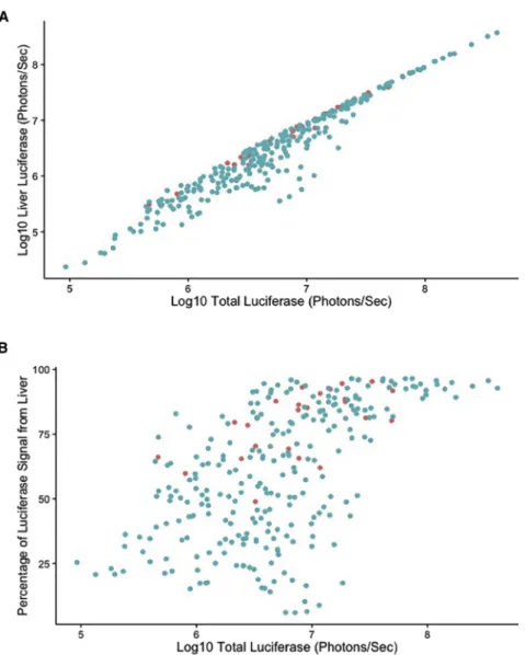
andTable 1. Overall, modest increases in luciferase expression were seen through the experiment. However, some strains (e.g., CC008/ GeniUnc and CC024/GeniUnc) showed stable luciferase expression throughout the experiment, and one strain (CC021/Unc) showed reduced luciferase expression throughout the study. Ideally, gene therapeutic products are specifically targeted to a given tissue. We were able to assess the liver-specific expression of luciferase across our set of mouse strains through this kinetic time course. In general, total and liver-specific luciferase levels were highly correlated (r2= 0.96, Figure 3A) throughout the study, and, therefore, analysis of these data tightly mirrored the responses seen for total luciferase levels. In liver, we found evidence of strain effects (F11,234= 40.157;
p < 1 1010), and also a strain-by-time point interaction
(F11,234= 3.18; p < 0.001). However, although we identified a
moder-ate effect of time point on total luciferase levels, we did not identify an effect of time point (F1,234= 0.0012; p = 0.97) on liver-specific
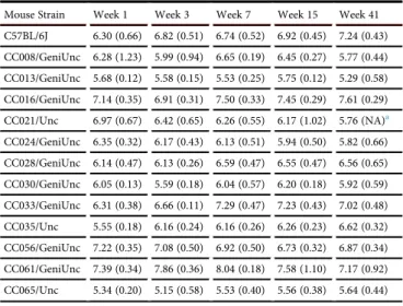
luciferase expression. Furthermore, at week 1, we found that as with total luciferase, liver-specific luciferase expression heritability was high, ranging from 46% to 64% (cgd = 0.466; icc = 0.636). Exam-ination of these data (Table 2) shows that in general, liver-specific
levels were stable through the study, with some strains showing modest changes in effect. Given these lines of evidence, especially the discordance between the temporal responses for total and liver-specific luciferase, wefinally asked whether the overall proportion of luciferase expression within individuals was under host genetic control. We first examined the correlation between total luciferase expression and the fraction of luciferase signal coming from the liver and found a modest correlation between these two traits (r2= 0.579,
Figure 3B). Importantly, samples having a broad range of total lucif-erase expression (105–107.3photons/s) could still exhibit low levels (20%) of liver-specific expression, suggesting that those individual animals with low fractions of liver-specific responses were not just those with low total levels (e.g., samples difficult to assess or incorrect attribution to spillover from the liver). Therefore, we assessed how this response changed throughout the study. As we expected, we iden-tified significant strain (F11,234= 56.3; p < 1 1010), time point
(F1,234= 27.9; p < 1106), and an interaction between these factors
(F11,234= 2.14; p = 0.018) driving the fraction of luciferase expression
coming from liver, with heritability estimates at week 1 ranging from 49% to 66% (cgd = 0.493; icc = 0.66). For example, CC033/GeniUnc and CC061/GeniUnc both maintain consistently high proportions of their total luciferase coming from liver similar to C57BL/6J, whereas CC013/GeniUnc showed a decreasing fraction of expression coming from the liver over time. Note that only C57BL/6J and CC035 showed increases in the fraction of their response coming from the liver, sug-gesting that although there might befluctuations between tissues or a silencing within the liver, in most strains, there was no expansion of the luciferase expression from this tissue.
Figure 2. Time Course of Luciferase Responses across Mouse Strains
Differential Success in Stable Vector Maintenance
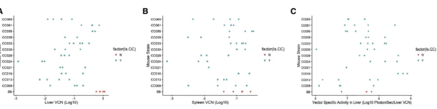
Following reverse transcription, lentiviral vectors efficiently import their genetic material to the host cells’nuclei, where it serves as a tem-plate for transcription of the relevant transgene of interest. Successful gene therapy with these vectors requires long-term persistence of vector-transduced cells. Following study termination, liver and spleen tissues were collected from these animals, and a qPCR assay was per-formed to identify the number of vector copies delivered into host cells’nuclei. Vector copy number per cell (VCN) in the liver differed by over 2 logs (min = 0.001; max = 1.12; mean = 0.22 copies/cell;
Figure 4A). C57BL/6J showed the highest level of VCN within the livers (0.85 ± 0.22 copies/cell), with all collaborative cross strains showing lower levels of VCN than C57BL/6J. Collaborative cross strains showed significant differences in liver VCN (F11,31= 8.023;
p = 2.1 106), and heritability estimates suggest that between 41% and 58% (cgd = 0.412; icc = 0.584) of the variation in vector copies/cell in the liver are under host genetic control. Levels of VCN within the spleen of most strains were reduced relative to liver (min < 0.001; max = 0.33; mean = 0.09 copies/cell), although there was a strong correlation between vector copies in the liver and vector copies in the spleen (r2= 0.78). C57BL/6J showed relatively high levels of vector copies/spleen (0.13± 0.08 vector copies per cell), but in contrast to liver-specific VCN, C57BL/6J was not the mouse strain with the highest levels of spleen VCN (Figure 4B). As with liver levels, there was an approximately 1.5-log difference between the highest and lowest collaborative cross strain, and these differences were sta-tistically significant (F11,29= 4.247; p = 0.0008). Spleen levels of vector
maintenance had a heritability of between 25% and 39% (cgd = 0.245; icc = 0.393) across these collaborative cross strains.
Relationship between VCN and Luciferase Expression
Given the highly significant variation in total and liver-specific lucif-erase expression between this collection of mouse strains, we sought
to assess the extent to which host genetic differences controlled these expression levels (that is, given the strain-specific VCN, are there dif-ferences in the efficiency of transgene expression). We determined the relative expression of luciferase at week 41 within the livers of each mouse relative to liver VCN. We found that vector-specific activity in mouse livers (F11,28= 2.81; p = 0.013) was impacted by the host
strain (Figure 4C). Heritability analysis showed that between 15% and 27% (cgd = 0.153; icc = 0.266) of the variation in vector-specific activity in the liver was explained by host genetic variation. We uti-lized a second analysis to assess the impact of host genetic variation on luciferase expression independent of host genetic variations’ impact on vector copy number. We used a partial F-test to assess the improvement infit when our model explaining luciferase levels included both strain and vector copy number as opposed to vector copy number alone. We found strong evidence for liver luciferase expression having other host factors driving expression (F27,38 =
3.26; p = 0.005), although the evidence for total luciferase expression was not significant (F26,36= 1.78; p = 0.11).
Mortality and Atypical Pathological Responses across This Treated Cohort
A total of 12/61 (19%) mice within this study died during the course of this experiment. This mortality impacted seven strains (Table S1), with CC021/Unc experiencing the highest mortality (4/5 mice). After sacrifice, six mice from four strains showed what appeared to be a macroscopic abnormality, including liver (CC024/GeniUnc#1) and spleen (CC035/Unc#5) tumors. Pathological analysis suggested these were common lesion types in aging animals and unlikely to be due to the presence of vector-related issues (Figure S2;Table S2).
DISCUSSION
Although gene therapy approaches have the power to alleviate se-vere congenital conditions, several aspects of viral-vector-delise-vered gene therapeutic regimens have raised causes for concern. Specif-ically, successful gene therapy requires optimal responses to a vari-ety of processes, all with a single delivery event. These include, but are not limited to, delivery of viral vectors to specific target cells, efficient transfer of viral genomes to host cells’nuclei, and mainte-nance of therapeutic levels of transgene expression without causing harmful adverse effects. Inefficiency in these processes as well as unforeseen adverse effects in some clinical trials have been the great-est impediments to broad adoption of gene therapy as a means to treat nonfatal diseases.20,22–24Although some studies have suggested that host genetic differences can play a role in gene therapy out-comes,31,32 there has been little research into the role that host genetic variation plays on the efficacy, maintenance, and safety of viral-vector-driven gene therapeutic approaches. We sought to assess the magnitude of the impact that host genetic variation could have on gene therapeutic efficacy. We used a set of strains from the collaborative cross genetic reference panel, and found that host genetic variation impacted multiple aspects of the gene therapy response within hosts, and, furthermore, that these responses were not always correlated across the strains (e.g., VCN and total lucif-erase expression).
Table 1. Mean±SD of Log10 Total Luciferase Activity Levels Measured in Photons/s through Time Course
Mouse Strain Week 1 Week 3 Week 7 Week 15 Week 41
C57BL/6J 6.41 (0.58) 6.88 (0.47) 6.86 (0.41) 7.01 (0.40) 7.36 (0.37)
CC008/GeniUnc 6.58 (0.97) 6.38 (0.73) 7.04 (0.17) 7.04 (0.21) 6.58 (0.48)
CC013/GeniUnc 5.94 (0.13) 5.89 (0.16) 6.00 (0.27) 6.21 (0.12) 5.97 (0.62)
CC016/GeniUnc 7.19 (0.31) 6.96 (0.31) 7.56 (0.33) 7.52 (0.28) 7.70 (0.25)
CC021/Unc 7.09 (0.60) 6.53 (0.58) 6.38 (0.51) 6.35 (0.82) 5.76 (NA)a
CC024/GeniUnc 6.75 (0.19) 6.69 (0.26) 6.77 (0.26) 6.91 (0.12) 6.63 (0.52)
CC028/GeniUnc 6.35 (0.43) 6.40 (0.23) 6.87 (0.38) 6.81 (0.31) 6.84 (0.55)
CC030/GeniUnc 6.36 (0.08) 5.97 (0.18) 6.41 (0.51) 6.59 (0.14) 6.22 (0.49)
CC033/GeniUnc 6.37 (0.37) 6.69 (0.11) 7.34 (0.42) 7.29 (0.39) 7.12 (0.44)
CC035/Unc 5.78 (0.11) 6.33 (0.18) 6.44 (0.21) 6.49 (0.15) 6.81 (0.29)
CC056/GeniUnc 7.27 (0.33) 7.22 (0.35) 7.12 (0.29) 7.00 (0.29) 7.07 (0.32)
CC061/GeniUnc 7.44 (0.33) 7.90 (0.35) 8.08 (0.18) 7.66 (1.04) 7.27 (0.87)
CC065/Unc 5.82 (0.22) 5.63 (0.51) 6.14 (0.35) 6.18 (0.36) 6.39 (0.58)
C57BL/6J is one of the most widely used mouse strains and is in fact the reference mouse genome. Various mouse models emulating hu-man diseases were established on a C57BL/6J genetic background. These include several lines of C57BL/6J-factor-IX-deficient mice, which modeled hemophilia.47–50Using adenoviral- and AAV-based vectors in gene therapy preclinical studies of hemophilia B, Fields et al. and Mingozzi et al. demonstrated that induction of immune tolerance to and long-term therapeutic levels of expression of vec-tor-delivered human factor IX were more readily achieved in C57BL/6 mice compared to BALB/c, C3H, and CD-1, although pre-cise assessment of the genetic contributions within these studies was not characterized.51,52In an earlier lentiviral-vector-based pre-clinical study, Follenzi et al. showed superior lentiviral vector long-term expression of and less prominent adaptive immune response to vector-delivered GFP reporter gene and human factor IX in C57BL/6 mice compared to FVB/N and BALB/c mice. Not surpris-ingly, C57BL/6 mice are considered the most permissive model for lentiviral-vector-based gene delivery.31In line with the Follenzi study
Figure 3. Relationships between Luciferase Expression Levels
(A) A strong correlation (r2= 0.96) was identified across all
time points between the liver-specific and total-body levels of luciferase expression. (B) A moderate (r2= 0.57)
corre-lation was identified between total luciferase levels and the percentage of luciferase signal coming from the liver. Each point is from a single mouse at a single time point, with C57BL/6J shown as red points and collaborative cross mice shown as blue points.
above, we found that peak luciferase expression in the C57BL/6J strain was higher (>1107.36) than in many collaborative cross strains in this study. Importantly, in this study, two collabora-tive cross strains (CC016/GeniUnc and CC061/ GeniUnc) exhibited equivalent or higher levels of transgene expression than the C57BL/6J strain at later time points. Furthermore, our study showed differences in peak luciferase expression across strains as >100-fold. These data suggest that significant differences in the efficacy of gene therapy protocols between human pa-tients can be anticipated solely due to genetic differences.
immune response directed to vector-transduced cells, adaptive immune responses to the vector-delivered gene products them-selves,23,31,56,59,60 transgene toxicity, and silencing of the vector expression cassette.61Previous clinical trials and other studies have suggested that immune responses against targeted cells can lead to adverse effects.23,24,60,62Although our study was not able to conclu-sively investigate whether this transgene silencing and mortality were connected and if they were the results of immune responses, these data suggest that there are host genotype-specific responses that can lead to suboptimal and adverse gene therapeutic responses.
Vector biodistribution is a key characteristic of all gene-delivery sys-tems. Lentiviral vector biodistribution is controlled by the efficiency at which reverse-transcribed vector genomes are nuclear imported and expressed in various host organs following systemic administra-tion. The ability of HIV-1 to transduce non-dividing cells, which facilitates in vivo gene delivery, is the hallmark of lentiviruses and lentiviral vector systems.63,64 Surprisingly, to date, the effects of host genetic variation on vector biodistribution has not been char-acterized. We were able to identify a difference in the success of VSV-G-pseudotyped lentiviral vectors to deliver reverse-transcribed genomes into both desired target (liver) and bystander (spleen) tis-sues (expressed as VCN) between the collaborative cross strains we assessed. With the exception of three mouse strains (C57BL/6J, CC056/GeniUnc, and CC061/GeniUnc), in which VCN levels in liver tissues were remarkably high, all other collaborative cross mouse strains exhibited comparable levels of VCNs in liver and spleen tis-sues. Furthermore, there was a strong correlation across all animals in the level of copies between tissues. These findings are in line with an earlier report by Pan et al. showing comparable VCN levels in liver and spleen tissues following intravenous administration of VSV-G-pseudotyped lentiviral vectors to BALB/c mice.65These re-sults suggest there are some variant host factors, which impact the
efficacy of delivery or maintenance of cells transformed by the vector. Nuclear import of vector DNA is a prerequisite for vector transcrip-tional activity and directly affects biodistribution of transgene expres-sion. However, vector design and host factors also have major effects on transgene expression in vector-transduced cells. In this study, a liver-specific promoter (hAAT) controlled vector-mediated luciferase expression. Indeed, most mouse strains exhibited predominantly hepatic expression. However, several mouse strains demonstrated relatively high extra-hepatic transgene expression. Furthermore, an earlier study reported on extra-hepatic transgene expression in non-hepatic cell lines in vitro and extra hepatic tissues in vivo and from lentiviral vectors carrying a liver-specific promoter.59 Although the mechanism involved in this phenomenon has not been completely elucidated, it was generally attributed to enhancer/ promoter effects of the host chromatin on liver-specific promoters in integrated lentiviral vectors.56,66In addition to VCN levels, specific activity of a lentiviral vector expression cassette as determined by total transgene activity per vector genome directly affects the therapeutic efficacy of a lentiviral vector as a therapeutic agent. Although vec-tor-specific activity was comparable across the collaborative cross mouse strains, significant high specific activity was exhibited by strain CC016/GeniUnc. Furthermore, and strikingly, both the prototypical C57BL/6J and CC061/GeniUnc strains showed much more modest specific activity than their overall luciferase expression levels would indicate, further highlighting the need for assessment of both trans-duction efficiency as well as transgene expression in considering the success of gene therapy. This phenomenon can be attributed to a single or a combination of strain-specific transcriptional and post-translational host factors, which enhanced luciferase activity in the above mouse strain. This observation is highly important in the gene therapyfield because it strongly suggests that therapeutic vector loads should be patient specific. This approach could potentially reduce transgene cytotoxicity on one hand and optimize overall vec-tor genomes required to achieve therapeutic transgene expression with minimal adverse effects on the other hand. Indeed, insertional mutagenesis is a major biosafety inherent to all integrating vectors, including lentiviral vectors. A previous study found some evidence for differential oncogenic potential.32We identified mice from four of the 12 collaborative cross strains used in this study (Table S2), which had macroscopic liver abnormalities following termination. The presence of these abnormalities did not appear to be tied to the lentiviral transduction, and the propensity to develop specific cancer types is known to be under genetic control.67,68However, more in-depth future studies in a more robust case-control framework will be required to both understand any potential exacerbation of tumor-igenesis driven by lentiviral vectors as well as to assess the distribution of integration into oncogenes and the extent to which host genetic differences directly (e.g., integration site biases) or indirectly (e.g., epigenetic and chromatin differences) might alter this. Here, we have demonstrated the strong effects of host genetic variation on the levels and kinetics of transgene expression, levels of VCN, and vector-specific activity. A larger number of collaborative cross strains should facilitate identification of putative genetic loci involved in the above strain-specific characteristics of in vivo lentiviral vector gene Table 2. Mean±SD of Log10Liver-Specific Luciferase Activity Levels
Measured in Photons/s through Time Course
Mouse Strain Week 1 Week 3 Week 7 Week 15 Week 41
C57BL/6J 6.30 (0.66) 6.82 (0.51) 6.74 (0.52) 6.92 (0.45) 7.24 (0.43)
CC008/GeniUnc 6.28 (1.23) 5.99 (0.94) 6.65 (0.19) 6.45 (0.27) 5.77 (0.44)
CC013/GeniUnc 5.68 (0.12) 5.58 (0.15) 5.53 (0.25) 5.75 (0.12) 5.29 (0.58)
CC016/GeniUnc 7.14 (0.35) 6.91 (0.31) 7.50 (0.33) 7.45 (0.29) 7.61 (0.29)
CC021/Unc 6.97 (0.67) 6.42 (0.65) 6.26 (0.55) 6.17 (1.02) 5.76 (NA)a
CC024/GeniUnc 6.35 (0.32) 6.17 (0.43) 6.13 (0.51) 5.94 (0.50) 5.82 (0.66)
CC028/GeniUnc 6.14 (0.47) 6.13 (0.26) 6.59 (0.47) 6.55 (0.47) 6.56 (0.65)
CC030/GeniUnc 6.05 (0.13) 5.59 (0.18) 6.04 (0.57) 6.20 (0.18) 5.92 (0.59)
CC033/GeniUnc 6.31 (0.38) 6.66 (0.11) 7.29 (0.47) 7.23 (0.43) 7.02 (0.48)
CC035/Unc 5.55 (0.18) 6.16 (0.24) 6.16 (0.26) 6.26 (0.23) 6.62 (0.32)
CC056/GeniUnc 7.22 (0.35) 7.08 (0.50) 6.92 (0.50) 6.73 (0.32) 6.87 (0.34)
CC061/GeniUnc 7.39 (0.34) 7.86 (0.36) 8.04 (0.18) 7.58 (1.10) 7.17 (0.92)
CC065/Unc 5.34 (0.20) 5.15 (0.58) 5.53 (0.40) 5.56 (0.38) 5.64 (0.44)
delivery. To avoid gender-specific effects on hepatic transduction, this study was premised on female mice only. Although more resource demanding, additional studies comprising male mice are needed to identify gender-specific effects on lentiviral vector trans-duction efficiency.
We anticipate that using the collaborative cross mouse system in an in vitro setting would further elucidate the molecular mechanisms by which host genetic variation affects specific steps in the HIV-1 life cycle. However, establishing the ability to genetically identify host factors affecting the efficacy of lentiviral vectors ferrying thera-peutic genetic cargos in the setting of a genetic disease model will be more challenging.
MATERIALS AND METHODS
Lentiviral Particles Production, Concentration, and Titration
Lentiviral vector (pTK979) harboring the firefly luciferase cDNA under the control of a liver-specific promoter (hAAT) was con-structed and used as described earlier.54Lentiviral vector particles were packaged with the packaging cassette,DNRF, in 293T cells using three-plasmid transient transfection as previously described.69Vector titer was determined by measuring the p24 capsid concentration us-ing ELISA as previously described.70All vector preps were tested for the absence of replication-competent retrovirus as described earlier.71
Animal Studies
All procedures involving animal study were performed in accordance with the Guide for the Care and Use of Laboratory Animals. The animal study protocol was approved by the University of North Carolina (UNC) Institutional Animal Care and Usage Committee. All mice were purchased from Jackson Laboratories (C57BL/6J) or the UNC Sys-tems Genetics Core Facility (collaborative cross strains listed inTable 1;
http://csbio.unc.edu/CCstatus/index.py?run=AvailableLines). Female mice were acquired at 5 to 6 weeks of age, and acclimated for 1 week in our experimental facility. Female animals (n = 4 to 5/strain) were intraperitoneally injected with 50 mg of p24gag lentiviral vectors
(in 250mL of phosphate-buffered solution). In vivo expression of vec-tor-deliveredfirefly luciferase in live animals was determined at weeks 1, 3, 7, 15, and 41 using the IVIS Lumina optical imaging system
(PerkinElmer). To this end, animals were intraperitoneally injected with 200mL (5 mg) of D-luciferin potassium salt reconstituted in phos-phate-buffered solution (Regis Technologies). Imaging was initiated 10 min after D-luciferin injection using a 5-min exposure time. The relative light counts obtained through a charge-coupled device (CCD) camera were converted to physical units of surface radiance (p/s/ cm2/sr) and displayed in luminescence radiance mode. All in vivo lucif-erase activities shown in this study were analyzed using Living Image Software (PerkinElmer) and reported in totalflux (photon/s). To mea-sure whole-body and liver-specific luciferase activity, identical regions of interest (ROIs) were measured. These comprised either the whole an-imal body (excluding head and tail) or the liver, respectively (Figure S1). Luciferase activity in the respective ROIs in each animal was measured as described above. Off-liver luciferase activity was calculated by sub-tracting liver-specific activity from whole-body luciferase activity.
Mouse Tissue DNA Preparation for Quantification of Vector Copy Number
At the end of the experiment, the viral copy number of liver and spleen tissues was determined. Genomic DNAs from tissues were iso-lated using the Blood & Tissue DNeasy kit (QIAGEN) according to the manufacturer’s instructions. RNAs were removed using RNase A (Fermentas). All samples were treated withDpnI(New England Biolabs). Total vector copy number of each sample was quantified us-ing multiplex qPCR, as previously described.55
Histopathology
Upon tissue harvesting, liver and spleen tissues with physical abnormalities in either size or appearance observed in six animals from four strains (Figure S2;Table S2) were sent to the UNC Animal Histopathology Core Facility for a histopathological examination. To this end, tissues were fixed in 10% neutral buffered formalin, embedded in paraffin, sectioned with a microtome, and stained with H&E. A board-certified veterinary pathologist analyzed these H&E-stained sections.
Statistical Analysis
All data were analyzed in the R programming environment (cran. r-project.org). Broad-sense heritability was assessed as either the Figure 4. Vector Copy Number and Luciferase Levels per Vector Copy Vary between Strains
interclass correlation or the coefficient of genetic determination.46 Briefly, each of these approaches uses the ANOVA model fit and then assesses the relative ratios of mean square explained by strain differences relative to the mean square error in the data. Both ap-proaches then apply a scaling factor relative to the number of samples (n) in each treatment class as follows:
(1) Interclass correlation: (MSstrain MSerror)/(MSstrain+ (n 1)
MSerror)
(2) Coefficient of genetic determination:
ðMSstrainMSerrorÞ=ðMSstrain +ð2n1ÞMSerrorÞ
In order to assess the significance of strain differences on given phenotypic traits, data were normalized and ANOVA was used to identify significance. For the model fitting assessing vector and strain effects driving luciferase expression, we used a partial F-test framework.
Data Availability
All data from this study, including calculated luciferase, vector copy number levels, and raw images for luciferase calculation, are available upon request. The 12 collaborative cross strains used within this study are available from the Systems Genetics Core Facility (http://csbio. unc.edu/CCstatus/index.py?run=AvailableLines), and both genotype files and haplotype reconstructions are available for these strains on that site.
SUPPLEMENTAL INFORMATION
Supplemental Information includes twofigures and two tables and can be found with this article online athttp://dx.doi.org/10.1016/j. omtm.2017.03.009.
AUTHOR CONTRIBUTIONS
Conceptualization and design of the study, T.K., and F.P.-M.d.V.; Acquisition of data, T.S., P.H., S.A.M., and T.G.; Analysis and inter-pretation of data, T.K., M.T.F., and F.P.-M.d.V.; Writing–Original Draft, T.K., M.T.F., and T.S.; Statistical analysis, M.T.F.; Critical revi-sion of the manuscript, T.K., M.T.F., F.P.-M.d.V., and T.S.
CONFLICTS OF INTEREST
T.K. is an inventor of a technology which was licensed by UNC to a commercial entity.
ACKNOWLEDGMENTS
The following reagent was obtained through the National Institutes of Health (NIH) AIDS Research and Reference Reagent Program, Divi-sion of AIDS, the National Institute of Allergy and Infectious Dis-eases: the HIV-1 p24 monoclonal antibody (183-H12-5C) from Bruce Chesebro and Kathy Wehrly. The study was supported by the UNC Center for AIDS Research and by the NIH grants R01-HL128119 (to T.S., M.T.F., P.H., T.G., F.P.-M.d.V., and T.K.), R01-DK058702 (to T.S., P.H., T.G., and T.K.), and U19-AI100625 (to M.T.F. and F.P.-M.d.V.). Histopathology analysis was performed in the UNC
Animal Histopathology Core Facility, which is supported in part by an NCI Center Core Support Grant (2P30CA016086-40) to the UNC Lineberger Comprehensive Cancer Center. This study is dedi-cated to the U.S. Marine Corps and the Gold Star families. In memory of Boaz Kofman, the epitome of a mensch.
REFERENCES
1.Cockrell, A.S., and Kafri, T. (2007). Gene delivery by lentivirus vectors. Mol. Biotechnol.36, 184–204.
2.Bieniasz, P.D. (2004). Intrinsic immunity: a front-line defense against viral attack. Nat. Immunol.5, 1109–1115.
3.Borsotti, C., Borroni, E., and Follenzi, A. (2016). Lentiviral vector interactions with the host cell. Curr. Opin. Virol.21, 102–108.
4.Goff, S.P. (2007). Host factors exploited by retroviruses. Nat. Rev. Microbiol.5, 253–263.
5.Sheehy, A.M., Gaddis, N.C., Choi, J.D., and Malim, M.H. (2002). Isolation of a human gene that inhibits HIV-1 infection and is suppressed by the viral Vif protein. Nature
418, 646–650.
6.Suzuki, Y., and Craigie, R. (2007). The road to chromatin - nuclear entry of retrovi-ruses. Nat. Rev. Microbiol.5, 187–196.
7.Taltynov, O., Desimmie, B.A., Demeulemeester, J., Christ, F., and Debyser, Z. (2012). Cellular cofactors of lentiviral integrase: from target validation to drug discovery. Mol. Biol. Int.2012, 863405.
8.Towers, G.J., Hatziioannou, T., Cowan, S., Goff, S.P., Luban, J., and Bieniasz, P.D. (2003). Cyclophilin A modulates the sensitivity of HIV-1 to host restriction factors. Nat. Med.9, 1138–1143.
9.Dochi, T., Nakano, T., Inoue, M., Takamune, N., Shoji, S., Sano, K., and Misumi, S. (2014). Phosphorylation of human immunodeficiency virus type 1 capsid protein at serine 16, required for peptidyl-prolyl isomerase-dependent uncoating, is mediated by virion-incorporated extracellular signal-regulated kinase 2. J. Gen. Virol.95, 1156–1166.
10.Fricke, T., White, T.E., Schulte, B., de Souza Aranha Vieira, D.A., Dharan, A., Campbell, E.M., Brandariz-Nuñez, A., and Diaz-Griffero, F. (2014). MxB binds to the HIV-1 core and prevents the uncoating process of HIV-1. Retrovirology11, 68.
11.Sayah, D.M., Sokolskaja, E., Berthoux, L., and Luban, J. (2004). Cyclophilin A retro-transposition into TRIM5 explains owl monkey resistance to HIV-1. Nature430, 569–573.
12.Shah, V.B., Shi, J., Hout, D.R., Oztop, I., Krishnan, L., Ahn, J., Shotwell, M.S., Engelman, A., and Aiken, C. (2013). The host proteins transportin SR2/TNPO3 and cyclophilin A exert opposing effects on HIV-1 uncoating. J. Virol.87, 422–432. 13.Yuan, T., Yao, W., Tokunaga, K., Yang, R., and Sun, B. (2016). An HIV-1 capsid
bind-ing protein TRIM11 accelerates viral uncoatbind-ing. Retrovirology13, 72.
14.Ambrose, Z., and Aiken, C. (2014). HIV-1 uncoating: connection to nuclear entry and regulation by host proteins. Virology454-455, 371–379.
15.Borrenberghs, D., Dirix, L., De Wit, F., Rocha, S., Blokken, J., De Houwer, S., Gijsbers, R., Christ, F., Hofkens, J., Hendrix, J., et al. (2016). Dynamic oligomerization of inte-grase orchestrates HIV nuclear entry. Sci. Rep.6, 36485.
16.Hilditch, L., and Towers, G.J. (2014). A model for cofactor use during HIV-1 reverse transcription and nuclear entry. Curr. Opin. Virol.4, 32–36.
17.Delelis, O., Carayon, K., Saïb, A., Deprez, E., and Mouscadet, J.F. (2008). Integrase and integration: biochemical activities of HIV-1 integrase. Retrovirology5, 114.
18.Poeschla, E.M. (2008). Integrase, LEDGF/p75 and HIV replication. Cell. Mol. Life Sci.
65, 1403–1424.
19.Mbonye, U., and Karn, J. (2014). Transcriptional control of HIV latency: cellular signaling pathways, epigenetics, happenstance and the hope for a cure. Virology
454-455, 328–339.
21.Gaspar, H.B., Cooray, S., Gilmour, K.C., Parsley, K.L., Adams, S., Howe, S.J., Al Ghonaium, A., Bayford, J., Brown, L., Davies, E.G., et al. (2011). Long-term persis-tence of a polyclonal T cell repertoire after gene therapy for X-linked severe combined immunodeficiency. Sci. Transl. Med.3, 97ra79.
22.Hacein-Bey-Abina, S., Von Kalle, C., Schmidt, M., McCormack, M.P., Wulffraat, N., Leboulch, P., Lim, A., Osborne, C.S., Pawliuk, R., Morillon, E., et al. (2003). LMO2-associated clonal T cell proliferation in two patients after gene therapy for SCID-X1. Science302, 415–419.
23.Manno, C.S., Pierce, G.F., Arruda, V.R., Glader, B., Ragni, M., Rasko, J.J., Ozelo, M.C., Hoots, K., Blatt, P., Konkle, B., et al. (2006). Successful transduction of liver in hemo-philia by AAV-Factor IX and limitations imposed by the host immune response. Nat. Med.12, 342–347.
24.Raper, S.E., Yudkoff, M., Chirmule, N., Gao, G.P., Nunes, F., Haskal, Z.J., Furth, E.E., Propert, K.J., Robinson, M.B., Magosin, S., et al. (2002). A pilot study of in vivo liver-directed gene transfer with an adenoviral vector in partial ornithine transcarbamylase deficiency. Hum. Gene Ther.13, 163–175.
25.Aiuti, A., Biasco, L., Scaramuzza, S., Ferrua, F., Cicalese, M.P., Baricordi, C., Dionisio, F., Calabria, A., Giannelli, S., Castiello, M.C., et al. (2013). Lentiviral hematopoietic stem cell gene therapy in patients with Wiskott-Aldrich syndrome. Science341, 1233151.
26.Biffi, A., Montini, E., Lorioli, L., Cesani, M., Fumagalli, F., Plati, T., Baldoli, C., Martino, S., Calabria, A., Canale, S., et al. (2013). Lentiviral hematopoietic stem cell gene therapy benefits metachromatic leukodystrophy. Science341, 1233158.
27.Cartier, N., Hacein-Bey-Abina, S., Bartholomae, C.C., Veres, G., Schmidt, M., Kutschera, I., Vidaud, M., Abel, U., Dal-Cortivo, L., Caccavelli, L., et al. (2009). Hematopoietic stem cell gene therapy with a lentiviral vector in X-linked adrenoleu-kodystrophy. Science326, 818–823.
28.Frey, N.V., and Porter, D.L. (2016). CAR T-cells merge into the fast lane of cancer care. Am. J. Hematol.91, 146–150.
29. Ghosh, S., Thrasher, A.J., and Gaspar, H.B. (2015). Gene therapy for monogenic dis-orders of the bone marrow. Br. J. Haematol., Published online June 5, 2015.http://dx. doi.org/10.1111/bjh.13520.
30.Kalos, M., Levine, B.L., Porter, D.L., Katz, S., Grupp, S.A., Bagg, A., and June, C.H. (2011). T cells with chimeric antigen receptors have potent antitumor effects and can establish memory in patients with advanced leukemia. Sci. Transl. Med.3, 95ra73.
31.Follenzi, A., Battaglia, M., Lombardo, A., Annoni, A., Roncarolo, M.G., and Naldini, L. (2004). Targeting lentiviral vector expression to hepatocytes limits transgene-spe-cific immune response and establishes long-term expression of human antihemo-philic factor IX in mice. Blood103, 3700–3709.
32.Zhao, Y., Keating, K., and Thorpe, R. (2007). Comparison of toxicogenomic profiles of two murine strains treated with HIV-1-based vectors for gene therapy. Toxicol. Appl. Pharmacol.225, 189–197.
33.Duggal, P., Thio, C.L., Wojcik, G.L., Goedert, J.J., Mangia, A., Latanich, R., Kim, A.Y., Lauer, G.M., Chung, R.T., Peters, M.G., et al. (2013). Genome-wide association study of spontaneous resolution of hepatitis C virus infection: data from multiple cohorts. Ann. Intern. Med.158, 235–245.
34.Everitt, A.R., Clare, S., Pertel, T., John, S.P., Wash, R.S., Smith, S.E., Chin, C.R., Feeley, E.M., Sims, J.S., Adams, D.J., et al.; GenISIS Investigators; MOSAIC Investigators (2012). IFITM3 restricts the morbidity and mortality associated with influenza. Nature484, 519–523.
35.Ferris, M.T., and Heise, M.T. (2014). Quantitative genetics in the study of virus-induced disease. Adv. Virus Res.88, 193–225.
36.Chick, J.M., Munger, S.C., Simecek, P., Huttlin, E.L., Choi, K., Gatti, D.M., Raghupathy, N., Svenson, K.L., Churchill, G.A., and Gygi, S.P. (2016). Defining the consequences of genetic variation on a proteome-wide scale. Nature534, 500–505.
37.Elbahesh, H., and Schughart, K. (2016). Genetically diverse CC-founder mouse strains replicate the human influenza gene expression signature. Sci. Rep.6, 26437.
38.Xiong, H., Morrison, J., Ferris, M.T., Gralinski, L.E., Whitmore, A.C., Green, R., Thomas, M.J., Tisoncik-Go, J., Schroth, G.P., Pardo-Manuel de Villena, F., et al. (2014). Genomic profiling of collaborative cross founder mice infected with respira-tory viruses reveals novel transcripts and infection-related strain-specific gene and isoform expression. G3 (Bethesda)4, 1429–1444.
39.Collaborative Cross Consortium (2012). The genome architecture of the Collaborative Cross mouse genetic reference population. Genetics190, 389–401.
40.Ferris, M.T., Aylor, D.L., Bottomly, D., Whitmore, A.C., Aicher, L.D., Bell, T.A., Bradel-Tretheway, B., Bryan, J.T., Buus, R.J., Gralinski, L.E., et al. (2013). Modeling host genetic regulation of influenza pathogenesis in the collaborative cross. PLoS Pathog.9, e1003196.
41.Graham, J.B., Swarts, J.L., Wilkins, C., Thomas, S., Green, R., Sekine, A., Voss, K.M., Ireton, R.C., Mooney, M., Choonoo, G., et al. (2016). A mouse model of chronic West Nile virus disease. PLoS Pathog.12, e1005996.
42.Gralinski, L.E., Ferris, M.T., Aylor, D.L., Whitmore, A.C., Green, R., Frieman, M.B., Deming, D., Menachery, V.D., Miller, D.R., Buus, R.J., et al. (2015). Genome wide identification of SARS-CoV susceptibility loci using the collaborative cross. PLoS Genet.11, e1005504.
43.Leist, S.R., Pilzner, C., van den Brand, J.M., Dengler, L., Geffers, R., Kuiken, T., Balling, R., Kollmus, H., and Schughart, K. (2016). Influenza H3N2 infection of the collaborative cross founder strains reveals highly divergent host responses and iden-tifies a unique phenotype in CAST/EiJ mice. BMC Genomics17, 143.
44.Rasmussen, A.L., Okumura, A., Ferris, M.T., Green, R., Feldmann, F., Kelly, S.M., Scott, D.P., Safronetz, D., Haddock, E., LaCasse, R., et al. (2014). Host genetic diver-sity enables Ebola hemorrhagic fever pathogenesis and resistance. Science 346, 987–991.
45.Zheng, C.L., Wilmot, B., Walter, N.A., Oberbeck, D., Kawane, S., Searles, R.P., McWeeney, S.K., and Hitzemann, R. (2015). Splicing landscape of the eight collabo-rative cross founder strains. BMC Genomics16, 52.
46.Petkova, S.B., Yuan, R., Tsaih, S.W., Schott, W., Roopenian, D.C., and Paigen, B. (2008). Genetic influence on immune phenotype revealed strain-specific variations in peripheral blood lineages. Physiol. Genomics34, 304–314.
47.Jin, D.Y., Zhang, T.P., Gui, T., Stafford, D.W., and Monahan, P.E. (2004). Creation of a mouse expressing defective human factor IX. Blood104, 1733–1739.
48.Kundu, R.K., Sangiorgi, F., Wu, L.Y., Kurachi, K., Anderson, W.F., Maxson, R., and Gordon, E.M. (1998). Targeted inactivation of the coagulation factor IX gene causes hemophilia B in mice. Blood92, 168–174.
49.Lin, H.F., Maeda, N., Smithies, O., Straight, D.L., and Stafford, D.W. (1997). A coag-ulation factor IX-deficient mouse model for human hemophilia B. Blood90, 3962– 3966.
50.Wang, L., Zoppè, M., Hackeng, T.M., Griffin, J.H., Lee, K.F., and Verma, I.M. (1997). A factor IX-deficient mouse model for hemophilia B gene therapy. Proc. Natl. Acad. Sci. USA94, 11563–11566.
51.Fields, P.A., Armstrong, E., Hagstrom, J.N., Arruda, V.R., Murphy, M.L., Farrell, J.P., High, K.A., and Herzog, R.W. (2001). Intravenous administration of an E1/E3-deleted adenoviral vector induces tolerance to factor IX in C57BL/6 mice. Gene Ther.8, 354–361.
52.Mingozzi, F., Liu, Y.L., Dobrzynski, E., Kaufhold, A., Liu, J.H., Wang, Y., Arruda, V.R., High, K.A., and Herzog, R.W. (2003). Induction of immune tolerance to coagulation factor IX antigen by in vivo hepatic gene transfer. J. Clin. Invest.111, 1347–1356.
53.Zhang, T.P., Jin, D.Y., Wardrop, R.M., 3rd, Gui, T., Maile, R., Frelinger, J.A., Stafford, D.W., and Monahan, P.E. (2007). Transgene expression levels and kinetics determine risk of humoral immune response modeled in factor IX knockout and missense mutant mice. Gene Ther.14, 429–440.
54.Bayer, M., Kantor, B., Cockrell, A., Ma, H., Zeithaml, B., Li, X., McCown, T., and Kafri, T. (2008). A large U3 deletion causes increased in vivo expression from a non-integrating lentiviral vector. Mol. Ther.16, 1968–1976.
55.Suwanmanee, T., Hu, G., Gui, T., Bartholomae, C.C., Kutschera, I., von Kalle, C., Schmidt, M., Monahan, P.E., and Kafri, T. (2014). Integration-deficient lentiviral vectors expressing codon-optimized R338LhFIX restore normal hemostasis in hemophilia B mice. Mol. Ther.22, 567–574.
56.Brown, B.D., Venneri, M.A., Zingale, A., Sergi Sergi, L., and Naldini, L. (2006). Endogenous microRNA regulation suppresses transgene expression in hematopoietic lineages and enables stable gene transfer. Nat. Med.12, 585–591.
can be broadly exploited to regulate transgene expression according to tissue, lineage and differentiation state. Nat. Biotechnol.25, 1457–1467.
58.Matsui, H., Hegadorn, C., Ozelo, M., Burnett, E., Tuttle, A., Labelle, A., McCray, P.B., Jr., Naldini, L., Brown, B., Hough, C., et al. (2011). A microRNA-regulated and GP64-pseudotyped lentiviral vector mediates stable expression of FVIII in a murine model of Hemophilia A. Mol. Ther19, 723–730.
59.Follenzi, A., Sabatino, G., Lombardo, A., Boccaccio, C., and Naldini, L. (2002). Efficient gene delivery and targeted expression to hepatocytes in vivo by improved lentiviral vectors. Hum. Gene Ther.13, 243–260.
60.Nathwani, A.C., Tuddenham, E.G., Rangarajan, S., Rosales, C., McIntosh, J., Linch, D.C., Chowdary, P., Riddell, A., Pie, A.J., Harrington, C., et al. (2011). Adenovirus-associated virus vector-mediated gene transfer in hemophilia B. N. Engl. J. Med.
365, 2357–2365.
61.Ellis, J. (2005). Silencing and variegation of gammaretrovirus and lentivirus vectors. Hum. Gene Ther.16, 1241–1246.
62.Raper, S.E., Chirmule, N., Lee, F.S., Wivel, N.A., Bagg, A., Gao, G.P., Wilson, J.M., and Batshaw, M.L. (2003). Fatal systemic inflammatory response syndrome in a ornithine transcarbamylase deficient patient following adenoviral gene transfer. Mol. Genet. Metab.80, 148–158.
63.Lewis, P., Hensel, M., and Emerman, M. (1992). Human immunodeficiency virus infection of cells arrested in the cell cycle. EMBO J.11, 3053–3058.
64.Naldini, L., Blömer, U., Gallay, P., Ory, D., Mulligan, R., Gage, F.H., Verma, I.M., and Trono, D. (1996). In vivo gene delivery and stable transduction of nondividing cells by a lentiviral vector. Science272, 263–267.
65.Pan, D., Gunther, R., Duan, W., Wendell, S., Kaemmerer, W., Kafri, T., Verma, I.M., and Whitley, C.B. (2002). Biodistribution and toxicity studies of VSVG-pseudotyped lentiviral vector after intravenous administration in mice with the observation of in vivo transduction of bone marrow. Mol. Ther.6, 19–29.
66.De Palma, M., Montini, E., Santoni de Sio, F.R., Benedicenti, F., Gentile, A., Medico, E., and Naldini, L. (2005). Promoter trapping reveals significant differences in inte-gration site selection between MLV and HIV vectors in primary hematopoietic cells. Blood105, 2307–2315.
67.Bai, L., Yang, H.H., Hu, Y., Shukla, A., Ha, N.H., Doran, A., Faraji, F., Goldberger, N., Lee, M.P., Keane, T., et al. (2016). An integrated genome-wide systems genetics screen for breast cancer metastasis susceptibility genes. PLoS Genet.12, e1005989.
68.Winter, J.M., Gildea, D.E., Andreas, J.P., Gatti, D.M., Williams, K.A., Lee, M., Hu, Y., Zhang, S.; NISC Comparative Sequencing Program, and Mullikin, J.C., et al. (2016). Mapping complex traits in a diversity outbred F1 mouse population identifies germ-line modifiers of metastasis in human prostate cancer. Cell Syst4, 31–45.e6.
69.Xu, K., Ma, H., McCown, T.J., Verma, I.M., and Kafri, T. (2001). Generation of a stable cell line producing high-titer self-inactivating lentiviral vectors. Mol. Ther.3, 97–104.
70.Kantor, B., Ma, H., Webster-Cyriaque, J., Monahan, P.E., and Kafri, T. (2009). Epigenetic activation of unintegrated HIV-1 genomes by gut-associated short chain fatty acids and its implications for HIV infection. Proc. Natl. Acad. Sci. USA106, 18786–18791.