CrossMark
Published by DiscoverSys ABSTRACT
Rabies virus is found in large quantities in the saliva of infected animals, and transmission occurs almost exclusively through inoculation of the infected saliva through a bite or scratch from a rabid mammal. Initial aggressive management with adequate supportive therapy may help in the survival of the patient. Rabies is generally fatal, and neither rabies immunoglobulin (RIG) nor rabies vaccine provides benefit once symptoms have appeared. The three-pronged approaches to prevent death caused by rabies in humans are stray animal control, post-exposure prophylaxis, and prior vaccination of people with a higher risk of exposure. Modern rabies vaccines have been proven to be safe, well- tolerated, and highly effective in preventing rabies, even if administered after exposure to bites. PEP consists of wound
cleaning, rabies vaccination, and passive immunization with rabies immune globulin (RIG), of which the most important treatment is rabies vaccination. Several regimens of rabies vaccination approved by WHO have shown to be immunogenic. Smaller doses and more advanced processing techniques have a relatively higher safety for the patients, especially for the young children. No significant differences in safety and immunogenicity between PVRV and PCECV both in Zagreb and Essen regimens. WHO recommends completing PEP against rabies with the same cell culture or embryonated egg rabies vaccine and with the same route of administration and any deviation from this shall be an exception. PEP was safe and effective despite changes in the route of administration and brand/ or type of rabies vaccine.
Keywords:
Cite This Article: Rampengan, N.H. 2017. Rabies post exposure prevention. Bali Medical Journal 6(2): 449-455. DOI:10.15562/bmj.v6i2.799
Rabies post exposure prevention
Dr. dr. Novie H. Rampengan, Sp.A(K), DTM&H, MCTM(TP)
INTRODUCTION
Rabies has an important place in the history of humankind. It may be the oldest infectious disease known. The first definitive references to rabies appear in Greek literature when in 400 BC Aristotle wrote that “rabies makes the dog mad. It is fatal to the dog itself, and to all animals it bites.”1 Rabies is a
zoonotic disease caused by the rabies virus.2 Rabies
virus is a bullet-shaped, negative-sense, single-stranded, enveloped RNA virus from the family Rhabdoviridae, genus Lyssavirus. There currently are 12 known genotypes of Lyssavirus, but only 7 Lyssavirus genotypes are associated with rabies in humans (genotype 1 is the majority of cases).1
EPIDEMIOLOGY
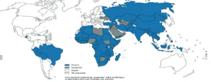
Rabies is present on all continents except Antarctica (Figure 1).2,3 Rabies predominantly afflicts
under-age, poor, and geographically isolated populations.3
As many as 95% of cases are reported in Asia and Africa.2,3 Approximately 50,000 cases of human
rabies occur in Africa and Asia annually.3
Almost half of documented rabies exposures worldwide occur in children under 15 years of age. Those at highest risk are 5–10 years old.1
Theoretically, rabies virus can infect any mammal (which then can transmit disease to humans), but true animal reservoirs that maintain the presence of rabies virus in the population are limited to
terrestrial carnivores and bats.3 Worldwide,
trans-mission from dogs accounts for >90% of human cases.2,3
TRANSMISSION
Rabies virus is found in large quantities in the saliva of infected animals, and transmission occurs almost exclusively through inoculation of the infected saliva through a bite or scratch from a rabid mammal.2-4
Approximately 35%-50% of people bitten by a known rabies infected animal do not receive post-ex-posure prophylaxis (PEP). The transmission rate is increased if the victim has suffered multiple bites and if the inoculation occurs in highly innervated parts of the body such as the face and the hands. Rabies from inhalational exposure can occur during laboratory accidents.3,4 Rabies virus strain from bat is reported
to be more infectious than dog rabies strains.4 Department of Child Health, Sam
Ratulangi University Medical School, Manado
*Correspondence to: Novie H.
Rampengan, Department of Child Health, Sam Ratulangi University Medical School, Manado
[email protected] and [email protected]
Received: 2017-08-08 Accepted: 2017-8-25 Published: 2017-8-26
Volume No.: 6
Issue: 2
First page No.: 449
P-ISSN.2089-1180
E-ISSN.2302-2914
Doi: http://dx.doi.org/10.15562/bmj.v6i2.799
ORIGINAL ARTICLE
The only well-documented cases of rabies caused by human-to-human transmission are those acquired through corneal and organ transplants.4
Mother-to-child transmission of rabies is possible, but rare, because rabies virus is not present in blood and exposure of the baby’s mucosa to maternal infectious fluids and tissue seems limited.5
PATHOGENESIS
After inoculation, rabies virus replicates slowly and at low levels in muscle or skin. This slow initial step likely accounts for the disease’s long incubation period. The virus then enters the peripheral motor nerve, utilizing the nicotinic acetylcholine receptor and possibly several other receptors for entry. Once the virus entered the nerve, the virus travels by fast axonal transport, crossing synapses roughly every 12 hours. Rapid dissemination occurs throughout the brain and spinal cord before symptoms appear. Infection of the dorsal root ganglia is apparently futile but causes characteristic radiculitis. The infection concentrates in the brainstem, accounting for autonomic dysfunction and relative sparing of cognition.3 Bites on the face, head, neck, and
hands are associated with the highest risk of rabies encephalitis with short incubation period.4
CLINICAL MANIFESTATIONS
The incubation period for rabies ranges from 1 to 3 months.1,3 In severe wounds to the head, symptoms
may occur within five days after exposure and occa-sionally the incubation period can extend to longer than six months.3 There are two clinical
manifesta-tions of rabies – furious (classical or encephalitic) and paralytic. Furious rabies is most common form of human rabies, accounting for approximately 80% of cases.2
DIFFERENTIAL DIAGNOSIS
The differential diagnosis of rabies encephalitis includes all forms of severe cerebral infections, tetanus, and some intoxications or envenomations. Rabies can be confused with autoimmune enceph-alitis, psychiatric illness, drug abuse, and conver-sion disorders. Paralytic rabies is most frequently confused with Guillain-Barré syndrome (GBS).3
DIAGNOSIS
The Centers for Disease Control and Prevention (CDC) require some tests to confirm a clinically suspected rabies case. Reverse transcription polymerase chain reaction (RT-PCR) is the most
sensitive assay available for the diagnosis of rabies when done iteratively. Rabies virus RNA has been detected in saliva, skin, and brain by the RT-PCR. The virus can be grown both in cell culture or injected animal host, but identification of rabies by these methods is slow. Rabies antigen is detected by immunofluorescence of saliva or biopsies of hairy skin or brain. MRI findings of rabies in the brain are usually appearing in the late phase. 3
TREATMENT AND PROGNOSIS
Rabies is generally fatal. Currently, no therapy has been shown to unequivocally improve survival in rabies.6 Initial aggressive management with
adequate supportive therapy may help in the survival of the patient.4 Conventional critical care
yielded one survivor from 74 attempts since 1990. Five of 16 patients survived without the use of critical care (including 3 milder cases) and 7 with use of the Milwaukee Protocol. Survival using the Milwaukee Protocol is estimated at 20%; neuro-logic outcomes are poor in one-third of patients. Neither rabies immunoglobulin (RIG) nor rabies vaccine provides benefit once symptoms have appeared. Among 11 survivors of rabies after use of biologics (What does biologics refer to? Suggestion: vaccines), 6 had poor neurologic outcomes. Among 5 vaccine-naïve survivors, 1 patient had a poor outcome.3 Survivors demonstrated a robust
immune response as indicated by peripheral viral clearance and very high serum and cerebrospinal fluid antibody titers. Immunologically-mediated virus clearance, therefore, appears to be a prereq-uisite for survival.6 Antiviral treatments for rabies
have not been effective.3
PREVENTION
Currently, there is a three-pronged approach to prevent deaths caused by rabies in humans. Firstly, if dog rabies could be fully prevented, there would be almost zero human cases. Thus, there has been a significant global drive to control canine rabies through dog vaccination and control of stray animals.7,8 However, in many environments,
wound, post-exposure vaccination with cell-culture vaccines and administration of equine or human rabies immunoglobulin. This important strategy is hampered by the tentative availability of vaccine and particularly rabies immunoglobulin in health services serving endemic areas and lack of aware-ness amongst some affected communities of the need to seek care immediately. The third effective measure is vaccinating people who are likely to be exposed to rabid animals prior to their exposure. This method has particularly been embraced by travelers from developed countries visiting canine
rabies endemic areas, or by individuals, including veterinarians, wildlife professionals, and labora-tory staff, whose employment places them at a higher risk of exposure compared with the general population.7
Immunization and Fertility Control of Animal Reservoirs
Control measures targeted at the dog population have been recommended by the World Health Organisation (WHO) and World Organization for Animal Health (OIE). These measures include mass vaccination of dogs, euthanasia of infectious dogs, and dog management actions, such as birth control and leashing or keeping dogs inside the house area.9
Post-exposure Prophylaxis (PEP)
Because of the very high case-fatality ratio (nearly 100%) of rabies, prophylaxis by vaccination is the only protection against rabies, including pre-expo-sure and post-expopre-expo-sure prophylaxis (PEP).11 The
development of the first rabies vaccine, long before recognition of the nature of viruses, represented an important milestone in public health history. In July 1885, Emile Roux successfully treated a human bite victim, Joseph Meister, who had been severely bitten by a rabid dog, with the vaccine developed by Louis Pasteur. For more than 70 years, only vaccines containing nerve tissue were available. In spite of improvements in the production and inactivation process over Pasteur’s method, these vaccines are poorly immunogenic and require numerous pain-ful injections. As they are not pain-fully free of myelin, they may be responsible for serious adverse reac-tions. Another major step forward was the devel-opment, in the 1960s, of ‘modern’ rabies vaccines, prepared from rabies virus grown in cell culture or embryonated eggs that are free of neuronal tissue. They have been proven to be safe, well-tolerated, and highly effective in preventing rabies. Uniquely among vaccines, human rabies vaccines are usually administered after exposure to the virus. PEP is possible because the exposure event, usually a dog bite, is easily identifiable, and the incubation period is usually long enough for vaccination to induce a protective immune response before the rabies virus reaches the central nervous system.1
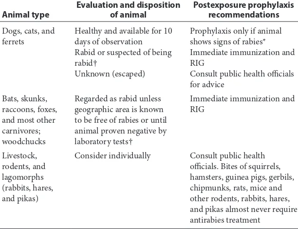
The guide for administering rabies PEP for most pediatricians centers is summarized in Table 1. No case of rabies has been documented to surface in a person receiving the recommended schedule of PEP since the introduction of modern cellular vaccines in the 1970s.3 Given the incubation period
for rabies, PEP is a medical urgency, not an emer-gency. Algorithms have been devised to aid prac-titioners in deciding when to initiate rabies PEP
Table 1 Rabies postexposure prophylaxis guide3
Animal type Evaluation and disposition of animal Postexposure prophylaxisrecommendations
Dogs, cats, and
ferrets Healthy and available for 10 days of observation Rabid or suspected of being rabid†
Unknown (escaped)
Prophylaxis only if animal shows signs of rabies* Immediate immunization and RIG
Consult public health officials for advice
Bats, skunks, raccoons, foxes, and most other carnivores; woodchucks
Regarded as rabid unless geographic area is known to be free of rabies or until animal proven negative by laboratory tests†
Immediate immunization and RIG
Livestock, rodents, and lagomorphs (rabbits, hares, and pikas)
Consider individually Consult public health
officials. Bites of squirrels, hamsters, guinea pigs, gerbils, chipmunks, rats, mice and other rodents, rabbits, hares, and pikas almost never require antirabies treatment
*During the 10-day observation period, at the first sign of rabies in the biting dog, cat, or ferret, treatment of the exposed person with RIG (human) and vaccine should be initiated. The animal should be euthanized immediately and tested.
†The animal should be euthanized and tested as soon as possible. Holding for observation is not recommended. Immunization is discontinued if immunofluorescent test result for the animal is negative.
RIG, rabies immunoglobulin.
(Figure 2). The decision to issue PEP ultimately depends on the local epidemiology of animal rabies as determined by active surveillance programs, information that can be obtained from local and state health departments. In general, bats, raccoons, skunks, coyotes, and foxes should be considered rabid unless proven otherwise through euthana-sia and testing of brain tissue, whereas bites from small herbivorous animals (squirrels, hamsters, gerbils, chipmunks, rats, mice, and rabbits) can be discounted. The response to bites from a pet, particularly a dog, cat, or ferret, depends on local surveillance statistics and on whether the animal is available for observation.3
The approach to bat exposures with no bite reported is controversial. In response to the obser-vation that most cases of rabies in the United States have been caused by bat variants and that the majority of affected patients had no recollection of a bat bite, the CDC has recommended that rabies PEP be considered after any physical contact with bats and when a bat is found in the same room as persons who may not be able to accurately report a bite, assuming that the animal is unavailable for testing. Such people include young children, the mentally disabled, and intoxicated individuals. Other nonbite contacts (e.g., handling a carcass, exposure to an animal playing with a carcass, or coming into contact with blood or excreta from a potentially rabid animal) usually do not require PEP.3
In all instances of a legitimate exposure, effort should be made to recover the animal for quaran-tine and observation or brain biopsy after eutha-nasia. Testing removes the need for PEP more than half the time. In most instances, PEP can be deferred until the results of observation or brain histology are known. In dogs, cats, and ferrets, symptoms of rabies always occur within several days of viral shedding; therefore, in these animals, a 10-day observation period is sufficient to eliminate the possibility of rabies.3
No duration of time between exposure and onset of symptoms should preclude rabies prophylaxis. Rabies PEP is most effective when applied promptly. Nevertheless, the series should be initiated in the asymptomatic person as soon as possible, regardless of the length of time since the bite. The vaccine and RIG are contraindicated once symptoms develop.3
PEP consists of wound cleaning, rabies vaccina-tion, and passive immunization with RIG, of which the most important treatment is rabies vaccina-tion.10 The first step in rabies PEP is to wash the
wound with soapy water thoroughly. Soapy water is sufficient to inactivate an enveloped virus, and its
effectiveness is well-supported. Other commonly used disinfectants, such as iodine-containing preparations, are virucidal and should be used in addition to soap when available. The most import-ant aspect of this component is that the wound is cleansed with abundant volumes of disinfectant. Due to the primary closure of rabies wound is avoided, such wounds may have a higher risk to be bacterially infected. Thus, the cosmetic repair should follow. Antibiotics and tetanus prophylaxis should be applied using the usual wound care criteria.3
The second component of rabies PEP consists of passive immunization with RIG. Most failures of PEP are attributed to not using RIG. Human RIG, the formulation used in industrialized countries, is administered at a dose of 20 IU/kg. As much of the dose is infused around the wound as possible, and the remainder is injected intramuscularly in a limb distant from the limb injected with the killed vaccine. Like other immunoglobulin prepa-rations, RIG interferes with the take of live viral vaccines for at least four months after administra-tion of the RIG dose. Human RIG is not available in many parts of the developing world. Equine RIG serves as a substitute for the human immu-noglobulin preparation in some areas. Current preparations of equine RIG are associated with fewer side effects than prior products composed of crude horse serum. Unfortunately, for a large segment of the world’s population, passive immu-nization product is unavailable.3 Immunoglobulin
should be given in a single dose of 20 IU per kg of body weight for human anti-rabies immunoglob-ulin, and 40 IU per kg of body weight for heter-ologous (equine) immunoglobulin; the first dose of vaccine should be inoculated at the same time as the immunoglobulin, but in a different part of the body.2
The third component of rabies PEP is immu-nization with inactivated vaccine. In most of the world, cell-based vaccines have replaced previous preparations.3 With nerve-tissue
vaccines replaced with cell-culture-derived vaccines (CCVs), adverse reactions induced by rabies vaccines have been immensely reduced. To date, a great number of CCVs are available worldwide, including mainly human diploid cell vaccine (HDCV), purified chick embryo cell vaccine (PCECV), and purified Vero cell rabies vaccine (PVRV).10
• Category I – touching or feeding animals, licks on the skin
• Category II - nibbling of uncovered skin, minor scratches or abrasions without bleeding, licks on broken skin
• Category III – single or multiple transdermal bites or scratches, contamination of mucous membrane with saliva from licks; exposure to bat bites or scratches
For category I contact, no treatment is required, whereas for category II contact, immediate vacci-nation is recommended, and for category III contact,immediate vaccination and administration of RIG are recommended in addition to imme-diate washing and flushing of all bite wounds and scratches. Depending on vaccine type, the post-exposure schedule prescribes intramuscular doses of 1 ml or 0.5 ml given as four to five doses over four weeks. For rabies-exposed patients who have previously undergone complete pre-exposure vaccination or post-exposure treatment with cell-derived rabies vaccines, two intramuscular doses of a cell-derived vaccine separated by three days are sufficient. RIG treatment is not necessary in such cases. The same rules apply to persons vaccinated against rabies who have demonstrated neutralizing antibody titers of at least 0.5 IU/ml.2
Treatment may be discontinued if the animal involved (dog or cat) remains healthy throughout an observation period of 10 days; or if the animal is killed humanely and found to be negative for rabies by laboratory examination. Any biting animal suspected of being rabid should be immediately killed humanely and tissues examined using appro-priate laboratory techniques. Modification of the recommended procedures would be indicated in a rabies-free area where animal bites are encoun-tered. In areas where canine or wildlife rabies is epizootic, adequate laboratory and field experience, indicating that there is no infection in the species involved, may justify local health authorities in not recommending specific anti-rabies treatment.2
Several schedules of rabies vaccination approved by WHO have shown to be immunogenic, includ-ing the 5-dose Essen regimen and the 4-dose Zagreb regimen (via the intramuscular route), and the Thai Red Cross 2-site regimen (via the intradermal route).10 In WHO 5-dose Essen regimen, one dose
of the vaccine should be administered on days 0, 3, 7, 14 and 30. In the abbreviated multisite schedule (Zagreb regimen), the 2-1-1 regimen, the vaccine is administered intramuscularly in a 1 mL volume, one dose is given in the right deltoid and one dose in the left deltoid at day 0, and one dose applied in the deltoid muscle on days 7 and 21 or 28. The 2-1-1
may be particularly effective when post-exposure treatment does not include administration of RIG.2
Injection into the gluteal area is associated with a blunted antibody response, so this area should not be used.3
To increase the cost-effectiveness of PEP, intra-dermal multi-site regimens using a fraction of the intramuscular volume per intradermal inoculation site have been developed. WHO recommended the following intradermal regimen and vaccines for use by the intradermal route: 2-site intradermal method (2-2-2-0-1-1) for use with PVRV (Verorab TM, Imovax TM, Rabies vero TM, TRC Verorab TM) and PCECV (Rabipur TM). The volume per intradermal site is 0.1 ml.2
Purified Vero cell vaccine has been given intra-dermally to more than 70,000 recipients in Thailand, where it has been in routine use for several years. Intradermal rabies vaccination is also recom-mended by the ministries of health of Sri Lanka (since 1995) and the Philippines (since 1997). In each of these countries, the introduction of this route for post-exposure treatment has permitted the discontinuation of the local production of vaccines prepared from brain tissue. Only the cell-derived vaccines that meet the WHO requirements regard-ing safety, potency, and efficacy for this application may be considered for intradermal use. Although rabies vaccines are usually administered under qualified medical supervision, field experience from routine infant immunization programs with other intradermally injected vaccines highlights the potential difficulties in assuring proper deliv-ery. This emphasizes the need for appropriate staff training to ensure correct storage, reconstitution, and injection. Provided that a correct sterile tech-nique is used, the remaining doses may be kept in the vial at 2°C–8°C and used for another patient within six hours after reconstitution.2,3
Salahuddi et al. documents the cost savings in using the Thai Red Cross intradermal regimen with cell culture vaccine instead of the customary 5-dose Essen intramuscular regimen for eligible bite victims. TRC-id regimen reduced the cost of vaccine to 1/5th of Essen regimen and is strongly recommended for institutions with large through-put. Training ED staff would save lives through a safe, effective, and affordable technique.11
Accessibility and lower economic status were the major factors associated with a delay in initia-tion of PEP for rabies preveninitia-tion.12 Shankaraiah et
and for intradermal rabies vaccination 77.0%. The major constraints were the loss of wages, forgotten dates, the cost incurred, and distance from the hospital. Hence, the study showed that the compli-ance to anti-rabies vaccination for PEP is low and is a cause of concern, as animal bite victims who do not complete the full course of vaccination are still at risk of developing rabies.13
One major barrier to PEP continues to be the high cost of RIG which can be solved by injecting RIG into the wounds only and omitting intramus-cular administration of the remaining part that was not used for wound injection. In India, injecting RIG into wounds only could save lives. Only a small quantity of equine rabies immunoglobulin (eRIG) was available from the government owned Central Research Institute (CRI) Kasauli. This available eRIG was used in 269 patients as an emergency response and only for local infiltration of severe bite wounds by suspected rabid dogs. This was followed by rabies vaccination, using the WHO approved intra-dermal Thai Red Cross Society vaccination schedule. A subgroup of 26 patients was later iden-tified who had been severely bitten by laboratory confirmed rabid dogs. They were followed for more than one year and all were found to be alive.5
Peng et al. evaluate the safeties of 4 types of rabies vaccines for patients with WHO category II animal exposure, especially in different age groups. A total of 4000 patients with WHO category II animal expo-sure were randomly divided into 4 vaccine groups, and were respectively given with Vaccines A (Chick embryo cell, 1 mL, lyophilized, Bioreactor produc-tion), B (Vero cell, 0.5 mL, lyophilized, Bioreactor production), C (Vero cell, 0.5 mL, liquid, Bioreactor production), and D (Vero cell, 0.5 mL, lyophilized, Roller bottle production). Consequently, except for vaccine B, patients under the age of 5 in Groups A, C, and D suffered from more adverse reactions than those in other age groups. Furthermore, for the children aged less than 5 years, incidence of adverse events (AEs) following administration of Vaccine B, with the dose of 0.5 mL and production of bioreac-tor systems, was significantly lower than Vaccines A and D. Their data showed that rabies vaccines with smaller doses and more advanced processing techniques have a relatively higher safety for the patients, especially for the young children.10
Fang et al. showed no significant differences in safety and immunogenicity between PVRV and PCECV both in Zagreb and Essen regimens. However, when compared with other age groups, most systemic AEs (36/61) occurred in <5-year-old patients, and <5-year-old patients have significant lower RVNA titer and seroconversion rate (RVNA ≥0.5 IU/ml) at day seven both in Zagreb and Essen regimens or PVRV and PCECV groups.14
The vaccine is generally well tolerated; most adverse effects are related to booster doses. Pain and erythema at the injection site occur commonly. Local adenopathy, headache, and myalgias occur in 10-20% of patients. Approximately 5% of patients who receive the HDCV experience an immune complex–mediated allergic reaction, including rash, edema, and arthralgias, several days after a booster dose.3 Ramezankhani et al. compare the adverse
reactions of PVRV with PCECV vaccination for the PEP in 5-dose regimen. The most common local adverse reaction in both groups was pain at the injection site (4%). Most of the reported systemic adverse reactions were headache (2.5%) and fever (1.9%) in PCECV and PVRV group, respec-tively. There was no significant difference between two groups regarding systemic adverse reactions.15
Sari et al. investigated side effects developed in patients following administration of rabies PEP. Out of a total of 1685 patients vaccinated, 265 patients (15.7%) and 1420 patients (84.2%) were adminis-tered the Essen regimen with equine rabies immu-noglobulin the Zagreb regimen respectively. A total of 761 (45.2%) patients was vaccinated with a vero-cell vaccine; Verorab and 924 patients (54.8%) were vaccinated with Abhayrab. Female patients have a higher over-all side effects than those of males. The patients with chronic illness also had significantly, increased side effects; headache (12.4%), pain at the site of administration (11.3%), and arthralgia (10.5%) compared to the patients without chronic illness. Side effects are significantly higher with the 2-1-1 scheme and Abhayrab trade mark vaccine, particularly following the first doses.16
Fulbright et al. describe a case of immune throm-bocytopenic purpura (ITP) occurring 15 days after the first dose of a 4-dose rabies vaccination series. This is the third reported case of ITP associated with rabies vaccination. ITP is thought to be an immune-mediated process triggered by an infec-tion or toxin. There is little evidence in the literature beyond case reports of an association of ITP with vaccines other than with the measles, mumps, and rubella vaccine. Because of the rare occurrence of this adverse event relative to the severity of rabies infection, the benefits of rabies vaccination, when indicated, outweigh the low and possible risk of ITP.17
Ozbagcivan et al. presented the first report of a case of lichen planus developing after administra-tion of the rabies vaccine.18
in 90 animal bite cases that had interchangeability of rabies vaccines either by route of administration or brand/type and such changes had occurred due to logistical/financial problems. Among them, 47 cases had changed in route of administration from intramuscular to intradermal or vice versa, and 43 cases had changed in the brand or type of cell culture rabies vaccine. All of them had cate-gory III rabies exposure and received equine rabies immunoglobulin along with the rabies vaccine. None of the study subjects had any adverse reac-tions. The rabies virus neutralizing antibody titers was assessed by rapid fluorescent focus inhibition test and all the vaccines had titered ≥0.5 IU per mL on day 14 which is considered as adequate for protection against rabies. Thus, the present study showed that rabies PEP was safe and effective despite changes in the route of administration and brand or type of rabies vaccine.19
REFERENCES
1. Dodet B, Durrheim DN, Rees H. Rabies: underused vac-cines, unnecessary deaths. Vaccine. 2014;32:2017-9. 2. World Health Organization. Rabies. Available from: http://
www.who.int/rabies/about/en/
3. Willoughby RE. Rabies. In: Kliegman RM, Stanton BF, Schor NF, Geme JW 3rd, Behrman RE, editors. Nelson textbook of paediatrics, 20th ed. Philadelphia: Elsevier Saunders; 2016. H. 1641-4.
4. Netravathi M, Udani V, ManiRS,Gadad V, Ashwini MA, Bhat M, et al.. Unique clinical and imaging findings in a first ever documented PCR positive rabies survival patient: A case report. J Clin Virol. 2015;70:83-8.
5. Aguèmon CT, Tarantola A, Zoumènou E, Goyet S, Assouto P, Ly S, et al.. Rabies transmission risks during peripartum--Two cases and a review of the literature. Vaccine. 2016;34:1752-7.
6. De Souza A, Madhusudana SN. Survival
from rabies encephalitis. J Neurol Sci. 2014;339:8-14. 7. Durrheim DN, Rees H, Briggs DJ, Blumberg LH. Mass
vaccination of dogs, control of canine populations and post-exposure vaccination--necessary but not sufficient for achieving childhood rabies elimination. Trop Med Int Health. 2015;20:682-4.
8. Lankester F, Hampson K, Lembo T, Palmer G, Taylor L, Cleaveland S. Infectious disease, implementing Pasteur’s vision for rabies elimination. Science. 2014;345:1562-4.
9. Wera E, Mourits MC, Hogeveen H. Intention of dog own-ers to participate in rabies control measures in Flores Island, Indonesia. Prev Vet Med. 2016;126:138-50. 10. Peng J, Lu S, Zhu Z, Zhang M, Hu Q, Fang Y. Safety
compar-ison of four types of rabies vaccines in patients with WHO category II animal exposure: An observation based on dif-ferent age groups. Medicine (Baltimore). 2016;95:e5049. 11. Salahuddin N, Gohar MA, Baig-Ansari N. Reducing
cost of rabies post exposure prophylaxis: experience of a tertiary care hospital in Pakistan. PLoS Negl Trop Dis. 2016;10:e0004448.
12. Joseph J, Sangeetha N, Khan AM, Rajoura OP. Determinants of delay in initiating post-exposure prophy-laxis for rabies prevention among animal bite cases: hospi-tal based study. Vaccine. 2013;32:74-7.
13. Shankaraiah RH, Rajashekar RA, Veena V, Hanumanthaiah AN. Compliance to anti-rabies vaccina-tion in post-exposure prophylaxis. Indian J Public Health. 2015;59:58-60.
14. Fang Y, Chen L, Liu MQ, Zhu ZG, Zhu ZR, Hu Q. Comparison of safety and immunogenicity of PVRV and PCECV immunized in patients with WHO category II ani-mal exposure: a study based on different age groups. PLoS Negl Trop Dis. 2014;8:e3412.
15. Ramezankhani R, Shirzadi MR, Ramezankhani A, Poor Mozafary J. A comparative study on the adverse reac-tions of Purified Chick Embryo Cell Vaccine (PCECV) and Purified Vero Cell Rabies Vaccine (PVRV). Arch Iran Med. 2016;19:502-7.
16. Sari T, Tulek N, Bulut C, Oral B, Tuncer Ertem G. Adverse events following rabies post-exposure prophylaxis: a com-parative study of two different schedules and two vaccines. Travel Med Infect Dis. 2014;12:659-66.
17. Fulbright JM, Williams SE, Pahud BA. A case of immune thrombocytopenic purpura after rabies vaccination. J Pediatr Hematol Oncol. 2015;37:e427-8.
18. Ozbagcivan O, Akarsu S, Ilknur T, Ozer E, Fetil E. Lichen planus after rabies vaccination. Int J Dermatol. 2015;54:e558-9.
19. Ravish HS, Sudarshan MK, Madhusudana SN, Annadani RR, Narayana DH, Belludi AY, et al.. Assessing safety and immunogenicity of post-exposure prophylaxis following interchangeability of rabies vaccines in humans. Hum Vaccin Immunother. 2014;10:1354-8.