IJPAR |Vol.8 | Issue 1 | Jan – Mar - 2019 Journal Home page: www.ijpar.com
Research article Open Access
Formulation and evaluation of sustained release tablets of indinavir by
using natural polymers
M.Aruna*, D.Karthikeyan
1*1
Sri Krupa Institute of Pharmaceutical Sciences, Siddipet, T.S.
*Corresponding Author: M.ArunaABSTRACT
The main objective of this research work was to formulate and evaluate of sustained release tablets of Indinavir by using natural polymers. It is having a short biological half-life (1.5 h) so it is considered as a suitable drug for the formulation of sustained release tablets to prolong its therapeutic action. Sustained release tablets were prepared by wet granulation technique, using synthetic and natural polymers at different ratios. Granules were prepared and evaluated for bulk density, tapped density, Hausner’s ratio, compressibility index. The Fourier-transform infrared spectra of the indinavir and different natural polymers alone show the compatibility of the drug with excipients. The physicochemical properties of tablets were found within the limits. The prepared tablets were evaluated for weight variation, thickness, hardness, % friability, % drug contents, and in vitro release. In vitro dissolution studies (USP dissolution rate test apparatus II, 50 rpm, 37°C ± 0.5°C) was carried out for the first 2 h in 0.1 N HCl (1.2 pH) and followed 6.8 phosphate buffer for 12 h as a dissolution medium. The optimized formulation F7 was shown maximum drug release 97.3±0.22% in 12 h of dissolution.
Keywords:
Indinavir, Sustained release tablets, Guar gum, Xanthan gum.INTRODUCTION
Sustained release dosage form is a modified dosage form that prolongs the therapeutic activity of the drug. Sustained release products provide an immediate release of drug that promptly produces the desired therapeutic effect which is followed by gradual release of additional amounts of drug to maintain this effect over a predetermined period of time. sustained release products often times eliminates the need for night dosing, which benefits
not only the patients but the care given as well because of the sustained plasma drug levels [1].
of convenience and ease of administration, greater flexibility in dosage form design, ease of production and low cost [2]
Most conventional oral drug products, such as tablets and capsules, are formulated to release the active drug immediately after oral administration, to obtain fast and complete systemic drug absorption. Such immediate-release products results in rapid drug absorption and onset of action. Plasma drug concentration reduces according to the drug's pharmacokinetic profile after absorption of the drug from the dosage form is absolutely complete. Plasma drug concentrations fall below the minimum effective plasma concentration (MEC), resulting in failure of therapeutic efficacy. Before this point is attained, the other dose is usually given if a sustained therapeutic outcome is desired. The other way of administering dose is to utilize a dosage form that will provide sustained release of drug by maintaining the plasma drug concentrations [3]
Indinavir is a protease inhibitor with activity against Human Immunodeficiency Virus Type 1 (HIV-1). Protease inhibitors block the part of HIV called protease. HIV-1 protease is an enzyme required for the proteolytic cleavage of the viral polyprotein precursors into the individual functional proteins found in infectious HIV-1. Indinavir binds to the protease active site and inhibits the activity of the enzyme. This inhibition prevents cleavage of the viral polyproteins resulting in the formation of immature non-infectious viral particles. Protease inhibitors are
almost always used in combination with at least two other anti-HIV drugs [4]
MATERIALS AND METHODS
Indinavir was a gift sample obtained from Chandra labs, Hyderabad, Guar gum, Xanthan Gum, Polyvinyl pyrrolidone, Isopropyl alcohol, Microcrystalline cellulose, Magnesium stearate, Talc, Di‐sodium hydrogen phosphate, potassium dihydrogen phosphate were obtained as a gift sample from S.D. Fine Chem. Ltd, Mumbai, India. Other materials used were of analytical grade, and procured from commercial sources.
FORMULATION DEVELOPMENT
Formulation of SR tablets
This sustained release tablets was prepared by wet granulation method. The active ingredient was passed through the sieve#40 followed by the other ingredients were passed the same sieve. Indinavir, Micro Crystalline Cellulose and natural polymers were taken in a poly bag and mixed for 5minutes to ensure uniform mixing of the ingredients with the drug. Preparation of binder solution. Weigh PVP K-30 accurately and it is mixed with IPA to form a solution is used as binder solution and kept separately. Then the granulation, drying and sieving were followed by lubrication for final compression [5]
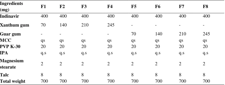
Table No.1: Formulation table for sustained release tablets Ingredients
(mg) F1 F2 F3 F4 F5 F6 F7 F8
Indinavir 400 400 400 400 400 400 400 400
Xanthum gum 70 140 210 245 - - - -
Guar gum - - - - 70 140 210 245
MCC qs qs qs qs qs qs qs qs
PVP K-30 20 20 20 20 20 20 20 20
IPA q.s q.s q.s q.s q.s q.s q.s q.s
Magnesium
stearate 2 2 2 2 2 2 2 2
Talc 8 8 8 8 8 8 8 8
EVALUATION OF TABLETS
Physical Appearance
The general appearance of a tablet, its identity and general elegance is essential for consumer acceptance, for control of lot-to-lot uniformity and tablet-to-tablet uniformity. The control of general appearance involves the measurement of size, shape, colour, presence or absence of odour, taste etc [6]
Hardness test
Hardness indicates the ability of a tablet to withstand mechanical strength while handling. The hardness of the tablets was determined using Monsanto Hardness tester. It is expressed in
kg/cm2. 10 tablets were randomly picked from each formulation and the mean and standard Deviation values were calculated [7]
Friability test
It is the phenomenon whereby tablet surfaces are damaged and/or show evidence of lamination or breakage when subjected to mechanical shock or attrition. The friability of tablets was determined by using Roche Friabilator. It is expressed in percentage (%). 10 tables were initially weighed (Wt. initial) and transferred into friabilator. The friabilator was operated at 25 rpm for 4 min or run up to 100 revolutions. [11] The tablets were weighed again (Wt. final). The percentage friability was then calculated by,
% F = (loss in weight/initial weight) × 100
% Friability of tablets less than 1% are considered acceptable [8]
Weight variation test
The tablets were selected randomly from each formulation and weighed individually to check for weight variation. The US Pharmacopoeia allows a little variation in the weight of a tablet. To study weight variation, 20 tablets of each formulation were weighed using an electronic balance Aqua and the test was performed according to the official method [9]
Drug content (Assay)
Drug content of the tablets was determined by UV Spectrophotometrically.
Uniformity of thickness
Thickness and diameter of tablets were important for uniformity of tablet size. Thickness and diameter was measured using vernier caliper [10]
In vitro
Dissolution Studies
In vitro drug release studies were carried out using USP XXIV dissolution apparatus type II, with 900ml of dissolution medium maintained at 37±1°C for 12 hr, at 50 rpm, pH 6.8 phosphate buffer for 12 hrs for sustained release tablets. 5ml of sample was withdrawn at predetermined time intervals replacing with an equal quantity of drug free dissolution fluid. The samples withdrawn were filtered through 0.45µ membrane filter, and drug content in each sample was analyzed at 265.5nm
after suitable dilution by UV/Vis
Spectrophotometer and cumulative percent drug release was calculate [11]
Drug release kinetics and mechanism
To analyze the mechanism of drug release from the formulation, the dissolution profile of all the batches were fitted to zero order, first order, Higuchi and Peppas models to ascertain the kinetic modelling of drug release [12].
Zero order
First order
Korsmeyer-Peppas model
Higuichi model
Zero Order
In many of the modified release dosage form particularly controlled or sustained release dosage form (those dosage forms that release the drug in planned, predictable and slower than normal manner) is zero order kinetics.
Q = K0 t
Where, Q is the amount of drug release at time, t and Ko is the release rate constant.
First order
This type of models to analyze the drug dissolution study was first proposed by Gibalgi and Feldman and later by Wagner. The relation expressing this model.
Log Qt = Log Qo + K1t / 2.303
Where Qt is the amount of drug released in
time t, Qo is intial amount of drug in the solution
way a graphical relationship between log percent drug remaining versus time to get the first order constant from the slope.
Peppas model
The peppas model is widely used, when the release mechanism is not well known or more than one type of release could be involved. The semi-empirical equation shown as equation:
Mt/M∞ = ktn
Where, Mt/M∞ is fraction of drug released at time‘t’, k represents a constant, and n is the diffusional exponent, which characterizes the type of release mechanism during the dissolution process. For non fickian release, the value of n falls between 0.5 and 1.0; while in case of fickian diffusion, n = 0.5; for zero-order release (case II transport), n = 1; and for super case II transport, n > 1.
Higuichi model
A Large number of modified release dosage form contain some sort of matrix system is such instances the drug dissolves from this matrix. The dissolution pattern of the drug is dictated by water penetration rate (diffusion control) and thus the following relationship applies.
Q = K2 t1/2
Where, Q is the percentage of drug release at time t and K2 is the diffusion rate constant.
RESULTS AND DISCUSSION
Drug
excipient
compatibility
studies
-
Fourier- transform infrared (FTIR)
Drug-excipient compatibility studies by FTIR revealed no interaction between drug and the n a t u r a l polymers used in the formulation thus showing compatibility.
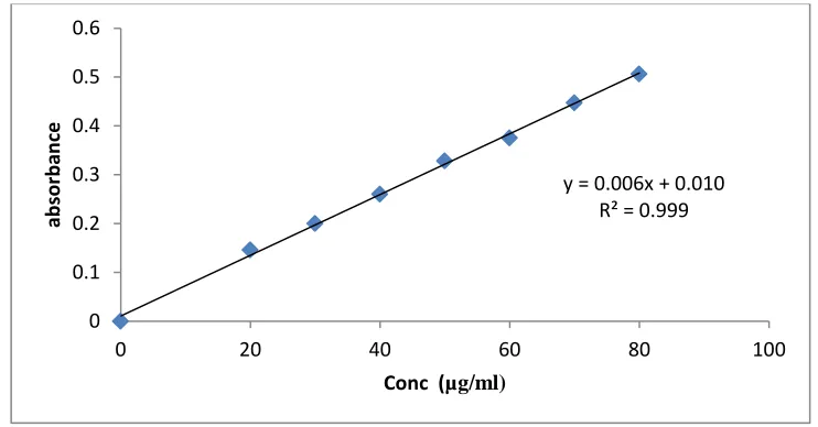
Fig No.1: Calibration curve of Indinavir in 6.8 pH Phosphate buffer
Pre Compression Parameters
Table No.2: Pre compression parameters for SR tablets
Formulations Angle of
Repose (θ)
Loose Bulk Tapped
Bulk %
Compressibility
Hausner’s ratio Density
(g/ml)
Density (g/ml)
F1 27°.30¹±0.06 0.578±0.02 0.661±0.21 12.56±0.16 1.14±0.11
F2 25°.10¹±0.04 0.612±0.04 0.721±0.12 15.12±0.21 1.18±0.24
F3 28°.15¹±0.03 0.653±0.15 0.749±0.18 12.82±0.24 1.13±0.51
y = 0.006x + 0.010 R² = 0.999
0 0.1 0.2 0.3 0.4 0.5 0.6
0 20 40 60 80 100
ab
sor
b
an
ce
F4 27°.16¹±0.06 0.603±0.09 0.694±0.14 13.11±0.11 1.14±0.11
F5 26°.28¹±0.07 0.667±0.21 0.768±0.51 13.15±0.12 1.15±0.11
F6 27°.20¹±0.07 0.660±0.22 0.759±0.16 13.13±0.12 1.16±0.21
F7 26°.19¹±0.06 0.684±0.13 0.784±0.11 12.76±0.12 1.12±0.10
F8 25°.10¹±0.02 0.672±0.14 0.775±0.21 13.29±0.12 1.18±0.18
From the above pre-compression parameters it was clear evidence that granules have good flow
properties. It is shown in Table No.2.Post Compression Parameters.
Tablet No.3: Post Compression Parameters for Sustained Release Tablets
Formulations Hardness
(kg/cm2)
Thickness (mm)
Weight variation (mg)
Friability (%)
F1 6.12±0.02 2.41±0.03 700±1.16 0.38±0.01
F2 6.51±0.02 2.29±0.03 699±1.25 0.27±0.05
F3 6.44±0.01 2.39±002 701±1.18 0.36±0.08
F4 6.30±0.03 2.39±0.01 698±1.89 0.41±0.11
F5 6.56±0.03 2.27±0.01 702±1.75 0.39±0.05
F6 6.85±0.02 2.19±0.01 697±1.45 0.33±0.16
F7 6.92±0.05 2.14±0.02 701±2.14 0.38±0.05
F8 6.80±0.06 2.23±0.03 700±2.15 0.35±0.18
The Post compression parameters such as Weight variation, Thickness, Hardness, Friability, Drug content of the all formulations (F1-F8) results
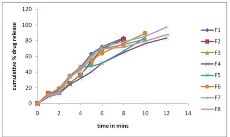
Fig No.2: In vitro Dissolution studies graph of indinavir sustained release formulations F1-F8)
The tablets were evaluated for in vitro dissolution studies in acid buffer (pH-1.2) for 2 hours followed by pH 6.8 buffer for 12 hours. The results of in-vitro drug release revealed that the indinavir was released in a controlled manner from all the formulations where formulation F7 showed maximum drug release i.e. 97.3±0.22% at the end of 12th hour. It is shown in Fig No.2
It was confirmed that the F7 formulation SR tablets fulfill the sustained release theory, In that the Guar gum was used separately in the formulations, but increasing the polymer concentration, it was clearly identified that the drug release was retarded. It was also confirmed that the formulation made with guar gum (F4 and F8) showed sustained drug release compared to the formulations made with xanthum gum (F1 to F4).
CONCLUSION
The Sustained released tablets containing Indinavir SR tablets were successfully prepared by
wet granulation method. The physiochemical evaluation results for the granules of all trials pass the official limits in angle of repose, compressibility index. The prepared granules were also maintained the physiochemical properties of tablets such as thickness, hardness, weight variation, friability. The optimized formulation contains the average thickness of 2.14±0.02 mm, average hardness of 6.92±0.05 Kg/cm2, average weight of 700 mg, and friability of 0.35. The optimized formulation F7 which releases the Indinavir in sustained manner in 1st hour it releases 10.1% but the remaining drug release was sustained up to 12 hours.
“Hence it may be summarized that the F7 tablets prepared by wet granulation method for sustained release tablets might be a perfect and effective formulation to treat the viral infections”
REFERENCES
[1]. Banker GS, Anderson NR. (1987). Tablets. In: Lachman L, Lieberman HA, Kanig JL. The theory and practice of industrial pharmacy. Mumbai: Varghese Publishing House. 3, 182-84, 296-303, 311-12. [2]. Oral controlled release matrix tablets: concept and review. 2005, 5 -30.
[3]. Applied Biopharmaceutics & Pharmacokinetics.Modified-Release Drug Products. 2004, .515-16.
[5]. Committee for veterinary medicinal products (CVMP). Note for guidance on, the quality of modified release dosage forms for veterinary use. European agency 2, 2003, 1-10.
[6]. Saptarshi D, Gupta MS. Modified release dosage form and drug delivery. Journal of Pharmacy Research. 2(11), 2009, 1728-29.
[7]. Ansel HC, Allen LV, Popovich NC. Pharmaceutical dosage forms and drug delivery systems. Lippincott Williams and Wilkins; Baltimore 7, 2000, 231-75.
[8]. Sustained Release dosage form in Pharmaceutics and pharmaceutical jurisprudence, Piyush Publications., 3(3), 111-3.112.
[9]. Gendle R, Kaushik B, Verma L. Parameters required for sustained release drug delivery system, .13, 2009, 45-51.
[10].Luana Periloli, Valeria Ambrogi. Mucoadhesive Bilayered Tablets for Buccal Sustained Release of Flurbiprofen. AAPS Pharm. Sci. Tech, 8(3), 2007 54-62.
[11].Hiremath S. N. Formulation and Evaluation of sustained release matrix tablet of Metformin HCl. Indian drugs, 44(1), 2007, 52-53.
[12].Vishnu M. Patel, Bhupendra G. Mucoadhesive Bilayer Tablets of Propranolol Hydrochloride. AAPS Pharm. Sci. Tech., 8(3), 2007, 77-85.
[13].Juan Manuel Llabot, Ruben Hilario Manzo. Double-Layered Mucoadhesive Tablets Containing Nystatin. AAPS Pharm. Sci. Tech., 3(3), 2002, 22-29.