Transferable Multi-Model Ensemble for
Benign-Malignant Lung Nodule Classification on Chest CT
Yutong Xie 1, Yong Xia 1*, Jianpeng Zhang 1, David Dagan Feng 2, 5, Michael Fulham 2, 3, 4, and Weidong Cai 2
1 Shaanxi Key Lab of Speech & Image Information Processing (SAIIP), School of Computer Science and Engineering, Northwestern Polytechnical University, Xi’an, PR China, 710072 2 Biomedical and Multimedia Information Technology (BMIT) Research Group, School of
In-formation Technologies, University of Sydney, NSW 2006, Australia
3 Department of Molecular Imaging, Royal Prince Alfred Hospital, NSW 2050, Australia 4 Sydney Medical School, University of Sydney, NSW 2006, Australia 5 Med-X Research Institute, Shanghai Jiaotong University, Shanghai 200030, China
Corresponding Author’s Email: [email protected]
Abstract. The classification of benign versus malignant lung nodules using chest CT plays a pivotal role in the early detection of lung cancer and this early detec-tion has the best chance of cure. Although deep learning is now the most success-ful solution for image classification problems, it requires a myriad number of training data, which are not usually readily available for most routine medical imaging applications. In this paper, we propose the transferable multi-model en-semble (TMME) algorithm to separate malignant from benign lung nodules using limited chest CT data. This algorithm transfers the image representation abilities of three ResNet-50 models, which were pre-trained on the ImageNet database, to characterize the overall appearance, heterogeneity of voxel values and heteroge-neity of shape of lung nodules, respectively, and jointly utilizes them to classify lung nodules with an adaptive weighting scheme learned during the error back propagation. Experimental results on the benchmark LIDC-IDRI dataset show that our proposed TMME algorithm achieves a lung nodule classification accu-racy of 93.40%, which is markedly higher than the accuaccu-racy of seven state-of-the-art approaches.
Keywords: Lung nodule classification, deep learning, ensemble learning, com-puted tomography (CT)
1
Introduction
The 2015 global cancer statistics show that lung cancer accounts for approximately 13% of 14.1 million new cancer cases and 19.5% of cancer-related deaths each year [1] . The 5-year survival for patients with an early diagnosis is approximately 54 %, as compared to 4 % if the diagnosis is made late when the patient has the stage IV disease [2]. Hence, early diagnosis and treatment are the most effective means to improve lung cancer survival. The National Lung Screening Trial [3] showed that screening with CT
will result in a 20% reduction in lung cancer deaths. On chest CT scans, a “spot” on the lung, less than 3 cm in diameter, is defined as a lung nodule, which can be benign or malignant [4]. Malignant nodules may be primary lung tumors or metastases and so the classification of lung nodules is critical for best patient care.
Radiologists typically read chest CT scans on a slice-by-slice basis, which is time-consuming, expensive and prone to operator bias and requires a high degree of skill and concentration. Computer-aided lung nodule classification avoids many of these issues and has attracted a lot of research attention. Most solutions in the literature are based on using hand-crafted image features to train a classifier, such as the support vector machine (SVM), artificial neural network and so on. For instance, Han et al. [5] ex-tracted Haralick and Gabor features and local binary patterns to train a SVM for lung nodule classification. Dhara et al. [6], meanwhile, used computed shape-based, margin-based and texture-margin-based features for the same purpose.
More recently, deep learning, particularly the deep convolutional neural network (DCNN), has become the most successful image classification technique and it provides a unified framework for joint feature extraction and classification[7]. Hua et al. applied the DCNN and deep belief network (DBN) to separate benign from malignant lung nodules. Shen et al. [8] proposed a multi-crop convolutional neural network (MC-CNN) for lung nodule classification. Despite improved accuracy, these deep models have not achieved the same performance on routine lung nodule classification as they have in the famous ImageNet Challenge. The suboptimal performance is attributed mainly to the overfitting of deep models due to inadequate training data, as there is usually a small dataset in medical image analysis and this relates to the work required in acquiring the image data and then in image annotation. Hu et al. [9] proposed a deep transfer metric learning method to transfer discriminative knowledge from a labeled source domain to an unlabeled target domain to overcome this limitation.
A major difference between traditional and deep learning methods is that traditional methods rely more on the domain knowledge, such as there is a high correspondence between nodule malignancy and heterogeneity in voxel values (HVV) and heterogene-ity in shapes (HS) [10], and deep learning relies on access to massive datasets. Ideally, the advantages of both should be employed. Chen et al. [11] fused heterogeneous Haralick features, histogram of oriented gradient (HOG) and features derived from the deep stacked denoising autoencoder and DCNN at the decision level to predict nine semantic labels of lung nodules. In our previous work [12], we used a texture descriptor and a shape descriptor to explore the heterogeneity of nodules in voxel values and shape, respectively, and combined both descriptors with the features learned by a nine-layer DCNN for nodule classification. Although improved accuracy was reported, this method still uses hand-crafted features to characterize the heterogeneity of nodules, which are less effective. Recently, Hu et al. [9] reported that the image representation ability of DCNNs, which was learned from large-scale datasets, could be transferred to solving generic small-data visual recognition problems. Hence, we suggest transferring the DCNN’s image representation ability to characterize the overall appearance of lung nodule images and also the nodule heterogeneity in terms of voxel values and shape.
In this paper, we propose a transferable multi-model ensemble (TMME) algorithm for benign-malignant lung nodule classification on chest CT. The main uniqueness of
this algorithm include (1) three types of image patches are designed to fine-tune three pre-trained ResNet-50 models, aiming to characterize the overall appearance (OA), HVV and HS of each nodule slice, respectively; and (2) these three ResNet-50 models are used jointly to classify nodules with an adaptive weighting scheme learned during the error back propagation, which enables our model to be trained in an 'end to end' manner. We compared our algorithm to seven state-of-the-art lung nodule classification approaches on the benchmark LIDC-IDRI dataset. Our results suggest that the proposed algorithm provides substantial performance improvement.
2
Data and Materials
The benchmark LIDC-IDRC database [13] were used for this study, in which the nod-ules were evaluated over five levels, from benign to malignant, by up to four experi-enced thoracic radiologists. The mode of levels given by radiologists was defined as the composite level of malignancy. Same to [5-8, 14, 15], we only considered nodules
≥ 3 mm in diameter and treated 873 nodules with composite level of 1 or 2 as benign and 484 nodules with composite level of 4 or 5 as malignant.
3
Algorithm
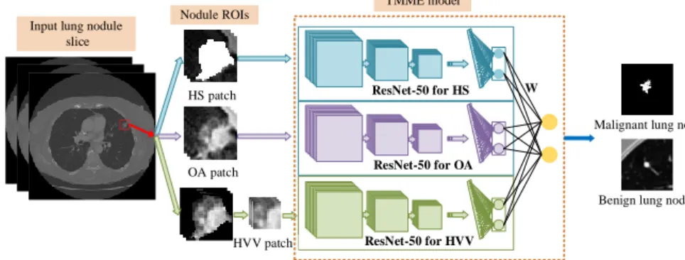
We have summarized our proposed TMME algorithm in Fi. 1. The algorithm has three steps: (1) extracting the region of interest (ROI) for preprocessing and data augmenta-tion, (2) building a TMME model for slice-based nodule classificaaugmenta-tion, and (3) classi-fying each nodule based on the labels of its slices.
HS patch OA patch HVV patch ResNet-50 for HS ResNet-50 for OA ResNet-50 for HVV W
Malignant lung nodule
Benign lung nodule TMME model
Input lung nodule slice
Nodule ROIs
Fig. 1. Framework of our proposed TMME algorithm
3.1 Preprocessing and Data Augmentation
A lung nodule is presented in multiple slices. On each slice, a square ROI encapsulating the nodule is identified using the tool developed by [16] to represent the nodule’s OA. To characterize the nodule’s HVV, non-nodule voxels outside the ROI are set to 0 and,
if the ROI is larger than 16×16, a 16×16 patch that contains the maximum nodule voxels is extracted. To describe the nodule’s HS, nodule voxels inside the ROI are set to 255. Then, the OA patch, HVV patch and HS patch are resized to 200×200, using the bicubic interpolation. Four augmented copies of each training sample are generated by using rotation, shear, horizontal or vertical flip and translation with random parameters to enlarge the size of the training set and are put in the enlarged training set.
3.2 TMME for Nodule Slice Classification
The ResNet-50 model [17] that has been pre-trained on the ImageNet dataset, is adopted. Two neurons in the last fully connected layer are randomly selected and other neurons, together with the weights attached to them, are removed. Three copies of this ResNet-50 are fine-tuned using all OA, HVV and HS patches in the enlarged training set, respectively, to adapt them to characterizing nodule slices. Denoting the prediction vector produced by each ResNet-50 by 𝑿𝑖= (𝑥𝑖1, 𝑥𝑖2) (𝑖 = 1,2,3), the ultimate predic-tion vector of the ensemble model can be calculated as
𝑃𝑘 = ∑ ∑2 𝜔𝑖𝑗𝑘𝑥𝑖𝑗
𝑗=1 3
𝑖=1 , 𝑘 = 1,2 (1)
where 𝑃𝑘 is the predicted likelihood of the input belonging to category 𝑘, and 𝜔𝑖𝑗𝑘 is the weight which connects the 𝑥𝑖𝑗 and 𝑃𝑘. Thus, the integrated loss of this ensemble model can be formulated as
𝐿(𝑦, 𝑷) = ln(∑2 𝑒𝑃𝑗
𝑗=1 ) − 𝑃𝑦, (2)
where 𝑦 ∈ {1,2} is the input’s true label, and 𝑷 = (𝑃1, 𝑃2). The change of weight 𝜔𝑖𝑗𝑘 in the ensemble model is in proportion to descend along the gradient, shown as follows
∆𝜔𝑖𝑗𝑘= −𝜂𝜕𝐿(𝑦,𝑷)
𝜕𝜔𝑖𝑗𝑘 =−𝜂𝑥𝑖𝑗(
𝑒𝑃𝑘
∑2 𝑒𝑃𝑚
𝑚=1 − 𝛿𝑘𝑦), (3)
where 𝜂 represents the learning rate, and, if 𝑘 = 𝑦, 𝛿𝑘𝑦= 1, otherwise, 𝛿𝑘𝑦= 0. Since our training data set is small, the learning rate is set to 0.00001 and the sto-chastic gradient descent with a batch size of 100 is adopted. Moreover, 10% of the training patches are chosen to form a validation set, and the training is terminated even before reaching the maximum iteration number of 50, if the error on the other 90% of training images continues to decline but the error on the validation set stops decreasing.
3.3 Nodule Classification
Let a lung nodule 𝜳 be contained in 𝑆 slices, denoted by 𝜳 = {𝛹1, 𝛹2, … , 𝛹𝑆}. Input the 𝑖-th slice 𝛹𝑖 into the TMME model, and we obtain a two-dimensional prediction vector 𝑯(𝛹𝑖). The class label of nodule 𝜳 is assigned based on the sum of the predic-tion made on each slice, shown as follows
𝑎𝑟𝑔 𝑚𝑎𝑥
𝑘
∑
𝐻𝑘(
𝛹𝑖)
𝑆
4
Results
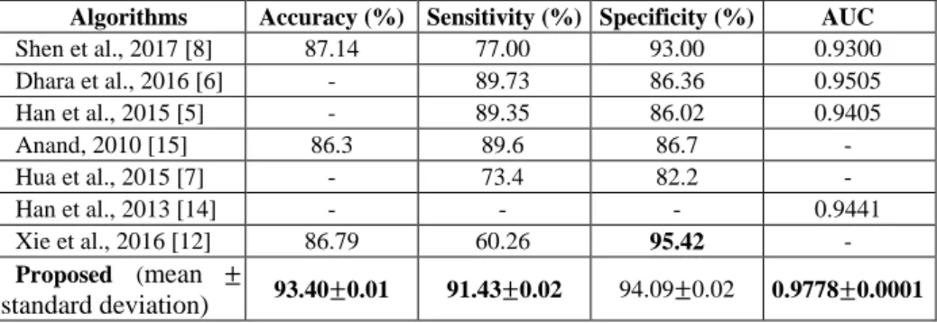
The proposed TMME algorithm was applied to the LIDC-IDRC dataset 10 times inde-pendently, with 10-fold cross validation. The mean and standard deviation of obtained accuracy, sensitivity, specificity and area under the receiver operator curve (AUC), to-gether with the performance of seven state-of-the-art methods on this dataset, were given in Table 1. It shows that our algorithm not only outperformed hand-crafted fea-ture-based traditional methods but also substantially improved upon Xie et al.’s method [12]. Our results indicate that the pre-trained and fine-tuned ResNet-50 model can ef-fectively transfer the image representation ability learned on the ImageNet dataset to characterizing the OA, HVV and HS of lung nodules, and an adaptive ensemble of these three models has superior ability to differentiate malignant from benign lung nodules.
Table 1. Performance of eight lung nodule classification methods on the LIDC-IDRC dataset
Algorithms Accuracy (%) Sensitivity (%) Specificity (%) AUC
Shen et al., 2017 [8] 87.14 77.00 93.00 0.9300 Dhara et al., 2016 [6] - 89.73 86.36 0.9505 Han et al., 2015 [5] - 89.35 86.02 0.9405 Anand, 2010 [15] 86.3 89.6 86.7 - Hua et al., 2015 [7] - 73.4 82.2 - Han et al., 2013 [14] - - - 0.9441 Xie et al., 2016 [12] 86.79 60.26 95.42 - Proposed (mean ± standard deviation) 93.40±0.01 91.43±0.02 94.09±0.02 0.9778±0.0001
5
Discussion
5.1 Data ArgumentationThe number of training samples generated by data augmentation plays an important role in applying a deep model to small-sample learning problems. On one hand, training a deep model requires as many data as possible; on the other hand, more data always lead to higher time cost. We re-performed the experiment using different numbers of augmented images and listed the obtained performance in Table 2.
Table 2. Performance of the proposed algorithm with different number of augmentatioin data.
Augmented
Data per Image Accuracy (%) Sensitivity (%) Specificity (%) AUC
Time for Training 0 89.84 83.85 93.43 0.9451 3 hours 2 92.24 88.74 93.99 0.9724 7 hours 4 93.40 91.43 94.09 0.9778 12 hours 6 93.66 91.65 94.90 0.9788 17.5 hours 8 93.73 91.90 94.90 0.9794 24 hours
It reveals that using four augmented images for each training sample achieved a trade-off between accuracy and time cost, since further increasing the number of aug-mented images only improved the accuracy slightly but cost much more time for train-ing. Meanwhile, it should be noted that our algorithm, without using data augmentation, achieved an accuracy of 89.84%, which is still superior to the accuracy of those meth-ods given in Table 1.
5.2 Ensemble Learning
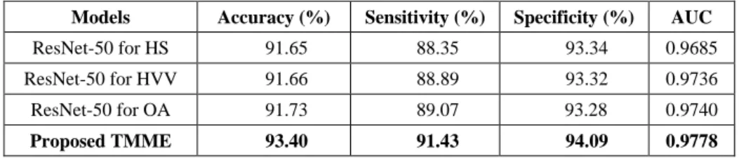
To demonstrate the performance improvement that results from the adaptive ensemble of three ResNet-50 models, we compared the performance of our algorithm to that of three component models, which characterize lung nodules from the perspective of OA, HVV and HS, respectively. As shown in Table 3, although each ResNet-50 model achieves a relatively good performance, an adaptive ensemble of them brings a further performance gain.
Table 3. Performance of each component ResNet-50 model and the proposed ensemble model
Models Accuracy (%) Sensitivity (%) Specificity (%) AUC
ResNet-50 for HS 91.65 88.35 93.34 0.9685 ResNet-50 for HVV 91.66 88.89 93.32 0.9736 ResNet-50 for OA 91.73 89.07 93.28 0.9740
ProposedTMME 93.40 91.43 94.09 0.9778
5.3 Other Pre-trained DCNN Models
Besides ResNet-50, GoogLeNet [18] and VGGNet [19] are two of the most successful DCNN models. Using each of those three models to characterize lung nodules from each of three perspectives, i.e. OA, HVV and HS, we have 27 different configurations. To evaluate the performance of using other DCNN models, we tested all 27 configura-tions and gave the accuracy and AUC of the top five configuraconfigura-tions in Table 4. It shows that ResNet-50 is very powerful and using three ResNet-50 results in the highest accu-racy and AUC. Nevertheless, it also suggests that GoogLeNet and VGGNet are good choices as well and using them to replace ResNet-50 may produce very similar accu-racy in some configurations.
Table 4. Performance of the top five out of 27 ensemble models
DCNN for HS DCNN for VVH DCNN for OA Accuracy (%) AUC
ResNet-50 ResNet-50 ResNet-50 93.40 0.9778
ResNet-50 GoogLeNet ResNet-50 93.30 0.9760 VGGNet ResNet-50 ResNet-50 93.28 0.9767 GoogLeNet ResNet-50 ResNet-50 93.21 0.9759 ResNet-50 ResNet-50 GoogLeNet 93.14 0.9765
5.4 Hybrid Ensemble of 27 TMME Models
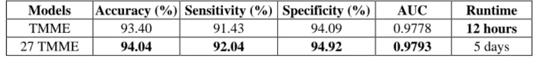
Using all possible combination of VGGNet, GoogLeNet and ResNet-50 to characterize the OA, HVV and HS of lung nodules, we can have totally 27 proposed TMME models, which can be further combined by using an adaptive weighting scheme learned in the same way. Table 5 shows that the ensemble of 27 TMME models can only slightly improve the classification accuracy, but with a major increase in the computational complexity of training the model.
Table 5. Performance of TMME and the ensemble of 27 TMME mdels
Models Accuracy (%) Sensitivity (%) Specificity (%) AUC Runtime
TMME 93.40 91.43 94.09 0.9778 12 hours
27 TMME 94.04 92.04 94.92 0.9793 5 days
5.5 Computational Complexity
In our experiments, it took about 12 hours to train the proposed model and less than 0.5 second to classify each lung nodule (Intel Xeon E5-2678 V3 2.50 GHz ×2, NVIDIA Tesla K40c GPU ×2, 128 GB RAM, 120 GB SSD and Matlab 2016). It suggests that the proposed algorithm, though computation very complex during the training process that can be performed offline, is very efficient for online testing and could be used in a routine clinical workflow.
6
Conclusion
We propose the TMME algorithm for benign-malignant lung nodule classification on chest CT. We used three pre-trained and fine-tuned ResNet-50 models to characterize the OA, HVV and HS of lung nodules, and combined these models using an adaptive weighting scheme learned during the back-propagation process. Our results on the benchmark LIDC-IDRC dataset suggest that our algorithm produces more accurate re-sults than seven state-of-the-art approaches.
Acknowledgments. This work was supported in part by the National Natural Science Foundation of China under Grants 61471297, in part by the Seed Foundation of Inno-vation and Creation for Graduate Students in Northwestern Polytechnical University under Grants Z2017041, and in part by the Australian Research Council (ARC) Grants. We acknowledged the National Cancer Institute and the Foundation for the National Institutes of Health, and their critical role in the creation of the free publicly available LIDC/IDRI Database used in this work.
Reference
1. Siegel, R. L., Miller, K. D., Jemal, A.: Cancer statistics, 2015. CA. Cancer J. Clin. 65(1), 5– 29 (2015)
2. Bach, P. B., Mirkin, J. N., Oliver, T. K., Azzoli, C. G., Berry, D. A., Brawley, O.W., Byers, T., Colditz, G.A., Gould, M.K., Jett, J.R.: Benefits and harms of CT screening for lung can-cer: a systematic review. Jama J. Am. Med. Assoc. 307, 2418-2429 (2012)
3. Abraham, J.: Reduced lung-cancer mortality with low-dose computed tomographic screen-ing. New Engl. J. Med. 365, 395-409 (2011)
4. American Thoracic Society: What is a Lung Nodule? Am. J. Respir. Crit. Care Med. 193, 11-12 (2016)
5. Han, F., Wang, H., Zhang, G., Han, H., Song, B., Li, L., Moore, W., Lu, H., Zhao, H., Liang, Z.: Texture Feature Analysis for Computer-Aided Diagnosis on Pulmonary Nodules. J. Dig-ital Imaging 28(1),99-115 (2015)
6. Dhara, A. K., Mukhopadhyay, S., Dutta, A., Garg, M., Khandelwal, N.: A Combination of Shape and Texture Features for Classification of Pulmonary Nodules in Lung CT Images. J. Digital Imaging 29(4), 466–475 (2016)
7. Hua, K. L., Hsu, C. H., Hidayati, S. C., Cheng, W. H., Chen, Y. J.: Computer-aided classi-fication of lung nodules on computed tomography images via deep learning technique. On-cotargets Ther. 8,2015-2022 (2015)
8. Shen, W., Zhou, M., Yang, F., Yu, D., Dong, D., Yang, C., Zang, Y., Tian, J.: Multi-crop Convolutional Neural Networks for lung nodule malignancy suspiciousness classification. Pattern Recogn. 61,663-673 (2017)
9. Hu, J., Lu, J., Tan, Y. P.: Deep transfer metric learning. In: CVPR 2015, pp. 325-333. IEEE Press, New York. (2015)
10. Metz, S., Ganter, C., Lorenzen, S., Marwick, S. V., Holzapfel, K., Herrmann, K., Rummeny, E. J., Wester, H. J., Schwaiger, M., Nekolla, S. G.: Multiparametric MR and PET Imaging of Intratumoral Biological Heterogeneity in Patients with Metastatic Lung Cancer Using Voxel-by-Voxel Analysis. Plos One 10(7), e0132386 (2014)
11. Chen, S., Qin, J., Ji, X., Lei, B., Wang, T., Ni, D., Cheng, J.Z.: Automatic Scoring of Multiple Semantic Attributes with Multi-task Feature Leverage: A Study on Pulmonary Nodules in CT Images. IEEE Transactions on Medical Imaging. 99,1-1 (2016)
12. Xie Y., Xia Y., Zhang J., Liu S., Cai W.: Lung Nodule Classification by Jointly Using Visual Descriptors and Deep Features. MICCAI Workshop on MCV (2016)
13. Iii, S. G. A., Mclennan, G., Bidaut, L., Mcnittgray, M. F., Meyer, C. R., Reeves, A. P., Zhao, B., Aberle, D. R., Henschke, C. I., Hoffman, E. A.: The Lung Image Database Consortium (LIDC) and Image Database Resource Initiative (IDRI): A Completed Reference Database of Lung Nodules on CT Scans. Med. Phys. 38, 915-931 (2011)
14. Han, F., Zhang, G., Wang, H., Song, B.: A texture feature analysis for diagnosis of pulmo-nary nodules using LIDC-IDRI database. In: ICMIPE 2013, pp. 14-18. IEEE Press, (2013) 15. Anand, S. K. V.: Segmentation coupled textural feature classification for lung tumor predic-tion. In: 2010 IEEE International Conference on Communication Control and Computing Technologies, pp. 518-524. IEEE Press, New York. (2010)
16. Lampert, T. A., Stumpf, A., Gançarski, P.: An Empirical Study into Annotator Agreement, Ground Truth Estimation, and Algorithm Evaluation. IEEE TIP. 25(6), 2557-2572 (2016) 17. He, K., Zhang, X., Ren, S., Sun, J.: Deep Residual Learning for Image Recognition. In:
CVPR 2016, pp. 770-778. IEEE Press, New York. (2016)
18. Szegedy, C., Liu, W., Jia, Y., Sermanet, P.: Going deeper with convolutions. In: CVPR 2015, pp. 1-9. IEEE Press, New York. (2016)
19. Simonyan, K., Zisserman, A.: Very Deep Convolutional Networks for Large-Scale Image Recognition. arXiv preprint arXiv:1409-1556 (2014)