THREE-DIMENSIONAL EXTRACELLULAR MATRIX
HYDROGEL ENVIRONMENTS FOR EMBRYONIC STEM CELL
GROWTH
A Thesis Presented to The Academic Faculty
by
Ima Mbodie Ebong
In Partial Fulfillment
of the Requirements for the Degree Master of Science in Bioengineering in the
Wallace H. Coulter Department of Biomedical Engineering/College of Engineering
Georgia Institute of Technology August 2007
THREE-DIMENSIONAL EXTRACELLULAR MATRIX
HYDROGEL ENVIRONMENTS FOR EMBRYONIC STEM CELL
GROWTH
Approved by:
Dr. Todd C. McDevitt, Advisor
Wallace H. Coulter Department of Biomedical Engineering
Georgia Institute of Technology
Dr. Julia E. Babensee
Wallace H. Coulter Department of Biomedical Engineering
Georgia Institute of Technology
Dr. Johnna S. Temenoff
Wallace H. Coulter Department of Biomedical Engineering
Georgia Institute of Technology
To my parents for their unconditional love and support.
iv
ACKNOWLEDGEMENTS
I wish to thank those persons who have assisted with various aspects of this thesis project, including Mr. Michael Mathews, Mr. Peter Yang, Mr. Johnafel Crowe, Ms. Sha’Aqua Asberry and Dr. Christopher Wilson. I wish to also thank my peers in the McDevitt Laboratory for their support.
TABLE OF CONTENTS
Page
ACKNOWLEDGEMENTS iv
LIST OF FIGURES viii
SUMMARY ix CHAPTER
1 INTRODUCTION 1
Background: Embryonic Stem Cells 1
Background: Extracellular Matrix 2
Hydrogels for In Vitro Studies 3
PDMS Mold Fabrication 4
PDMS Molds for Hydrogel Casting 5
Purpose for Thesis Project
6
2 MATERIALS AND METHODS 8
Polycarbonate Master and PDMS Mold Fabrication 8
Cell Culture 9
Mouse Embryonic Stem Cell Culture 9
NIH 3T3 Fibroblast Cell Culture 9
Hydrogel Construct Formation and Culture 9
Type-I Collagen Hydrogel Constructs 10
Fibrin Hydrogel Constructs 10
Oligo(polyethylene glycol fumarate)(OPF) Hydrogel Constructs 10
vi
Stereomicroscopic Imaging of Hydrogel Constructs 11
Rheology 11
Nuclei Density Analysis 12
Cell Viability Analysis 13
Analysis of Cells in 2D Culture 13
Analysis of Cells in Hydrogels (3D) 13
Histological Analysis 14
Analysis of Cells in 2D Culture 14
Analysis of Cells in Hydrogels (3D) 15
Morphometric Analysis 15
Immunofluorescence Staining 16
Serial Passaging of mESCs in 2D and 3D 17
3 RESULTS 18
Polycarbonate Master and PDMS Mold Fabrication 18
Stereomicroscopic Imaging of Hydrogels 20
Rheology analysis of acellular hydrogels cast in different molds 21
Histological Analysis 22
Viability Analysis 23
Rheology analysis of acellular hydrogels of various protein concentrations 24
Nuclei Density Analysis 25
Morphometric Analysis 27
Cell Viability Analysis 29
Immunofluorescent staining of mESC clusters 31
4 DISCUSSION 34
PDMS Molds for flexible hydrogel casting 34
PDMS can be fabricated into a mold to successfully cast various
types of hydrogels 34
Mold geometry affects viscoelastic properties of hydrogels 36 Differences in viscoelastic properties do not affect cell behavior 37
3D Hydrogel Matrices for mESC Maintenance 38
Benefit of simultaneous analysis of different hydrogels on ESC behavior38
Morphologies of mESC colonies in 2D vs. 3D 39
Serial passaging of mESCs from 3D results in homogeneity of cells 39
5 CONCLUSION 41
APPENDIX A: MATRIGEL HYDROGELS 43
viii
LIST OF FIGURES
Page
Figure 1: Polycarbonate master for PDMS mold fabrication. 18
Figure 2: Polydimethylsiloxane (PDMS) mold for hydrogel casting. 19 Figure 3: Macroscopic images of natural and synthetic hydrogels. 20 Figure 4: Viscoelastic properties of hydrogels cast in vinyl or PDMS molds. 21 Figure 5: Histological analysis of mESCs and 3T3 fibroblasts in hydrogels. 22
Figure 6: Viability (Live/Dead) analysis. 23
Figure 7: Rheology analysis of acellular hydrogels. 24
Figure 8: Nuclei density analysis. 26
Figure 9: Microscopic images of mESC clusters. 28
Figure 10: Size and shape factor distributions of mESC colonies/clusters. 29 Figure 11: Cell viability as determined by trypan-blue and live/dead analysis. 30
Figure 12: Oct-4 protein expression. 31
Figure 13: Serial passaging of cells in 2D and 3D. 32
SUMMARY
Embryonic stem cells (ESCs) are pluripotent cells derived from the inner cell mass of the blastocyst that can give rise to cells of the ectoderm, endoderm and mesoderm lineages. Once isolated from the blastocyst, ESCs can be cultured indefinitely
in vitro in an undifferentiated state or can be induced to differentiate. In the case of
mouse ESCs (mESCs), the cytokine leukemia inhibitory factor (LIF) is added to culture media to maintain pluripotency and is removed to induce differentiation. Although it is known that extracellular matrix (ECM) components influence stem cell maintenance, proliferation and differentiation, the precise effects of ECM environments on embryonic stem cell behavior have not been systematically studied. The main purpose of this thesis project was to investigate the behavior of undifferentiated mESCs cultured in different 3D hydrogel matrices and to determine whether viscoelastic and biochemical variations in the matrices differentially affect the ability of stem cells to self-renew; that is, retain their pluripotency or undifferentiated phenotype. Their behavior in 3D environments was compared to mESC behavior in traditional 2D culture. In addition, a new method of casting hydrogels in polydimethylsiloxane (PDMS) molds was developed in order to efficiently cast multiple hydrogels of varying sizes and shapes.
Undifferentiated D3 mESCs were cultured on gelatin-coated substrates and routinely passaged at 60-70% confluence. The cells were then cultured in 3-D constructs made of type I collagen, fibrin or oligo(polyethylene glycol fumarate) (OPF), and supplemented with complete mESC media containing LIF. Samples were obtained on days 2 and 4 of culture to assess cell viability and perform morphological analyses.
x
density analysis was performed and was correlated with rheology data. Viability was assessed and morphometric analyses were performed in order to qualitatively and quantitatively compare morphologies of mECSs in both 2D and 3D environments. Immunofluorescence staining was performed in order to detect the expression of the pluripotent protein Oct-4 by the cells in both 2D and 3D cultures.
Results showed that mESC constructs cast in PDMS molds possessed similar characteristics to those cultured in control disposable vinyl molds throughout the duration of the studies, suggesting that the casting method developed had no adverse effect on 3D mESC culture. Viability analysis indicated that both cells in 2D and cells in the 3D hydrogel environments remained viable throughout the duration of the experiment. The viscoelastic moduli of the collagen and fibrin hydrogels increased with increasing protein concentration. In both 2D and 3D environments mESCs grew as distinct independent clusters or colonies, however cell colonies in 2D were less uniform in size, shape and morphology. No significant differences in the nuclei density of the cell colonies were observed in the different matrices. In contrast, mESC colonies grown on 2D substrates were more spread and hence, the nuclei density was less. Colonies grown in 2D were also less uniform than those grown in 3D hydrogels, as indicated by broader distributions of shapes and sizes. The mESCs retained their undifferentiated morphology in 3D comparable to those in 2D and expressed Oct-4 in both fibrin and collagen hydrogels.
This thesis project suggests that 3D hydrogel microenvironments are able to support the growth of mESCs in vitro and may serve as the basis of hydrogel carriers for
CHAPTER 1
INTRODUCTION
Background: Embryonic Stem Cells
Embryonic stem cells (ESCs) are cells that are obtained from embryos, specifically from those that have been fertilized in vitro for research purposes 1. ESCs
are pluripotent cells derived from the inner cell mass of the four to five day old embryo called the blastocyst. These cells can give rise to cells of the three germ layers: ectoderm, endoderm and mesoderm lineages. Once isolated from the blastocyst, ESCs can be cultured indefinitely in vitro to maintain their pluripotency 2-5 or can be induced to
differentiate into cell lineages of the three germ layers 6-13. In the case of mouse embryonic stem cells (mESCs), the cytokine leukemia inhibitory factor (LIF) must be added to culture media in order for the cells to retain their undifferentiated state 14-16. Although researchers have shown that ESCs can be successfully sub-cultured indefinitely
in vitro, there remains some uncertainty of the quality of the cells during later passages.
Because of issues such as spontaneous differentiation, changes in phenotype, genotype, and/or karyotype, the pluripotency of late-passage ESCs have been shown to be affected 17,18. These changes seen may be attributed to the environment in which ESCs are
cultured in vitro. The blastocysts from which ESCs are derived are three-dimensional
(3D) structures composed of both internal (inner cell mass) and external (trophoblast) clusters of cells, a fluid-filled cavity (blastocoel) and other molecular cues that direct cell fate. Traditional two-dimensional (2D) ESC culture in vitro lacks the main components
2
Background: Extracellular Matrix
Extracellular matrix (ECM) is material that is not inherently a part of the cell and consists of proteins, glycoproteins, proteoglycans, polysaccharides, lipids and fluid. Its functions include providing structural support for cells, as well as cues necessary for regulating cell dynamics. It is known that ECM has many important roles in the growth and development of cells and tissues during embryogenesis and tissue regeneration. Not only can ECM components influence the differentiation of stem cells, the extracellular microenvironment can contribute to the growth and maintenance of undifferentiated cells as well 19,20 However, the precise effects of ECM environments on embryonic stem cell fate have not been systematically studied. What is known is that during in utero
embryonic development biochemical soluble signals as well as macromolecular ECM components play important roles during this process 21. The architecture of the ECM is essential for mechanical support of the cells. Also, ECM proteins, sugars and other essential biomolecules elicit cues that are able to control and direct cell proliferation, migration and differentiation 19. In vitro studies have shown that ECM components such
as proteins and other growth factors are able to regulate self-renewal and differentiation of stem cells in order to create in vitro models that support normal cell function and
structure and mimic physiological environments 19,22-26.
Current in vitro ESC culture techniques, however, do not take full advantage of
what is known about in utero cell development, hence is somewhat inefficient 27. While
development of the blastocyst takes place in a controlled three-dimensional environment, ESCs are typically grown in two-dimensions on tissue culture dishes. Continual sub-culturing of the cells in vitro often leads to loss of phenotype and spontaneous
differentiation 28,29. There has been use, however, of ECM-derived components as substrates for 2D stem cell culture. For example, during in vitro human embryonic stem
cell (hESC) culture, cells are routinely grown on surfaces coated with MatrigelTM (BD Biosciences), which is solubilized basement membrane matrix extracted from murine tumor 30. Matrigel is rich in ECM proteins, with the major components being laminin and collagen IV. Matrigel has been proven to support undifferentiated growth of hESCs. In differentiation studies, researchers have shown that tissue-specific matrices influence the developmental lineage and may further promote organ differentiation 22.
Hydrogels for In Vitro Studies
Hydrogels are water-swollen, cross-linked polymeric structures that have received significant attention over the past few decades because of their exceptional promise in biomedical applications 31,32. The physical properties of hydrogels are essential to a variety of biomedical applications; for example, many tissue-engineering applications rely on assembling cells in a 3D scaffold in an attempt to provide an environment reminiscent of natural ECM structure. A variety of different natural or synthetic polymers hydrogels have been developed to be used as scaffolds capable of supporting cell viability and function 31. For this thesis project, both naturally-derived and synthetic hydrogels were studied. The naturally-derived hydrogels studied were collagen (specifically type I) and fibrin, while the novel polymer oligo(polyethylene glycol fumarate), or OPF, was the synthetic hydrogel used.
Collagen was chosen because it is the most abundant protein found in mammals and is the major component of ECM. Moreover, type I collagen was chosen specifically
4
because it is the most abundant of all the collagens and plays an important role in tissue regeneration and repair. It is also commercially available as a solubilized material and forms a hydrogel at physiological temperatures. It has been used as the primary 3D matrix material in many biomedical and tissue engineering applications including orthopedic tissue engineering 33 and fabrication of tissue-engineered blood vessels 34.
Fibrin, another protein found in mammals, was chosen also because of its important role in tissue regeneration and repair. It is the major protein component of blood clots and is made via the cleavage of the protein fibrinogen by the enzyme thrombin. Like collagen, it can form a gel at physiological temperatures and has been used extensively in biomedical engineering applications, ranging from cardiovascular tissue engineering 34 to wound healing experiments 35.
OPF hydrogels, which were studied during the mold fabrication stage of this thesis project, is a synthetic polyethylene glycol (PEG) based hydrogel. The purpose of studying this type of hydrogel was to determine whether or not the molds fabricated during this project were able to cast hydrogels of both natural and synthetic origin. OPF is a novel hydrogel developed by the Mikos Laboratory at Rice University and has been used as a 3D matrix for soft tissue and orthopedic tissue engineering applications 36-39. Its properties, such as non-toxicity, biomcompatibility and rapid gelling make it a promising material for 3D cell culture.
PDMS Mold Fabrication
Since the hydrogels studied for this thesis project are thermoresponsive and undergo sol-to-gel transformation, they must be cast in molds where they acquire a
specific shape and size. In order to produce cell-laden hydrogels in vitro, a flexible mold was developed using polydimethylsiloxane (PDMS).
PDMS Molds for Hydrogel Casting
There are many issues associated with creating hydrogels for biomedical applications, specifically for the 3D culture of cells. Currently, most hydrogels are cast within sterile and biocompatible multi-well plates or similar mass-produced materials, such that the geometry of hydrogel constructs is dictated by the choice of pre-fabricated “mold”. The downfall to this casting method, however, is that the user has little control over macroscopic properties of the hydrogel such as size and shape. In many instances it may be advantageous to use a flexible system capable of producing a broad range of hydrogel geometries without having to make any significant changes to the casting method. Photolithography and soft lithography techniques have been used to create flexible molds to encapsulate live cells within microscale polymeric hydrogels (microgels) on 2D surfaces 40-45. Polydimethylsiloxane (PDMS) is an elastomeric polymer that has been used extensively in these applications to create molds for microgel formation and microcell patterning on 2D substrates 40,41,46. In this study, an attempt was made to extend the advantages of PDMS for creating flexible molds to 3D cultures. Properties such as easy replica molding, ability to seal with hard surfaces, non-toxicity to cells and biocompatibility make it a suitable material for molding hydrogel scaffolds 42,47. PDMS replicas are typically fabricated by casting the pre-cured PDMS solution on rigid, patterned molds or masters 48. In the formation of PDMS microfluidic devices and PDMS molds for microgel formation, masters were typically made of silicon or
6
glass. 42,45,49. However these materials are not easily machined and can be expensive 42. In our studies, the master is fabricated using polycarbonate. Polycarbonate is very good, general purpose plastic that is inexpensive and can be easily machined into objects of various geometries 50,51. Once pre-cured PDMS prepolymer is fully polymerized, it is peeled off the surface of the master, resulting in an inverse, replica mold that possesses wells in the shapes of the solid master. The PDMS can be easily sterilized with ethanol then exposure to ultra-violet (UV) light. The mold is also reusable, as once hydrogels are formed and removed, the mold can again be washed and re-sterilized.
Purpose of Thesis Project
There are two main purposes to this thesis project. The first was to develop a flexible polydimethylsiloxane mold for simple and economic casting of 3D hydrogels. The ability to cast synthetic and naturally 3D hydrogels containing cells using PDMS molds was examined. The hydrogels studied were collagen and fibrin, which are both naturally derived, and the synthetic hydrogel oligo(polyethylene glycol fumarate) (OPF). Rheology was used to study the effects of PDMS properties on hydrogel viscoelastic properties. In addition, the behavior of mouse embryonic stem cells (mESCs), as well as fibroblast cells, encapsulated in the various hydrogels was studied in order to determine whether PDMS had any effect on cell viability, morphology and pluripotent protein expression.
The second was to investigate the behavior of undifferentiated mESCs cultured in different 3-D matrices and to determine whether viscoelastic and biochemical variations in the matrices differentially affect the ability of stem cells to self-renew or differentiate.
Although other groups have studied the effects of 3-D environment on ESC culture, few have studied the effects of different ECM environments simultaneously, and almost all of looked at 3-D effects on embryoid bodies (aggregates of differentiated stem cells) as opposed to stem cells in their undifferentiated state 23,25-27,52. We hypothesize that both viscoelastic and biochemical variations in the 3-D protein matrices may differentially affect the ability of stem cells to self-renew or differentiate. The behavior of ESCs in 3D environments was compared to that of cells cultured using traditional 2D methods. During these studies, mESCs were cultured in two different hydrogels in the presence of LIF. The naturally-derived protein hydrogels chosen were type I collagen and fibrin. The naturally-derived protein hydrogels may offer mESCs cues for both self-renewal and differentiation in addition to structural support provided by the 3D architecture. Secondly, each matrix type is distinctly different from the other in terms of biochemical make up, microstructure and intrinsic mechanical properties; thus, it would be interesting to observe whether or not these differences may affect stem cell behavior. Finally, extensive studies have been performed on these materials in other areas of tissue engineering and regenerative medicine and it would be interesting to see how these materials contribute to stem cell growth and development 23,27. This study utilized rheology, viability analysis, morphometry, and immunofluorescence of the mESC hydrogels to elucidate the effect of mESC maintenance and/or differentiation in 3D environments.
8
CHAPTER 2
MATERIALS AND METHODS
Polycarbonate Master and PDMS Mold Fabrication
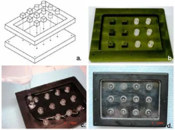
Polycarbonate blocks, cylindrical and rectangular rods were purchased from McMaster-Carr and were used to create the master mold shown in Figure 1a and 1b. Briefly, the polycarbonate master was designed so that removable protruding circular (dimensions: diameter = height = 10mm) and square (dimensions: length = width = height = 10mm) shapes could be screwed into a rectangular polycarbonate base. SolidEdge software was used to design the blueprints for the masters.
PDMS molds were created by mixing Sylgard® 184 Silicone Elastomer curing agent (Dow Corning) with Sylgard® 184 Silicone Elastomer base (Dow Corning) in a 1:10 w/v ratio respectively. Once thoroughly mixed, the solution was poured carefully into the polycarbonate master, ensuring that the base was filled evenly. To eliminate air bubbles that were formed due to mixing, the polycarbonate mold containing the pre-cured PDMS mixture was placed in a vacuum desiccator and degassed for approximately 1 hour; air was released at 10 minute intervals. Once the air bubbles were removed, the mold was transferred to an oven set at 100°C and cured for 45 minutes. Once hardened and cooled, the PDMS mold was carefully peeled away from the polycarbonate master and sterilized by rinsing five times with 70% ethanol solution (5 minutes each) then five times with deionized water (5 minutes each). The mold was then dried in a sterile laminar flow hood under ultra-violet (UV) light overnight.
Cell Culture
Mouse Embryonic Stem Cell Culture
Mouse embryonic stem cells (mESCs) from the D3 line were maintained on 0.1% gelatin coated dishes in Dulbecco’s Modification of Eagle’s Medium or DMEM (Mediatech), 2mM L-glutamine (Mediatech), 1x non-essential amino acids (Mediatech), 15% FBS (Hyclone), 0.1 mM β-mercaptoethanol (Disher), penicillin-streptomycin (GIBCO) and 103U/mL leukemia inhibitory factor (ESGRO®, Chemicon). Cells were routinely passaged every 2-3 days at 60-70% confluence.
NIH 3T3 Fibroblast Cell Culture
NIH 3T3 cell fibroblasts were cultured in DMEM 2mM L-glutamine (Mediatech), 1x non-essential amino acids (Mediatech), 10% FBS (Hyclone), and penicillin-streptomycin (GIBCO). Cells were passaged at no less than 90% confluence.
Hydrogel Construct Formation and Culture
Hydrogels were either naturally-derived or synthetic. The naturally-derived hydrogel constructs were made of type-I collagen or fibrin, while the synthetic hydrogels were made of oligo(polyethylene glycol fumarate), or OPF. For all cell-laden construct types mESCs or fibroblasts were passaged as stated above. Cells were counted using a hemacytometer in order to obtain a desired cell density of 1.5 x 10⁵ cells per hydrogel construct. The following methods were used to develop the constructs.
10 Type-I Collagen Hydrogel Constructs
In order to make collagen constructs (final collagen concentration ranged from 1mg/mL to 3 mg/mL), 1x DMEM was added to the centrifuge tube containing the cells. In another test tube 5x DMEM, 1M sodium hydroxide and type I rat-tail collagen (BD Biosciences) were mixed together so that the final concentration of the mixture equaled 1x. The collagen mixture was then added to the tube containing 1x DMEM and cells.
Fibrin Hydrogel Constructs
In order to make fibrin constructs, a 50-50 mixture of bovine fibrinogen (Sigma) and thrombin (Sigma-Aldrich) was made so that the final fibrinogen concentration ranged from 2mg/mL to 6 mg/mL and the final thrombin concentration was 0.1 U/mg of fibrinogen; the thrombin stock was reconstituted in 0.1% bovine serum albumin solution. The mixture remained on ice to slow the cleavage of fibrinogen by thrombin, and cells were added to the cold mixture.
Oligo(polyethylene glycol fumarate) (OPF) Hydrogel Constructs
In order to make OPF 3K hydrogels (PEG initial Mn = 3,350 g/mol), OPF was dissolved in phosphate-buffered saline (pH 7.4) and mixed with the cross linker poly(ethylene glycol) diacrylate (PEG-DA; Mn = 3400g/mol, Laysan Bio) (20 wt % initial polymer, 3 OPF/2 PEG-DA by weight). To initiate polymerization, ammonium persulfate (APS; Sigma-Aldrich) and N,N,N',N'- tetramethylethylenediamine (TEMED; Sigma-Aldrich) were introduced (final concentrations of both APS and TEMED were 18
mM). In some cases, cells were added to the OPF solution prior to addition of the cross-linker.
Hydrogel Construct Culture and Maintenance
Using a micro pipet, 500 µL samples of each hydrogel type were dispensed into disposable vinyl molds or PDMS molds and then incubated at for one hour at 37°C to allow for gelation. Upon gelation, hydrogels were removed from the molds and transferred to 12-well plates (Corning) containing 3mL of culture media. Media was replenished every two days and the hydrogels were kept in the incubators until they were retrieved for further analysis.
Stereomicroscopic Imaging of Hydrogel Constructs
A Nikon SMZ1500 Zoom Stereomicroscope (Nikon USA) with attached digital camera (Sony Cyber-Shot 5.1 MP) was used to take macroscopic images of constructs on days 2 and 4 of culture. Special care was taken to observe the edges of the constructs in order to determine whether or not constructs maintained their defined shapes after removal from the PDMS mold.
Rheology
The viscoelastic properties of acellular fibrin and collagen hydrogels were determined using a CVO 120 Rheometer (Bohlin Instruments). Triplicate samples of fibrin and collagen constructs were tested 24 hours after gelation. A 6-mm biopsy punch of each construct was analyzed using a parallel plate set up with the following
12
parameters: gap width = 1500µm; temperature = 37°C; 0.01Hz ≤ frequency ≤ 0.1Hz; shear stress = 1.8 Pa; % strain = 0.5%. The elastic moduli of the constructs were used as a basis for comparison for the purposes of this study.
Nuclei Density Analysis
Nuclei density analysis was performed on mESCs cultured on both 2D dishes and in hydrogels. On day 4 of culture, triplicate samples of collagen and fibrin hydrogels, as well as cells cultured on 2D plates were fixed with formalin solution. The samples were washed three times with phosphate buffered saline (PBS) then once with distilled water.
The constructs samples were placed in histological cassettes and transferred to a container containing 70% ethanol where they remained until they were dehydrated with an alcohol gradient and paraffin processed. The processed samples were embedded in paraffin then sectioned into 5 µm slices using a rotary microtome. The sections were put on microscope slides and were allowed to dry overnight at 37°C. The sections were deparaffinized using a Leica AutoStainer then treated with a 1:100 solution of Hoechst dye in 2% normal goat serum in PBS for 5 min at room temperature. The samples were then washed twice in 1x PBS for 5 min and once in diH2O for 5 min, then were mounted and cover-slipped.
The 2D samples were stained directly in the culture dish with Hoechst solution (1:100) immediately after fixing. The after washing three times with PBS, the samples remained submerged and hydrated with deionized water for imaging
Images of cell clusters/colonies from the 3D samples were taken using a Nikon 80i upright microscope, while images of the colonies grown on the 2D samples were
taken using a Nikon TE inverted microscope. The areas of the cell clusters were determined using NIH Image J Software while the number of nuclei in each cluster was determined using a counter.
Cell Viability Analysis
Analysis of Cells in 2D Culture
On days 2 and 4 of culture, cells were washed with 1x PBS then dissociated with 0.05% trypsin-EDTA solution for 2 minutes. Cells were neutralized with equal volumes of culture media and a 10 µL sample of the cell suspension was added to 10 µL of trypan blue solution. The components were thoroughly mixed by pipetting and then transferred to a hemacytometer where viability was determined.
In addition to the trypan blue assay, Live/Dead Cytotoxicity Assay (Molecular Probes) was performed. Cells were plated on gelatin-coated 60mm culture dishes at a density comparable to 3D cell-seeding studies. On days 2 and 4 of culture, cells were washed with 1x D-PBS solution then incubated for 30 minutes at room temperature in a solution containing 2µM calcein AM (Component B of kit) and 4 µM ethidium homodimer-1 (Component A of kit) in DPBS. Cells were washed with DPBS and remained in DPBS until imaging. Fluorescent images were obtained using a Zeiss LSM 510 Confocal Microscope
Analysis of Cells in Hydrogels (3D)
Cell viability was analyzed on days 2 and 4 of culture. Briefly, collagen and fibrin hydrogels were enzymatically digested at 37°C with either 1 mg/mL collagenase
14
solution in 1x DMEM or 20 mg/mL papain solution in 1x PBS, respectively. Once the hydrogels were completely digested, the enzyme solutions were inactivated by adding equal volume of DMEM containing 15% FBS and cells were pelleted via centrifugation. To obtain a single cell suspension, the cell pellet was dissociated with 0.05% trypsin/EDTA solution for 2 min at 37°C. The trypsin was inactivated with an equal volume of complete media and the cell mixture was centrifuged at 1000 rpm for 5 min. The supernatant was aspirated and the cells were resuspended in 1x PBS. A 10 µL sample of the cell suspension was added to 10 µL of trypan blue solution. The components were thoroughly mixed by pipetting and then transferred to a hemacytometer where viability was determined.
In addition to trypan-blue assay, cell viability was spatially analyzed on days 2 and 4 of culture using a Live/Dead viability/cytotoxicity kit (Invitrogen, Molecular Probes). Briefly, collagen, fibrin and OPF constructs were washed in DPBS for 5 minutes. Constructs were then incubated in the dark for 30 min at 37°C in a solution containing 2µM calcein AM (Component B) and 4 µM ethidium homodimer-1 (Component A) in DPBS. Constructs were washed in DPBS and remained in DPBS until imaging. Fluorescent images were obtained using a Zeiss LSM 510 Confocal Microscope.
Histological Analysis
Analysis of Cells in 2D Culture
Cells were plated on gelatin-coated cover slips at a density comparable to 3D cell-seeding studies. Cells were fixed for 30 minutes with formalin solution after 2 and 4
days of culture. The cells were then stained manually with hematoxylin solution for 10 seconds, followed by subsequent washes with water, acid alcohol and Scott’s Solution. The cover slips were then mounted onto microscope slides and the cells were imaged using a Nikon Eclipse 80i upright microscope and camera.
Analysis of Cells in Hydrogels (3D)
Cell-laden hydrogel constructs were fixed for two hours with formalin solution after 2 and 4 days of culture. The constructs were placed in histological cassettes and transferred to a container containing 70% ethanol where they remained until they were dehydrated with an alcohol gradient and paraffin processed. The processed samples were embedded in paraffin then sectioned into 5µm slices using a rotary microtome. The sections were put on microscope slides and were allowed to dry overnight at 37°C. The sections were stained using a Leica AutoStainer XL with hematoxylin and eosin (H&E). The samples were then imaged using a Nikon Eclipse 80i upright microscope and camera.
Morphometric Analysis
Images of the histological samples were analyzed using NIH Image J software. For each group (n=3), approximately one hundred cell clusters from each sample were taken into account. The averages and distributions of the stem cell cluster area and shape factor on days 2 and 4 of culture were calculated for both 2D and 3D samples.
16
Immunofluorescence Staining
Immunofluorescence analysis was performed on cells cultured both on 2D plates and within hydrogels. On days 2 and 4 of culture, samples of collagen and fibrin constructs and cells grown on 2D substrates were fixed with formalin solution for 30 min at room temperature. Samples were washed twice with 1x PBS for 5 min then washed once with deionized water.
Hydrogel samples were stored in 70% ethanol at 4°C until paraffin processing. The processed samples were then embedded in paraffin then sectioned into 5µm slices using a rotary microtome. The sections were put on microscope slides and were allowed to dry overnight at 37°C. The sections were deparaffinized using a Leica AutoStainer XL then transferred to 1x PBS solution for 5 min.
All samples (both 2D and 3D) were simultaneously blocked and permeablized with 0.05% Triton X solution in 2% normal goat serum for 45 min at room temperature. The solution was removed and the samples were then incubated at room temperature for 2 hours with a 1:100 dilution of Oct-4 mouse monoclonal antibody (Chemicon) in 2% normal goat serum. The samples were washed thrice for five minutes in 1x PBS. Samples were then incubated at room temperature for 1 hour with a 1:100 dilution of FITC conjugated goat anti-mouse secondary antibody (SouthernBiotech) in 2% normal goat serum. Again, the samples were washed thrice for five minutes in 1x PBS. Finally, samples were counterstained by incubating with a 1:100 dilution of Hoechst dye for 5 min at room temperature. After two 5 min rinses with 1x PBS and one final 5 min rinse with deionized water, the samples were covered with aqueous mounting media and glass
cover slips. Fluorescent images were obtained using a Nikon Eclipse 80i upright microscope and camera.
Serial Passaging of mESCs in 2D and 3D
ES cells from the same passage were sub-cultured and used to make fibrin and collagen constructs (as stated above) or re-plated on tissue culture dishes at approximately the same density as those in hydrogel constructs (n = 3). On day 4 of culture, cell clusters were obtained from collagen and fibrin constructs digested with 1 mg/mL collagenase type 2 (from Clostridium histolyticum) solution (Worthington Biochemical Corporation) and 20 mg/mL papain solution, respectively, and were either plated on tissue-culture plates for imaging, or were further treated with trypsin/EDTA to obtain single cell suspensions and encapsulated in new constructs. Simultaneously, cells originally cultured on 2D plates were imaged and sub-cultured. These steps were continued for three passages in order to observed cell morphology in 2D sub-culturing in comparison to 3D subculturing.
18
CHAPTER 3
RESULTS
Polycarbonate Master and PDMS Mold Fabrication
The design or blueprint of the master to create PDMS molds was created using SolidEdge software (Figure 1a) and was fabricated out of polycarbonate materials according to the original design (Figure 1b).
The prototype design demonstrates the ability to fabricate an organized array of different size and shape molds in an interchangeable format. Pre-cured PDMS prepolymer (consisting of a mixture of the elastomer base and curing agent) can be poured directly into the master (Figure 1c). Due to the mixing of the elastomer and curing agent, air bubbles form in the liquid and can adversely affect the subsequent PDMS mold, especially if the bubbles occur around the edges of the wells. In order to remove air bubbles prior to curing of the PDMS mold, the master containing the liquid
Figure 1. Polycarbonate master for PDMS mold fabrication. a. SolidEdge drawing that was generated
during the design stage. Cubic and cylindrical pegs could be screwed tightly to the base of the polycarbonate mold. b. Photograph of the finished mold. c. Pre-cured PDMS being poured into master. (Note how pegs can be interchanged from the array seen in Figure 1b). d. PDMS in master after curing.
pre-cured PDMS was placed in a vacuum desiccator and gas was removed at 10 minute intervals. Once all air bubbles were extracted, the master containing the uncured PDMS was placed in an oven set to 100°C for 45 min to accelerate the curing process (Figure 1d).
After curing the PDMS polymer in the polycarbonate master, the resulting PDMS mold could be easily removed from the polycarbonate base peeling it away from the base without tearing or leaving behind any residue (Figure 2a). The mold can be rinsed thoroughly with 70% ethanol and deionized water before being placed under UV light for further sterilization.
The PDMS mold fits in the center of a standard 150mm round tissue culture dish and forms a tight seal with the surface of the dish so that the liquid hydrogel does not leak prior to gelation (Figures 2b and 2c). Because of the flexible nature of PDMS, the molds can be cut per the user’s desire and can fit into other size culture dishes such as sterile 100mm culture plate (Figure 2d). Upon hydrogel gelation the PDMS mold can be easily
Figure 2, Polydimethylsiloxane (PDMS) mold for hydrogel casting. a. Photograph of PDMS mold being
peeled from polycarbonate master. b. The PDMS surface forms a tight seal with the surface of a standard tissue culture dish (150mm BD Falcon plate shown here). This may allow for sterile casting and culture of hydrogels. c. Hydrogels poured in mold. This image shows that the seal between the culture dish and the PDMS is tight enough to prevent leakage. d. Peeling away the PDMS mold. e. Hydrogels are formed and retain the shape of the mold. f. Hydrogels can be easily manipulated using forceps and can be transferred to a 12-well plate containing media for long-term culture.
20
removed, leaving completely formed gels that retain the shape of the mold (Figure 2d and 2e) and can be transferred to another culture dish with media for longer-term culture (Figure 2f).
Stereomicroscopic Imaging of Hydrogels
The shapes of hydrogels cast in PDMS molds were well-retained by original mold geometry over time (Figure 3). The sharply defined edges of hydrogels created within PDMS molds recapitulated the original features of the polycarbonate material, whereas the geometries of hydrogels cast in vinyl molds were not as clearly defined. Synthetic (OPF) hydrogels had more defined features than the natural collagen or fibrin gels. This was observed particularly in the hydrogels with the square geometries. Synthetic hydrogels also swelled more than natural hydrogels. This was observed for both the different molds and shapes.
Figure 3. Macroscopic images of natural (collagen) and synthetic (OPF) hydrogels. Images show
magnified hydrogels on days 2 and 4 of culture. The edges of the hydrogels cast in square PDMS molds were retained over the course of the study. However, the edges of hydrogels cast in the square vinyl mold seemed to be rounded. The hydrogels cast in the circular PDMS molds also retained their definitive shapes. (Scale = millimeter ).
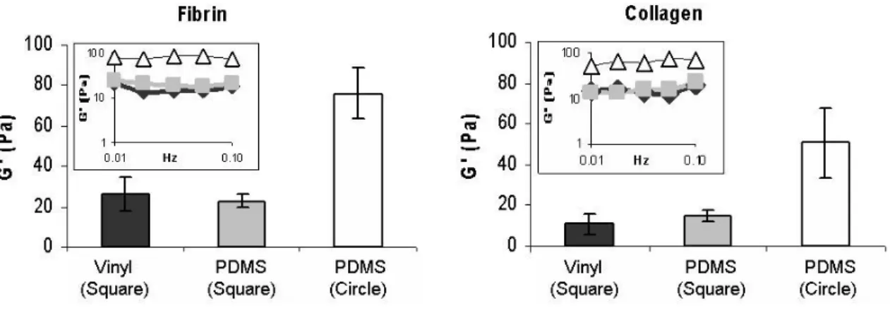
Rheology analysis of acellular hydrogels cast in different molds
The viscoelastic properties of acellular collagen and fibrin hydrogels were determined using rheology. Elastic modulus (G’) was chosen as a basis for comparison because it signifies hydrogel stiffness and has been used by other researchers for similar analysis 53,54. For both collagen and fibrin, viscoelastic parameters (elastic modulus, viscous modulus, and tan delta) remained constant over the frequency range between 0.01 Hz and 0.1 Hz (inset graphs, Figure 4). Although the hydrogels evaluated were distinctly different from one another in biochemical make-up, the values and trends of elastic moduli were similar for both collagen and fibrin (Figure 4).
For both hydrogel types, casting in the circular PDMS mold resulted in higher elastic moduli than square molds. Casting hydrogels in square geometries, using either disposable vinyl molds or PDMS molds resulted in similar elastic moduli, suggesting that the type of mold did not influence viscoelastic properties of hydrogels cast with similar geometries.
Figure 4. Viscoelastic properties of hydrogels cast in disposable vinyl or PDMS molds. Average
elastic modulus for fibrin and collagen constructs respectively over the frequency range 0.01 Hz to 0.1 Hz Insets. Data collected from rheometer showing that G’ remained constant over the frequency range tested.
22
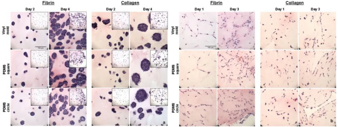
Histological Analysis
Cell morphology within the different hydrogel materials was assessed by hematoxylin and eosin staining of fixed samples. When cultured in either collagen or fibrin hydrogels mESCs grew in generally round clusters in both control disposable vinyl molds and PDMS molds (Figure 5a). The cells also seemed to maintain their undifferentiated morphology, suggesting that casting method had no effect on this aspect of cell phenotype.
In order to show that not only hydrogels consisting of one cell type can be successfully cast using this technique, constructs made of NIH 3T3 fibroblast cells were cultured and histological analysis was performed on days 1 and 3 of culture. 3T3 cells remained viable in both collagen and fibrin hydrogels (Figure 5b). Unlike mESCs which tended to form round cell clusters in the same hydrogel materials, fibroblasts tended to elongate and spread independently in the 3D environments, similar to behavior observed in control 2D experiments (not shown). These observations indicate that the cells appear
Figure 5.Histological analysis of mESCs and 3T3 fibroblasts in hydrogels. a. mESCs formed generally
round clusters that increased in size over time in fibrin and collagen hydrogels. mESC clusters were distributed throughouy the hydrogels (inset images). This behavior was independent of mold type. b.3T3
fibroblasts remained viable and elongate and spread throughout both fibrin and collagen hydrogels. These characteristics are indicative of fibroblasts cells and are independent of casting method. Purple – hematoxylin, Pink – eosin. (Scale = 100 µm).
to retain their normal phenotype in 3D hydrogels independent of the type of mold used for casting.
Viability Analysis
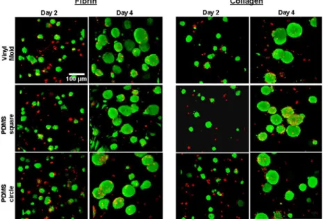
Results from Live/Dead viability assay indicated that cells remained mostly viable in naturally-derived hydrogels throughout the duration of the four-day study. Intense green fluorescence throughout the mESC clusters indicated that the cells were mostly viable not only along the periphery of the cell clusters, but also well into the center of the clusters (Figure 6). There were, however, few dead cells which could be seen toward the central core of the cell clusters and other isolated areas throughout the constructs. The 3D projections allows for viability analysis through the depth of the constructs (Z-stacks). This indicated that there were viable cells clusters throughout the constructs, as opposed to just in one arbitrary 2D plane.
Figure 7. Viability (Live/Dead) analysis. Confocal microscope images of fibrin and collagen hydrogels
were obtained on days 2 and 4 of culture. Green (calein AM) dye indicated viable (live) cell populations, while ref (ethidium homodimer) dye indicated dead cells. On day 2, mESCs were mostly alive with some
24
Live/Dead viability assay was also performed on cells seeded in the OPF hydrogels. Results showed that although mESCs were able to be encapsulated in the OPF hydrogel in the same manner as collagen and fibrin hydrogels, they did not remain viable over time when cultured in OPF (not shown). One potential conclusion is that OPF materials may lack the biological motifs to support the survival of mouse embryonic stem cells.
Rheology analysis of acellular hydrogels of various protein concentrations
Rheology experiments were conducted on acellular fibrin and collagen hydrogels immediately upon gelation (inset photographs, Figure 7) and the elastic moduli of the hydrogels were used as a basis for comparison for the purposes of this study.
During preliminary experiments, protein concentrations of the hydrogels were chosen based on published data and rates of gelation 53,54. Hence, 1 mg/mL collagen and 2 mg/mL fibrin hydrogels were chosen for initial studies based on the aforementioned criteria. However, rheology analysis indicated that these hydrogels had significantly different viscoelastic properties represented by elastic modulus or hydrogel stiffness
Figure 6.Rheology analysis of acellular hydrogels. Rheology was performed on hydrogels immediately
after gelation. Prior to testing, 2 mg/mL fibrin and 1 mg/mL collagen were used in preliminary studies. However, rheology tests indicated that these hydrogels had very different viscoelastic properties. In an attempt to normalize stiffness (elastic modulus, G’) a range of protein concentrations were evaluated and it was determined that 6 mg/mL fibrin and 3 mg/mL collagen have similar G’ values. Inset images were taken of hydrogels upon gelation (n = 3).
(Figure 7). In an attempt to normalize for the viscoelastic properties of the different hydrogels, a range of hydrogels composed of varying protein concentrations were tested. From the analysis, it was determined that 3 mg/mL collagen hydrogels and 6 mg/mL fibrin hydrogels had more similar viscoelastic properties (Figure 7). The elastic moduli of these hydrogels were all in the 70 – 80 Pa range and these values were independent of frequency. The elastic moduli values are comparable to previously reported data on similar gels 53,54.
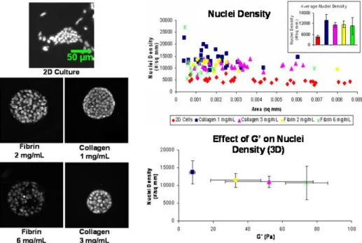
Nuclei Density Analysis
Nuclei density analysis was performed to indirectly measure the effects of the mechanical and biochemical properties of the hydrogels on cell cluster size (growth). The rationale behind this method is that the intrinsic mechanical properties of the hydrogels may influence cell behavior in the form of forces experienced by the cells. Similarly, the different biochemical properties of the hyrdrogels may have some effect on cell cluster growth and size. Microscopic images of nuclei of representative cell colonies from mESCs cultured on 2D substrates as well as cell clusters from each of the construct types were analyzed (Figure 8). It was observed that while mESCs grown on 2D substrates tended to spread and form irregular, loosely-packed colonies, mESCs in 3D hydrogels form uniform, spherical clusters as seen in both fibrin and collagen hydrogels, regardless of protein concentration and gel stiffness.
A scatter plot showing the distribution of nuclei density with respect to cell cluster area indicated that although cell cluster area (size) may differ for each independent cluster in the hydrogel, the nuclei density, or number of nuclei per unit area,
26
remains relatively constant for each cluster. However, nuclei densities for colonies grown on 2D substrates were significantly lower even though the areas of the colonies are similar to those in 3D. The inset bar graph in the top right corner of the scatter plot reiterates the average nuclei density was lower for colonies cultured in 2D, while they were higher and relatively constant for colonies in each construct type. It was determined from this analysis that for 3D mESC cultures, nuclei density is constant and independent of matrix type.
In order to correlate nuclei density with viscoelastic properties, average nuclei density for each construct type was plotted against the corresponding average elastic modulus (Bottom graph, Figure 8). Both 6 mg/mL fibrin and 3 mg/mL collagen
Figure 8. Nuclei density analysis. Fluorescent micrographs: Representative fluorescent microscopy
images of mESC nuclei on a 2D surface and in various fibrin and collagen matrices. Top graph: Scatter plot relating nuclei density and cell cluster area. (Inset bar graph showing average nuclei density for each group.) Nuclei density remained relatively constant and is independent of matrix type. However, nuclei densities of mESCs cultured in 2D were less than their 3D counterparts Bottom graph: This indirect measurement of the effects of hydrogel mechanical properties on nuclei density indicated that nuclei density is independent of hydrogel viscoelastic properties (Scale = 50 µm, n = 3).
hydrogels have similar viscoelastic properties, as shown previously in Figure 7. They also have overlapping nuclei densities. However, it was interesting to note that although the 2 mg/mL fibrin and 1 mg/mL collagen hydrogels all have significantly different elastic moduli, there are no significant differences in nuclei density. Hence, it was determined that nuclei density is independent of the viscoelastic properties of the hydrogels.
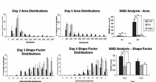
Morphometric Analysis
Morphologies of mESCs seeded onto 2D substrates were compared to those cultured in 3D hydrogels. While mESCs formed colonies or clusters on both 2D plates and in 3D hydrogels, mESC colonies grown in 2D tended to be less uniform in shape and size while mESCs seeded in collagen and fibrin hydrogels formed round, uniform clusters that grew larger with time (Figure 9). By day 2 of culture, small cell clusters formed on the 2D substrate as well as within both the construct types and consist of about two to twenty cells. The clusters were viable and are dispersed evenly throughout the plates and the constructs. By day 4, the sizes of the colonies clusters had increased considerably and at higher magnification, more detail could be seen with regard to cell cluster morphologies. Like cells grown on 2D, the cells in fibrin and collagen seem to maintain undifferentiated morphology.
28
Further, more quantitative morphometric analysis was performed using NIH Image J software to determine cell cluster area (size) and shape factor distributions. In both 2D and 3D environments, mESCs grew as colonies or clusters that increase significantly in size over time. However, clusters in both fibrin and collagen hydrogels were more uniform in size on days 2 and 4 of analysis, as indicated by narrow distributions, while colonies grown on 2D plates were less uniform, as indicated by the broad distribution (Figure 10). Although there was significant difference in size distribution between the 2D colonies and those in 3D hydrogels, there were no significant differences in size distribution between construct types, as indicated by mean absolute deviation (MAD) analysis (Top row, Figure 10). Shape factor, which is the ratio of the
Figure 9. Microscopic images of mESC clusters. Cells grown on 2D substrates and in hydrogels were
stained with hematoxylin and eosin (H & E) and images were taken on days 2 and 4 of culture. Colonies grown in 2D varied in size and shape, while clusters are generally round and uniform in appearance and increase in size with time. (Scale = 100 µm).
minor and major axes of an ellipse, was used as an indicator of uniformity. A shape factor of one (1) indicates that an object is circular, while a shape factor of zero (0) indicates the object is linear. From the distributions, it is evident that the clusters in the fibrin and collagen hydrogels were more uniform than colonies grown on 2D culture plates on both day 2 and day 4. Clusters in 3D acquired a mostly round morphology, whereas colonies on 2D varied in shape due to cell spreading. The significance of the distributions was indicated by MAD analysis (Bottom row, Figure 10).
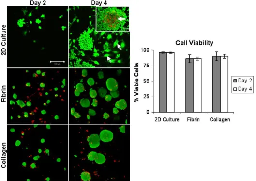
Cell Viability Analysis
In order to analyze cell viability throughout the duration of the study, both trypan-blue and Live/Dead analysis were conducted. Trypan-trypan-blue analysis of cells in 2D culture, as well as in both the collagen and fibrin hydrogels indicated that at least 90% of
Figure 10. Size and shape factor distributions of mESC colonies/clusters in both 2D and 3D cultures.
Area and shape factor distributions show that while cell colonies on 2D cultures have a broad size and shape distributions on both days of culture, clusters in both fibrin and collagen 3D hydrogel environments are more uniform. MAD analysis indicates significant differences in the size distribution between 2D colonies and 3D colonies. Shape factor distribution, an indicator of uniformity, indicated that the colonies in fibrin were less uniform than those in fibrin and collagen on both days.
Figure 10. Size and shape factor distributions of mESC colonies/clusters in both 2D and 3D cultures.
Area and shape factor distributions show that while cell colonies on 2D cultures have a broad size and shape distributions on both days of culture, clusters in both fibrin and collagen 3D hydrogel environments are more uniform. MAD analysis indicates significant differences in the size distribution between 2D colonies and 3D colonies (* p ≤ 0.05). Shape factor distribution, an indicator of uniformity, indicated that the colonies on 2D surfaces were less uniform than those in fibrin and collagen on both days (* p ≤ 0.05, n = 3).
30
was no significant difference in cell viability with respect to time, nor was there any significance in cell viability between the 2D method and the two 3D construct types.
Live/Dead cytotoxicity analysis using confocal microscopy was performed in order to visualize the spatial distribution of live and dead cells throughout the constructs as well as throughout the 2D culture plates. In 2D culture, cells were mostly viable. However, during this analysis it was observed that the cells were not uniform, as a number of cells of differentiated morphology could be seen throughout the sample (Figure 11, double-tailed arrows). Also, many of colonies grown on 2D seemed to grow extremely large in size and as a result, a necrotic core consisting of mostly dead cells could be observed in these colonies (Figure 11, solid arrow). In both fibrin and collagen
Figure 11. Cell viability as determined by trypan-blue analysis and live/dead staining. Graph: The
quantitative data shows that mESCs are mostly viable during the four-day study regardless of culture method. Image panel: Confocal microscopy images from qualitative live/dead analysis visualize cell viability. Cells in 2D formed colonies that were less homogeneous than cell clusters in 3D hydrogels. Some cells in 2D acquired differentiated morphologies that were not observed in 3D (double-tailed arrows). Also, cells in the center of large cell colonies in 2D culture were less viable than their 3D counterparts (solid arrow, inset) . (Green = live cells; Red = dead cells; Scale bar = 100 µm, n = 3).
hydrogels cells grew overtime into large clusters that were mostly viable (green), with fewer dead cells (red) seen on both days of culture. Unlike colonies grown in 2D, 3D cell clusters were more uniform or homogeneous and no obvious necrotic cores were observed in the centers of the clusters (Figure 11, micrographs).
Immunofluorescent staining of mESC clusters
ES cell colonies from 2D cultures as well as ES cell clusters in fibrin and collagen hydrogels were stained for expression of the pluripotent marker Oct-4 on days 2 and 4 of culture (Figure 12). 2D colonies stained positive for Oct-4. However, the differentiated-looking cells that contribute to the heterogeneity of the population in 2D cell culture did not seem to express this marker. Cell clusters from both hydrogels were Oct-4 positive on both days, indicating that cells retained their pluripotency while in 3-D environments.
Figure 12. Oct-4 protein expression. mESC colonies grown on 2D substrates, as well as in 3D hydrogels
32
Serial Passaging of mESCs in 2D and 3D
Serial passaging was performed in order to compare the effects of sub-culturing on mESCs morphology in both 2D and 3D. For the three subsequent passages, mESCs grew as colonies on both 2D substrates as well as within collagen hydrogels (Figure 13).
Colony growth is usually indicative of embryonic stem cell maintenance in vitro.
However, in 2D serial passaging, there were areas on the plates where cells did not form colonies; rather they tended to spread and took on a more differentiated phenotype. This was not observed in mESCs clusters obtained from collagen. The clusters obtained from fibrin, however, were not maintained over the course of the three passages. After the first
Figure 13. Serial passaging of cells from hydrogels in comparison to traditional 2D sub-culturing methods. Cells grown on both tissue culture plates (2D) and within collagen hydrogels form and retain
colonies for up to three consecutive passages. However, it can be noted from the photographs that there is a considerable number of grown on 2D that seem to spread and take on a differentiated morphology. This is seen less in the cells obtained from collagen. Although mESCs cultured in fibrin hydrogels seem to form colonies after one passage, they diminish in size by the second passage and seem to not form any by the third passage (images not shown).
passage from fibrin, it was observed that while the cells did form colonies, they were smaller in comparison to the colonies obtained from collagen or grown on 2D plates (not shown). Beyond the first passages, there was a decline in the number of colonies formed within the fibrin hydrogels, with none obtained by the third passage (not shown). A possible reason for this may be that papain, unlike collagenase digests not only the proteinaceous fibrin matrix, but may also digest cellular proteins, resulting in the decline in cells obtained from fibrin over time.
34
CHAPTER 4
DISCUSSION
PDMS Molds for flexible hydrogel casting
One objective of this research was to develop a more efficient method of casting cell-laden hydrogels for 3D culture applications. The results of this study indicate that a flexible mold made from the elastomer polydimethylsiloxane (PDMS) can be used to efficiently and economically cast multiple hydrogels of various shapes and sizes. In order to create PDMS molds of varying shapes, PDMS pre-polymer can be cast in a solid master fabricated from a machinable polymer, such as polycarbonate. In this case, the master was designed to contain polycarbonate pegs of varying shapes and sizes in an interchangeable format, enabling greater flexibility from a single master. Upon curing, the PDMS mold can be peeled off of the master, sterilized and sealed against a flat substrate before casting of different types of hydrogels. The geometric shape of the PDMS mold can affect mechanical properties of the hydrogels, such as viscoelasticity; however, the casting of the different hydrogels in PDMS molds did not appear to exert any detrimental effects on cell morphology, viability or phenotype.
PDMS can be fabricated into a mold to successfully cast various types of hydrogels
Although PDMS has been extensively used in photolithography and soft lithography applications in the creation of microgels 40,41,43-46,55,56, no significant data has been published about PDMS as a mold material for casting of macroscopic hydrogel constructs. Micromolding involves the manufacture of micron sized units of tissue that
have the potential to create hydrogel constructs of controlled microscale structure and architecture. This provides a potentially useful method for fabricating microstructures and possibly nanostructures 40,41. However, the creation of microgels using soft lithography and photolithography techniques may require various equipment and use of clean room facilities. In addition, the number of steps involved in these processes can become quite tedious and time-consuming 42,45.
For creation of PDMS molds used in this study, materials used for both the polycarbonate master and the PDMS mold itself are both relatively inexpensive and manufacturing of the molds requires no time-consuming steps. There has been reported use of silicone and glass raw materials for fabricating masters elsewhere 42; however, these materials can be brittle and can not be machined as easily as polycarbonate. Like the soft lithography and photolithography techniques involved with micromolding, we took advantage of the properties of PDMS for molding of our hydrogels. PDMS is durable, inexpensive, non-toxic and moldable 57. It can also be washed and sterilized easily with ethanol and exposure to UV light. This is important for the sterile culture of cells in hydrogels in vitro. The method discussed in this study, however, involves the
creation of hydrogel constructs that are orders of magnitude larger than microgels. Cells are encapsulated in the hydrogel prior to gelation and the liquid hydrogel is poured into the PDMS mold.
Also, in our study we showed that both naturally-derived (collagen and fibrin) and synthetic (OPF) hydrogels could be cast in PDMS molds. Although these diverse materials were observed to be compatible with the molding technique (i.e. collagen, fibrin and OPF formed hydrogels that maintained size and shape with time), only the
36
naturally-derived collagen and fibrin hydrogels sustained viable cells for the duration of the study. This observation was independent of the casting method. These preliminary results warrant further studies in order to determine the ability of OPF to potentially serve as 3D support for mouse embryonic stem cells.
Mold geometry affects viscoelastic properties of hydrogels
An unexpected result of this study was that PDMS mold shape had an affect on the elastic moduli of the hydrogels. In proteins, increasing gel stiffness (elastic modulus, G’) is known to be attributed to increasing protein concentration 58,59. However, for both collagen and fibrin samples, hydrogels of identical protein concentrations cast in circular molds had higher elastic moduli than those cast in the square PDMS mold and the control (square) disposable vinyl mold. One explanation for this may be increased tension in squares at the corners as opposed to the center. Because of this tension, hydrogels may gel differently at the four corners than in the center where the sample was taken for rheology. Circles, on the other hand, have no corners and thus hydrogels cast in this shape may gel more uniformly in all directions. A square has curvature gradients, with high curvature at the corners and zero curvature along the faces. The equilibrium shape for any object, when the surface tension is isotropic, is a circle 60. These properties give some plausibility to the observations that were made; a hydrogel in the shape of a square may indeed gel differently from that in the shape of the circle, even if the dimensions of the two geometries are similar. Another reason why there were differences seen may be due to how collagen and fibrin fibers may align when the respective hydrogels are cast in the varying shapes. It was important to note that regardless of these differences in
viscoelastic properties, observed cell behavior was similar in all hydrogels and independent of mold type. This suggests that the intrinsic mechanical properties of the hydrogels have no effect on mESC behavior during the time period in which the hydrogels were studied.
Differences in viscoelastic properties do not affect cell behavior
Finally, the results of these studies indicated that casting 3D hydrogels in PDMS molds do not compromise cell viability or phenotype. Viability analysis showed that mouse embryonic stem cells cultured in both naturally-derived collagen and fibrin hydrogels remain viable for the duration of the study. Although OPF hydrogels were successfully cast in the PDMS molds, the cells did not survive the environment. As stated previously, the reason why the cells did not survive were independent of hydrogel casting method; rather, mESCs may have died because of the synthetic nature of the hydrogel and its inability to provide the necessary biological cues essential for cell survival. In addition, studies were performed to show that hydrogels containing cells of possibly any cell type can be cast in PDMS molds. Histological results showed that 3T3 mouse fibroblast cells can survive in the hydrogels regardless of casting method. The 3T3 hydrogels maintained their in vitro phenotype; that is they spread and elongated
throughout the hydrogels. This again proves that cell characteristics are not compromised when constructs are set in the PDMS molds.
38
3D Hydrogel Matrices for mESC Maintenance
The effects of 3D hydrogel environments on mouse embryonic stem cell behavior were studied over the course of this thesis project. Undifferentiated mESCs were cultured either on 2D gelatin-coated substrates or in 3D fibrin or collagen hydrogel matrices. Cells were maintained in culture with media containing LIF and behavior was observed and analyzed during the first four days of culture. Although other groups have studied the effects of 3D hydrogel environments on embryonic stem cell behavior, many focus primarily on the differentiation of stem cells in 3D environments and, to our knowledge, none have compared their findings to standard 2D culture methods 4,8,19,20,22-25,27,52,61-63.
Benefit of simultaneous analysis of different hydrogels on ESC behavior
The study of the effects of 3D environments on embryonic stem cells is not a new venture in the field. Multiple groups have studied 3D culture for expansion and differentiation of mouse, human and rhesus monkey stem cells in both synthetic and naturally-derived hydrogel biomaterials 19,20,23,27. However, those groups who focused primarily on the expansion or self-renewal of stem cells only studied the effects of one specific matrix. One major advantage of simultaneously observing the effects of different 3D environments on ESC behavior is that one can make comparisons between biochemical, mechanical and physical properties of hydrogels and can adequately conclude whether or not these differences have effects on embryonic stem cell maintenance and/or differentiation. An attempt to simultaneously characterize the effects of various hydrogel matrices on ESC behavior was executed by this thesis project. As a
result, there is more insight on the influences of collagen, fibrin, and, to some extent, OPF 3D environments on ESC maintenance.
Morphologies of mESC colonies in 2D vs. 3D
Embryonic stem cells are characterized in culture by colony formation on 2D substrates. From these studies, it was observed that mESCs also form independent colonies when cultured in 3D hydrogel environments. The colonies formed in both collagen and fibrin hydrogels were more uniform in size and shape than those grown on 2D plates, which tended to spread over a broad range of sizes and shapes. These observations were quantified using image analysis techniques. It can be concluded from both the qualitative and quantitative analyses that 3D encapsulation of mESCs for in vitro
culture resulted in better control of cell colony/cluster size. This control may hinder the adverse effects of cell necrosis observed in the cores of very large colonies grown on 2D culture plates. 2D mESC cultures often consisted of cells with differentiated or undefined phenotypes, probably due to spontaneous differentiation of the cells. This was never observed in any of the 3D hydrogel cultures. From these observations, it seemed that 3D culture of embryonic stem cells maintained uniform embryonic stem cell morphology and inhibited spontaneous differentiation seen in standard 2D cultures.
Serial passaging of mESCs from 3D results in homogeneity of cells
Studies from this thesis project have shown that the culture of mESCs on 2D substrates results in heterogeneous cell populations consisting of colonies of mESCs and other cells that seem to take on differentiated morphologies or phenotypes with continued
40
subculturing. This heterogeneity was not observed when cells were serially passaged in the collagen matrices. mESCs retained their undifferentiated morphology for up to three subsequent passages when subcultured in collagen, and differentiated-looking cell populations were not observed.
Although it was observed that the mESCs subcultured in fibrin matrices did not survive over time, it is unfair for us to conclude at this time that this was due to the matrix type, as all our other studies prior to serial passaging showed the mESCs thrived in fibrin and expressed the Oct-4 marker. One possible reason why the cells obtained from fibrin constructs did not survive the serial passaging is because of the method in which the cells were obtained. Fibrin constructs have proven to be difficult to enzymatically digest in vitro. During these studies, various agents were used in an
attempt to digest the fibrin hydrogels, including plasminogen, trypsin, and collagenase. However, none of these were able to digest the fibrin hydrogels. A solution containing the enzyme papain was the only reagent that accomplished fibrin digestion. Unlike collagenase, which was successfully used to digest the collagen hydrogels without compromising cell viability, papain not only acts on the fibrin matrix, but possibly other proteins associated with the cells. This may be the main cause of the decline in cells from fibrin seen during the serial passaging studies.
CHAPTER 5
CONCLUSION
During the course of this thesis project an inexpensive, efficient method of casting multiple 3D hydrogels of varying shapes and sizes using PDMS molds was developed. Furthermore, the effects of different 3D hydrogel environments on mouse embryonic stem cell maintenance was assessed and compared to traditional 2D culture methods.
For the PDMS mold fabrication portion of the project, molds were fabricated by pouring pre-cured PDMS onto a patterned polycarbonate master consisting of removable pegs of varying shapes. The concept of creating a constant master with removable pegs of any imaginable shape is novel, as users would be able to control hydrogel size and geometry without having to create new masters for each specific shape. Casting of cells in hydrogels within PDMS molds does not appear to have any adverse effect on cell viability, morphology or phenotype and compared to hydrogels cast in control vinyl molds, cell constructs cast in PDMS molds exhibited similar cellular morphology. This method of simultaneously casting multiple hydrogels of both natural and synthetic origin is both efficient and inexpensive and has the potential to be used for any 3D cell culture applications.
From the analyses of hydrogel environments on embryonic stem cell behavior various conclusions can be made about mESCs cultured in 3D hydrogels. First, rheology, in addition to nuclei density analysis showed that nuclei density is unaffected by neither the viscoelastic properties of the hydrogels nor the hydrogel matrix composition. However, nuclei densities of cell clusters cultured in the 3D hydrogel environments were larger in comparison to mESC colonies cultured on 2D substrates. This is because while