Theodoros Papadopoulos
A Thesis in the Field of Machine Learning
Harokopio University
March 2019
ii Theodoros Papadopoulos (itp1611)
Supervisor: Iraklis Varlamis, Associate professor
Examining Committee: Iraklis Varlamis, Associate professor Dimitrios Michail, Associate professor Sophia Karagiorgou, Adjunct Lecturer
Master’s Thesis
iii Abstract
This thesis explores machine learning models for the analysis and classification of electroencephalographic (EEG) signals used in Brain-Computer Interface (BCI) systems. The goal is 1) to develop a system that allows users to control home-automation devices using their mind, and 2) to investigate whether it is possible to achieve this, using low-cost EEG equipment. The thesis includes both a theoretical and a practical part.
In the theoretical part, we overview the underlying principles of Brain-Computer Interface systems, as well as, different approaches for the interpretation and the classification of brain signals. We also discuss the emergent launch of low-cost EEG equipment on the market and its use beyond clinical research. We then dive into more technical details that involve signal processing and classification of EEG patterns using machine leaning. Purpose of the practical part is to create a brain-computer interface that will be able to control a smart home environment. As a first step, we investigate the generalizability of different classification methods, conducting a preliminary study on two public datasets of brain encephalographic data. The obtained accuracy level of classification on 9 different subjects was similar and, in some cases, superior to the reported state of the art.
Having achieved relatively good offline classification results during our study, we move on to the last part, designing and implementing an online BCI system using Python. Our system consists of three modules. The first module communicates with the MUSE (a low-cost EEG device) to acquire the EEG signals in real time, the second module process those signals using machine learning techniques and trains a learning model. The model is used by the third module, that takes control of cloud-based home automation devices. Experiments using the MUSE resulted in significantly lower classification results and revealed the limitations of the low-cost EEG signal acquisition device for online BCIs.
Keywords: Brain-Computer Interface, Machine Learning, Deep Learning, Signal Processing, Electroencephalography, Spatial Filtering, Python, Smart Home
iv Abstract (Greek) Η διπλωματική αυτή εργασία εξετάζει μοντέλα μηχανικής μάθησης για την ανάλυση και την κατηγοριοποίηση ηλεκτροεγκεφαλογραφικών σημάτων (EEG), που χρησιμοποιούνται σε συστήματα διεπαφής εγκεφάλου-υπολογιστή (BCI). Ο στόχος είναι 1) να αναπτυχθεί ένα σύστημα που να επιτρέπει στους χρήστες να ελέγχουν συσκευές «έξυπνου σπιτιού» χρησιμοποιώντας το μυαλό τους, και 2) να διερευνηθεί εάν αυτό μπορεί να επιτευχθεί χρησιμοποιώντας φθηνές εμπορικές συσκευές ηλεκτροεγκεφαλογραφίας. Η διπλωματική αυτή περιλαμβάνει τόσο θεωρητικό, όσο και πρακτικό μέρος. Στο θεωρητικό μέρος εξετάζουμε τις βασικές αρχές των συστημάτων διεπαφής εγκεφάλου-υπολογιστή, καθώς και διαφορετικές προσεγγίσεις για την ερμηνεία και την κατηγοριοποίηση των εγκεφαλικών σημάτων. Συζητάμε επίσης, την εμφάνιση καταναλωτικών φορητών συσκευών ηλεκτροεγκεφαλογραφίας, καθώς και την πιθανή χρήση τους πέρα από την κλινική έρευνα. Στη συνέχεια, αναλύουμε τεχνικές πτυχές που αφορούν την επεξεργασία και κατηγοριοποίηση σημάτων ηλεκτροεγκεφαλογραφίας. Σκοπός του πρακτικού μέρους είναι να αναπτυχθεί μια διεπαφή εγκεφάλου-υπολογιστή που θα είναι σε θέση να ελέγξει ένα περιβάλλον «έξυπνου σπιτιού». Για το σκοπό αυτό, αρχικά εξετάζουμε μεθόδους για την κατηγοριοποίηση των εγκεφαλικών νευρωνικών σημάτων και στη συνέχεια, διεξάγουμε μια προκαταρκτική συγκριτική μελέτη με την εφαρμογή τεχνικών προεπεξεργασίας και κατηγοριοποίησης σήματος σε δύο δημόσια σύνολα εγκεφαλογραφικών δεδομένων, επιτυγχάνοντας καλές αποδόσεις πρόβλεψης. Στο τελευταίο μέρος υλοποιούμε ένα σύστημα διεπαφής εγκεφάλου-υπολογιστή, που αποτελείται από τρεις μονάδες. Η πρώτη μονάδα επικοινωνεί με τη συσκευή ηλεκτροεγκεφαλογραφίας για την απόκτηση των σημάτων EEG σε πραγματικό χρόνο. Στη συνέχεια, η δεύτερη μονάδα επεξεργάζεται αυτά τα σήματα χρησιμοποιώντας τεχνικές μηχανικής μάθησης και εκπαιδεύει ένα μοντέλο που τροφοδοτείται στην τρίτη μονάδα, η οποία αναλαμβάνει τον έλεγχο των συσκευών αυτοματισμού του «έξυπνου σπιτιού». Λέξεις κλειδιά: Διεπαφή Εγκεφάλου-Υπολογιστή, Μηχανική Μάθηση, Βαθιά μάθηση, Επεξεργασία Σήματος, Ηλεκτροεγκεφαλογράφημα, Χωρικά Φίλτρα, Έξυπνο Σπίτι
v Any sufficiently advanced technology is indistinguishable from magic
vi Dedication
To my parents,
For the memories of a childhood in which I always return when I cannot stand a world faster than the speed of life.
For the long summers, the sea, the carefree and the love.
vii Acknowledgment
I would like to thank my supervisor, associate professor Iraklis Varlamis, for the continuous support throughout the development of my master thesis.
I would also like to thank all the staff of the secretariat because without them there would be no university.
My uncle, for buying me my first computer when I was 12 years old.
Eleni, who without her distractions, I would have traveled less and finished this thesis many months earlier, having nothing to do right now.
Table of contents
Abstract ... iii Abstract (Greek)... iv Dedication ... vi Acknowledgment ... vii Chapter 1. Introduction ...14 Brain-Computer Interfaces ...16 Thesis Objective...18 Thesis Structure ...19Chapter 2. Theoretical background and basιc concepts ...20
Introduction ...20
The Building blocks of the brain ...21
Methods for measuring brain functions ...22
Electroencephalography ...24
Chapter 3. EEG-based BCI Systems ...31
Introduction ...31
BCI Applications ...33
Major Components of a BCI system ...36
Types of BCI systems ...38
EEG devices ...40
EEG-based paradigms for BCI systems ...42
Chapter 4. Machine Learning ...45
9
Supervised Classification ...47
Supervised Classification Algorithms...50
Deep Learning and Artificial Neural Networks ...54
Chapter 5. The EEG Pipeline for MI-BCIs ...59
Introduction ...59 Data Recording ...61 Data Preprocessing...64 Feature Extraction ...68 Classification...72 Model Evaluation ...74
Chapter 6. Evaluation of Public Datasets ...76
Introduction ...76
Datasets ...77
Evaluation Design ...80
Analysis Results ...86
Discussion ...100
Chapter 7. Experiments with MUSE headband ...102
Introduction ...102
The MUSE Headband ...103
Software Tools & Frameworks ...104
Offline Analysis ...106
Experimental Online BCI using MUSE ...121
10 Summary ...124
Future Work ...126
11 List of Figures
Figure 1: A Brain-Computer Interface system... 16
Figure 2: A typical neuron ... 21
Figure 3: One of the first recordings of EEG signals made by Berger ... 24
Figure 4: Structure of a typical chemical synapse ... 25
Figure 5: Neural Oscillations ... 27
Figure 6: Brain-computer interfacing controlled Tetris game ... 34
Figure 7: A typical BCI pipeline ... 36
Figure 8: fMRI scans of actual and motor imagery ... 44
Figure 9: Layers of the MultiLayer Perceptron ... 55
Figure 10: The outline of a BCI training and evaluation pipeline ... 60
Figure 11: Four basic filter types ... 66
Figure 12: Filter Bank Common Spatial Patterns ... 71
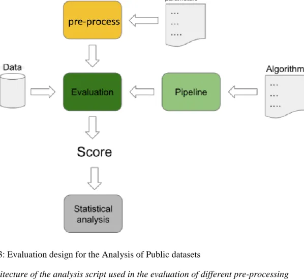
Figure 13: Evaluation design for the Analysis of Public datasets ... 80
Figure 14: BCI42B - optimization steps and results ... 86
Figure 15: BCI42B dataset – Cross-Subject performance (AUC) per subject using optimal pre-processing configuration and the best pipeline. ... 89
Figure 16: PhysionetMI optimization steps and results ... 90
Figure 17: Performance of pipelines with and without spectral filtering ... 93
Figure 18: PhysionetMI dataset – Cross validation score per subject using optimal pre-processing configuration and the best pipeline. ... 94
Figure 19: PhysionetMI - Average score per trial type for the best 5 pipelines. ... 96
12
Figure 21: PhysionetMI - The TPOT generated pipeline ... 98
Figure 22: The MUSE headband and the location of its electrodes ... 103
Figure 23: Data acquisition from MUSE (diagram) ... 107
Figure 24: Capturing data from the MUSE using Python... 107
Figure 25: MUSE – Motor Imagery data recording protocol ... 108
Figure 26: MUSE - The PSD of the Motor Imagery task ... 109
Figure 27: MUSE - The filtered PSD of Motor Imagery task ... 110
Figure 28: MUSE - The CSP components of the captured data ... 111
Figure 29: MUSE - Motor Imagery Classification results ... 113
Figure 30: The N170 ERP component ... 114
Figure 31: MUSE - ERP data recording protocol ... 115
Figure 32: MUSE - The PSD of the ERP task ... 116
Figure 33: MUSE - The filtered PSD of the ERP task ... 117
Figure 34: MUSE - averaged waveforms over each electrode for the N170 ... 118
Figure 35: MUSE - ERP Classification results ... 120
13 List of Tables
Table 1: Evaluation datasets ... 77
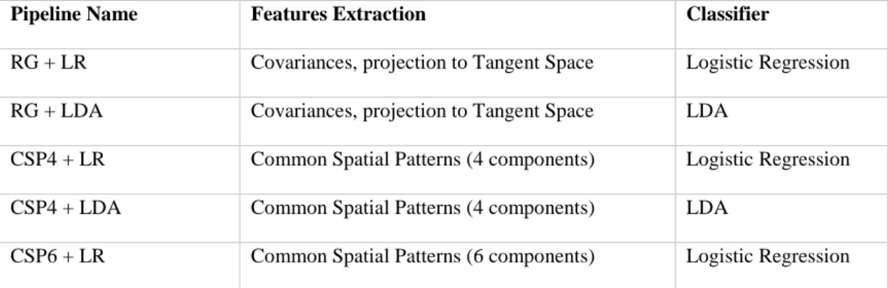
Table 2: The 25 pipelines that have been used to evaluate the 2 public datasets ... 82
Table 3: BCI42B – Cross-Subject classification performance for the top 5 pipelines using optimal pre-processing configuration. ... 87
Table 4: BCI42B – Cross-Subject mean classification score for all the pipelines over different pre-processing configuration ... 87
Table 5: BCI42B – Cross-Subject classification performance (AUROC) per subject ... 88
Table 6: BNCI - Cross-Subject vs Inter-Subject classification score (AUROC) ... 90
Table 7: PhysionetMI – Cross-Subject classification performance for the top 5 pipelines using optimal pre-processing configuration ... 91
Table 8: PhysionetMI - Cross validation score for all the pipelines ... 91
Table 9: PhysionetMI – mean score of features extraction ‘families’ ... 92
Table 10: PhysionetMI - Cross validation score per subject ... 94
Table 11: PhysionetMI - Hyper-parameters optimization for Logistic Regression Classifier ... 95
Table 12: PhysionetMI - results using a shallow convolutional neural network ... 99
Table 13: MUSE - Motor Imagery Classification results ... 113
14 Chapter 1.
Introduction
Over eons, biology via evolution and natural selection has solved the problem of processing, prioritizing and interpreting massive amount of noisy and highly redundant information, coming from a rapidly changing external environment. The brain, a constantly evolving network of billions of interconnecting neural cells, processes internal and external stimuli and makes decisions in a very short time. The input of the brain includes sensory cells (associated with seeing, hearing, tasting, touching etc.), motor/muscle cells, and even some cells within the brain. These cells pick up the stimulus provided and take it in for further processing. In fact, it's not only the brain but our entire nervous system that contributes towards the larger network, which is our consciousness.
During the last decades, humanity is developing artificial constructions, aiming to solve the exact same problems. Building a system that mirrored the simulations of the human brain, was the starting point of artificial neural networks (ANNs). But despite the recent technological advances that boosted the usage of artificial neural networks in multiple domains of human decision making and automation, and gave a thrive to artificial intelligence, there is still a huge gap in terms of abilities and effectiveness between the human brain and the artificial constructions of the man. There is also a big efficiency gap, which results in today’s supercomputers that run on megawatts to consume huge amount of power, while the human brain relies only on water and some calories in order to function.
In recent years, researches in the field of Artificial Intelligence are resuming the study of the models and the architecture of the human brain as a means for the development of AI, introducing structural and operational principles of the brain into the design of machine learning algorithms. Convolutional feedforward networks, which now dominate computer vision, take inspiration from the architecture of the primate visual hierarchy. Other research approaches are inspired by the recurrent connections in the human brain, and their key role for associative learning and pattern classification.
On the other side, neuroscientists use AI tools to learn more about the workings of the brain. In order to decode and understand the brain signals, ANNs and other machine
15 learning algorithms are being applied for pattern recognition, anomaly detection, and brain neural signals interpretation.
Recently, another research approach aiming to develop the technology for human enhancement by merging human with artificial intelligence is gaining attention. The ability to cooperatively use the two types of intelligence, by providing the appropriate interfaces that will allow human brain to communicate unobtrusively with a computer program and vise versa, will unlock the great potential of the human brain and will open new research opportunities in human-driven artificial intelligence. Brain-computer interfaces, combining knowledge and techniques both from neuroscience and Artificial Intelligence, are already used in medical applications but gradually, non-medical application paradigms also emerge. Translating thoughts into action, without acting physically or allowing direct brain-based communication between humans may seem to belong to the realm of science fiction today, and yet science fiction has a good track record for predicting inventions and developments we now take for granted.
Investments to the fields of human-driven intelligence and Brain-computer Interfaces (BCIs) are steadily increasing and more companies and research laboratories are entering the field. According to a market analysis conducted by OMR (OMR, 2018), global BCI market is expected to witness a significant growth rate at a CAGR of 22.8% during the forecasted period (2018-2023). Recently Elon Musk entered the industry, announcing a $27 million investment in Neuralink, a venture with the mission to develop a BCI that improves human communication in light of AI, and Facebook announced its plans for a Brain-Computer Interface technology, that would allow for more efficient digital communication between brains.
16 Brain-Computer Interfaces
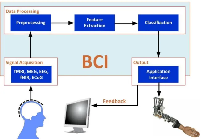
Human-computer interaction (HCI) is a research field focused on the interfaces between people (users) and computers. Humans interact with computers in many ways. Those include the prevalent graphical user interfaces (GUI) of today, the emerging voice user interfaces (VUI) which are used for speech recognition and synthesizing, as well as, other types of interaction mechanisms aiming to make human-computer interaction more natural, such as touch-screens, gesture-based interfaces, or eye-tracking driven interfaces. Over the last twenty years, neural engineering has emerged as a new field that merges neuroscience and information technology and has resulted in neurotechnology, able to link brain activity with man-made devices. A brain-computer interface (BCI) is a combination of hardware and software that permits the capture of cerebral activity associated with a user’s intent or emotions and the translation of this recorded activity to specific control signals. These control signals can be used to control an external device, such as a computer or an external smart-home device. In other words, a BCI allows direct communication between the brain and an external device.
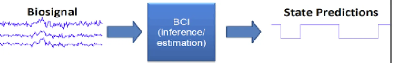
A modern definition defines BCI as a system which captures a biosignal measured from a person in real-time and predicts an abstract aspect of the person’s cognitive state.
Figure 1: A Brain-Computer Interface system
Image from BCILAB tutorials and presentations: ftp://sccn.ucsd.edu/pub/bcilab/
Brain-computer interfaces combine knowledge and techniques from neuroscience, signal processing, and machine learning domains.
The original development of BCI systems targeted severely paralyzed people and was focused on the ability to communicate with the external environment, without the
17 necessity of muscular control. Recently, the advent of the first commercial, inexpensive, dry-electrode devices, made it feasible to target new fields and apply this technology outside the laboratory. In the near future, it is possible to develop BCIs that will be able to facilitate hands-free applications, allowing the mind-controlling of machines.
Despite strong efforts, current BCIs are still facing several challenges that limit their usefulness for everyday applications. These challenges are related to increasing bit rates (Allison et al., 2012b), optimizing sensors, signal processing, and classification techniques, but also to the type of control signal and overall systems design. Many of these issues directly affect BCI performance, which is a field of active research and addressed at multiple levels of the BCI loop.
18 Thesis Objective
This thesis includes both a theoretical and a practical part.
The theoretical part has three main objectives. The first is to give an overview of the underlying functionality of BCI systems, focusing specifically to EEG systems. The second is to give an overview of the state of the art of machine learning algorithms, focusing on supervised classification algorithms and neural networks. The third is to evaluate different EEG pipelines, by conducting experiments on publicly available datasets of brain signals.
The practical part aims in designing and implementing an asynchronous active BCI system, using a commercially available EEG device able to control external home-automation devices. In our experiment, we employed Muse, a portable headband launched by InteraXon in 2014 to collect data of brain waves. We also developed an interface with the Amazon Echo (Alexa) device, in order to send control commands for home automation devices.
In order to implement the proposed BCI solution, we focus on the classification of EEG signals generated during motor imagery tasks (e.g. imagining the movement of a limb). This method has been reported as a reliable way for developing Motor Imagery (MI)-based BCI, which means BCIs that can recognize imagined movements, such as left or right-hand imagined movements. This is usually done by associating different limb movements to software commands.
In order to complete this thesis, we had to carry out several tasks such as:
• Review of the EEG decoding and BCI systems literature.
• Review of the Machine learning methodology.
• Develop, train, and evaluate different EEG decoding Pipelines in Python.
• Comparison of the obtained results with state-of-the art-results in literature. In addition, we performed an initial attempt to:
• Develop an EEG Pipeline using Deep Learning Neural Networks.
• Develop an active BCI system, able to control home-automation devices in a real-time scenario.
19 Thesis Structure
This thesis consists of 8 chapters structured as follows:
Chapter 2 provides a brief overview of the underlying functionality of BCI systems. This chapter begins by describing the building blocks of the human brain and the electrochemical interactions that produce its electrical signals. Furthermore, presents various methods for measuring brain activity, and discusses in more detail the functional principles of Electroencephalography.
Chapter 3 elaborates on EEG-based BCI systems and their fields of application. Different EEG-based activity patterns used in the development of BCI systems are presented and the emergence commercialization of EEG devices is discussed, along with a presentation of their basic characteristics and limitations. Finally, some examples of novel applications, which provide evidence for the promising potential of BCI technology both for medical and non-medical uses, are presented.
Chapter 4 gives an overview of the state-of-the-art machine learning algorithms, focusing on supervised learning used for EEG classification. Furthermore, it discusses deep learning approaches based on artificial neural networks.
Chapter 5 discusses in more depth the fundamental steps for the design of an EEG pipeline suitable for BCI systems, based on oscillatory EEG activity. It analyses the importance of proper data recording and data pre-processing, detailing the processing steps and providing an overview of the most important features extraction algorithms for EEG signals.
Chapter 6 presents our methodology for the evaluation of different EEG classification pipelines and discusses the results of experiments conducted on two publicly available datasets.
Chapter 7 presents the setup and methodology for the implemented BCI system and details on the hardware & software stack and the software architecture.
Chapter 8, summarizes and concludes the work presented on this thesis. This final chapter provides also ideas for future development of this work.
20 Chapter 2.
Theoretical background and basιc concepts
Introduction
A brain-computer interface (BCI) acquires brain signals and provides to the Human Neural System a new output that is not neuromuscular or hormonal. In order to design such a system, a thorough understanding of the complexities of the human brain, both in structure and in function, are required. It’s also important to investigate different approaches for acquiring the input signals and select the source that is more suitable for the use case of the designed BCI-system.
This chapter first presents the neurophysiological and anatomical basis of the function of the human brain. Next, we discuss various methods for capturing the brain activity and explain the advantages and drawbacks for each one. The chapter concludes with a brief introduction to Electroencephalography (EEG), the most commonly used method for brain signal acquisition and the one we are using for the development of our BCI system in this thesis.
21 The Building blocks of the brain
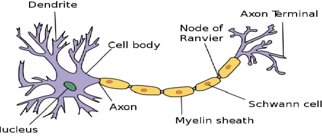
The human brain is the main organ of the human central nervous system (CNS). The brain consists of hundreds of thousands of cells, so-called neurons. Recent studies estimate that there are approximately 100 billion neurons in the human brain, which are all heavily interconnected.
A typical neuron consists of three parts: the soma or cell body, several dendrites, and the axon. The soma is usually compact; the axon and dendrites are filaments that extrude from it. The soma may give rise to numerous dendrites, but never to more than one axon. Dendrites typically branch profusely, getting thinner with each branching, and extending their farthest branches a few hundred micrometers from the soma.
Figure 2: A typical neuron
22 Methods for measuring brain functions
BCI must operate on observable effects of brain activity. Most BCI systems, operate on electrical effects of neural processes using Electroencephalography (EEG) and can detect large-scale neural dynamics (e.g. 10.000 neurons firing in near-synchrony). Other methods based on the metabolic changes of the brain during neural activity (e.g. PET, fMRI) can also be used, but are less suitable for BCI systems, because of their low temporal resolution.
From the methods for capturing signals that represent brain activity, Electroencephalography is the most commonly used and is the method used in this work. In order to give a more systematic and detailed description than the other methods, we dedicate the next section especially to it. Ιn the remainder of this section, we summarize the main methods for capturing brain activity.
Electroencephalography
Electroencephalography (EEG) is an electrophysiological monitoring method to record the electrical activity of the brain. It is typically noninvasive, with the electrodes placed along the scalp, although invasive electrodes are sometimes used such as in electrocorticography (ECoG). One important strength of EEG is its high temporal resolution, which is in the range of milliseconds (Nunez & Williamson, 1996). This highly temporal resolution of EEG allows the capture of underlying physiological changes of the cognitive processes, most of which, occur within tens of millisecond.
Magnetoencephalography
Magnetoencephalography (MEG) is a non-invasive technique that measures the magnetic fields generated by neural activity. Like EEG, MEG has excellent time resolution and is often considered to capture deeper neural activity much better than EEG. One important advantage of MEG over EEG is the fact that the measured signals are not distorted by the body. However, the signal strengths are extremely weak and specialized shielding is required to eliminate the magnetic interference of the external environment.
23 MEG scanners are large, stationary and expensive and they require heavy technical maintenance and training resources.
Functional Magnetic Resonance Imaging
Functional Magnetic Resonance Imaging (fMRI) measures the metabolic changes that take place in the active parts of the brain, and more specifically, the changes in blood flow associated with neural activity. Increased neural firing increases the need for oxygen, which is delivered by the neighboring blood vessels. Because the magnetic properties of oxygenated blood are different from those of non-oxygenated blood, the increase in oxygenated blood is measured by fMRI as a distortion of the magnetic field generated by protons. fMRI has excellent spatial resolution (a few millimeters), but poor time resolution (comparable to EEG). Apart from the low time resolution, the disadvantages of fMRI include the fact that it measures the brain function indirectly and that it requires large-scale non-portable and expensive equipment.
Positron emission tomography
Positron emission tomography (PET) is an invasive nuclear imaging technique based on gamma radiation of a decay, which is inserted into the body of the subject. Like fMRI, PET monitors the metabolic activity (for example, blood flow, oxygen, and glucose metabolism) of neurons, and therefore provides an indirect measure of neural activity. While PET has a high spatial resolution, in the order of few millimeters, it is lacking in time resolution. The temporal resolution of PET varies from minutes to hours (Nunez & Williamson, 1996). Its main drawbacks, beyond the low temporal resolution, is the injection of a radioactive substance into the bloodstream and that it requires a large-scale non-portable and expensive equipment.
24 Electroencephalography
Overview
Electroencephalography (EEG) measures electrical activity on the scalp. This activity is the sum of the post-synaptic potentials generated by thousands of neurons having the same radial orientation (typically pyramidal neuron cells) with respect to the scalp.
Electroencephalography has been discovered in 1929, by Hans Berger. In a set of experiments in which electrodes were placed on the scalp, Berger described the electroencephalogram (the plotting of the changes in voltage over time) and suggested that brain electrical currents reflected the functional status of the brain such as sleep, anesthesia, and epilepsy. Over the following decades, EGG has been widely used in both scientific and clinical applications (e.g. to evaluate neurological disorders or to monitoring depth of anesthesia during a surgery).
Figure 3: One of the first recordings of EEG signals made by Berger
By Hans Berger - Berger H. Über das Elektrenkephalogramm des Menchen. Archives für Psychiatrie. 1929; 87:527-70., Public Domain,
https://commons.wikimedia.org/w/index.php?curid=2900591
The electrical signals of the brain
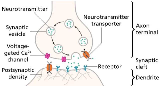
The neural cells exchange signals with each other via the synapses, the structures that permit a neuron to pass an electrical or chemical signal to another neuron or to the target effector cell. Synaptic signals from other neurons are received by the soma and dendrites; signals to other neurons are transmitted by the axon. A typical synapse, then, is a contact between the axon of one neuron and a dendrite or soma of another. Synapses can act as inhibitory or excitatory gateways, preventing or propagating impulses across neurons. The synaptic transmission is triggered by the release of neurotransmitters
25 (dopamine, epinephrine, acetylcholine, etc.), which causes a voltage change across the cell membrane.
Figure 4: Structure of a typical chemical synapse
Image from Wikipedia
There are two main types of electrical activity associated with neurons, action potentials, and postsynaptic potentials.
• Action potentials (AP) are discrete voltage spikes that travel from the beginning of the axon at the cell body to the axon terminals, where neurotransmitters are being released.
• Postsynaptic potentials (PSP) are the voltages that arise when the neurotransmitters bind to receptors on the membrane of the postsynaptic cell, causing ion channels to open or close and leading to a graded change in the potential across the cell membrane.
If the PSP reaches the threshold conduction level for the postsynaptic neuron, the neuron fires and an AP is initiated (Atwood and MacKay, 1989). Because neurons rarely
26 fire at precisely the same time, action potentials in different axons will typically cancel, and the only way to record the action potentials from a large number of neurons is to place the electrode near the cell bodies and to use a very high impedance electrode that is sensitive only to nearby neurons. Whereas the duration of an action potential is only about a millisecond, postsynaptic potentials typically last tens or even hundreds of milliseconds. In addition, postsynaptic potentials are largely confined to the dendrites and cell body and occur essentially instantaneously rather than traveling down the axon at a fixed rate. Under certain conditions, these factors allow postsynaptic potentials to summate rather than cancel, making it possible to record them at a great distance (i.e., at the surface of the scalp).
Not all electrical fields generated by the brain are strong enough to spread all the way through tissue, bone, and skull towards the scalp surface. Research indicates that it is primarily the synchronized activity of pyramidal neurons in cortical brain regions which can be measured from the outside. Pyramidal cells can be found in all cortical areas (occipital, temporal, parietal, frontal cortices), where they are always oriented perpendicular to the cortical surface. The cell body is heading away from the surface (towards the grey matter), while their dendrite is heading towards the surface (for more details see Luck, 2014 and Buzsáki et al., 2012). This unique orientation of the pyramid cells generates an electrical field with a very stable orientation. EEG activity, therefore, represents a sum of the activity of millions of neurons having a similar spatial orientation. By contrast, the electrical fields from cells in deeper brain structures (such as brain stem or cerebellum) that don’t have this specific orientation, are more likely to spread into various directions and cancel out instead of projecting in a stable way towards the scalp surface - even if hundreds of thousands of neurons in these deeper regions show synchronized activity.
Oscillatory activity
In general, EEG signals have a broad spectral content, but also expose oscillatory activity in specific frequency bands. Although the raw data obtained from EEG are formatted as a function of time, neural oscillations are usually visualized in terms of frequency using the Fourier transform and are measured in units of Hertz (Hz).
27 Figure 5: Neural Oscillations
Neural oscillations are visualized in terms of frequency.
From a diagnostic perspective, neural oscillations can be used as indicators of specific neurological phenomena such as sleep state, state of consciousness, perception and information processing, memory, abnormal neural function, such as epilepsy, and Parkinson’s. There are 5 frequency bands categorizing the EEG signals based on their frequencies, named after Greek letters:
• delta (1–4 Hz), which are high amplitude waves mostly linked with slow-wave sleep.
• theta (4–8 Hz), which are mostly observed when the subject is in a meditative, daydreaming or in early stages of sleep.
• alpha (7.5–12.5 Hz), which are associated with cognitive functions such as relaxation and disengagement and are therefore the most commonly used in mood/meditation applications.
• beta (13–30 Hz), which are commonly associated with concentration or attention or more generally when. a subject is actively engaged in an activity.
• gamma (> 30 Hz), which are thought to play a crucial role in information processing, concentration, and learning.
Electrode arrays and placement
EEG recordings are performed using electrode arrays, comprising of a varying number of electrodes, depending on the scope of the experiment. Conventional EEG
28 systems use a conductive gel or paste for the electrodes, but many cheaper commercial systems use dry electrodes, reducing the preparation time, and making EEG more suitable for generic use.
For faster application, and consistent results between different recordings, EEG electrodes are mounted in elastic caps, meshes or rigid grids, ensuring that the data can be collected from identical scalp positions across sessions or respondents. Electrode locations and names are specified by an international system, named ‘10–20 system’. This system ensures that the naming of electrodes is consistent across laboratories and experiments. In the 10-20 system, electrode names begin with one or two letters indicating the general brain region where the electrode is placed (F = frontal, Fp = frontopolar; C = central; P = parietal; O = occipital; T = temporal). Each electrode name ends with a number or letter indicating the distance to the midline. Odd numbers are used in the left hemisphere, even numbers in the right hemisphere. Larger numbers indicate greater distances from the midline, while electrodes placed at the midline are labeled with a “z” (for zero).
Because the EEG voltage reflects the potential (or current) between two sites, different montages can be used for the electrodes.
• In bipolar (or sequential) montage the potential difference between a pair of electrodes is measured.
• In unipolar (or referential) montage the potential of each electrode is compared to a common neutral (reference) electrode. Typical reference positions are the tip of the nose, the cheek, and the right and left mastoids (the bony part behind left/right ears).
• In an average referential montage, the average of all electrodes is used as a reference point.
Advantages of EEG
EEG has several benefits compared to other imaging techniques. The most central benefit of EEG is its excellent time resolution, that is, it can take hundreds to thousands of snapshots of electrical activity across multiple sensors within a single second. This makes
29 EEG an ideal technology to study the precise time course of cognitive and emotional dynamics most of which, occur within tens of millisecond. The second reason that justifies EEG as such an advantageous technique for the study of neurocognitive processes is that it allows the direct measure of neural activity. EEG signals directly reflect biophysical phenomena occurring on neuron populations. This is a clear advantage over other methods such as fMRI that do not directly measure neural activity but introduce an extra relationship between what is measured (changes in blood flow in the case of fMRI) and the actual neural activity. Finally, EEG is non-invasive, and the required equipment is relatively cheap, portable and relatively easy to operate.
Disadvantages of EEG
The main disadvantage of EEG is its poor spatial resolution. Neural activity is conducted through the brain volume to the scalp and electrodes by volume conduction (the transmission of electric or magnetic fields from an electric primary current source through biological tissue towards measurement sensors). The concept of volume conduction carries important implications for surface EEG measurements because it means that (a) currents are not restricted to the immediate neighborhood of the source, and (b) the electrical activity measured between electrodes has more to do with their orientation to the actual generator than with the proximity of the electrodes to the generator. Because the skull is a poor conductor current tends to “splash off of it” and each electrode receives signals from millions of neurons, reducing any potential spatial localization. This is exacerbated by the fact that the conductivities of the head tissues, varies across individuals and within the same individual due to variations in age, disease state, and environmental factors. The inference of the location of the current sources from electrode voltage measurements on the scalp is known as the EEG inverse problem and is comparable to reconstructing an object from its shadow: only generic features (the shape) are uniquely determined, others must be deduced on the ground of additional information.
EEG is also very sensitive to subject movement and external noise. Electrodes used in EEG recording do not discriminate the electrical signals they receive. The recorded activity which is not of cerebral origin is termed artifact. Artifacts are noncerebral signals that often contaminate the recordings in both temporal and spectral domains within a wide
30 frequency band. The internal source of artifacts may be due to physiological activities of the subject (e.g., eyes movement, electrocardiographic activity, sweat or muscle artifacts) or its movement. External sources of artifacts are environmental interferences such as electrical noise from mains interference, bad contacts between electrode and skin, or interferences from recording equipment and cable movement.
31 Chapter 3.
EEG-based BCI Systems
Introduction
A brain-computer interface is a direct communication pathway between a brain and an external device. There are four criteria that need to be met, for a system to function as a BCI system:
1. Direct: The system must rely on activity recorded directly from the brain. 2. Intentional control: At least one recordable brain signal, which can be
intentionally modulated, must provide input to the BCI (electrical potentials, magnetic fields or hemodynamic changes).
3. Real-time processing: signal processing must occur online and yield a communication or control signal.
4. Feedback: The user must obtain feedback about the success or failure of his/her efforts to communicate or control.
Although that, as we have already mentioned, there are various types of bio-signals that can be used to measure brain activity and act as an input for a BCI. Ιn the rest of this thesis, we will discuss and describe BCI systems that are based on EEG signals in order to function. The main advantage of using EEG as an input signal for a BCI is that EEG (using dry electrodes) is the easiest and least invasive method.
The most difficult task in designing real-time BCI systems is interpreting the recorded data. First, EEG data are a superposition of the brain signals of interest, with a plethora of other signals from other brain regions, muscles, and from non-biological artifacts. Second, brain activity exhibits a huge variability across subjects. Since neural responses are different across subjects even for the same stimulus, almost all EEG-based brain-computer interfaces need some labeled, subject-specific, data to calibrate a new subject. As a result, a major challenge in developing high-performance and user-friendly BCIs is to cope with such individual differences, so that the calibration can be reduced or even completely eliminated (He, Wu, & Member, 2018). In order to overcome these
32 problems, state of the art BCI systems need to use continuous adaptive signal processing and machine learning algorithms, to extract meaningful information from brain signals. Until recently, the requisite technology to adapt and analyze the EEG in real-time, either did not exist or was extremely expensive. The improvements in processing power along with the developments in machine learning algorithms allow the introduction of small portable BCI systems suitable for every-day use.
33 BCI Applications
The ability to control the world around us, using only our mind, has been a feature of some of the best science fiction stories. Still, a lot of research is taking place around the world for advanced brainwave-based digital interfaces and in recent years remarkable strides have been achieved toward this goal. Brain-computer interfaces are already been applied in various fields of research.
One of the main areas of applied research concerns the use of BCIs in medical applications. Such systems are mainly targeting people with severe disabilities such as tetraplegia, locked-in syndrome etc. The aim of these BCIs is to either restore movement of individuals with paralysis or provide some special devices to assist them (POSTELNICU, TALABĂ, & M.I, 2010). In other cases, the research targets the restoring of communication with the external environment. The brain-controlled speller is one of the most famous BCI paradigms as it allows communication disabled people to spell letters or words. MindDesktop (Ossmy et al., 2017) is a medical purpose BCI allowing people with severe disabilities to operate any Windows-based operating system. Another area of medical BCIs is focusing on motor neuroprosthetics. For example, artificial arms or legs are being controlled by a portable BCI system, thus allowing individuals with a paralyzed or missing body part to move or grasp.
Gaming is among the most promising applications for BCI systems mainly because the number of potential users in BCI games is very high. BCI can be used either as a primary controller of a game or as an extra channel for special actions. For example, the user may imagine body part movement or concentrate on a certain object to generate brain signals that reveal the user's intention. Studies have shown promise in achieving 2D or 3D navigation, including moving a computer cursor or walking in a virtual world. Several studies have demonstrated the use of BCI for controlling popular games such as Tetris (Pires, Torres, Casaleiro, Nunes, & Castelo-Branco, 2011) or World of Warcraft (Van De Laar, Gurkok, Plass-Oude Bos, Poel, & Nijholt, 2013). An example of use in Virtual environments was demonstrated by the He’s research group in studies that showed that human subjects could fly a virtual helicopter in a 3D virtual world using EEG signals recorded from the scalp (Doud, Lucas, Pisansky, & He, 2011). A different approach
34 promotes the use of BCI for mental state monitoring during gaming, in order to make adaptive and dynamic games.
Figure 6: Brain-computer interfacing controlled Tetris game
A volunteer is playing a BCI-controlled version of the Tetris computer game. He uses left- and right-hand motor imagery to move the falling pieces horizontally, mental rotation to rotate it clockwise and foot motor imagery to let it drop.
Another active field or research concerns BCIs that allows for continues mental state monitoring. There is a wide range of everyday application that can be enhanced by adding passive mental state monitoring. When aiming to optimize the design of user interfaces or, more generally, of a workflow, the mental state of a user during task execution can provide valuable information. This information can be exploited for the improvement of industrial production environments, the user interface of cars and for many other applications. Examples of these mental states are the levels of arousal, fatigue, emotion, workload or other variables the brain activity correlates of which are (at least partially) accessible by measurement (Blankertz et al., 2010).
More futuristic research proposals include the ability of “telepathic” communication between people, or the use of brain waves for user authentication. Published studies have demonstrated direct transmission of brain activity between two
35 humans (Rao et al., 2014), between two animals, and even between human and rat (Yoo, Kim, Filandrianos, Taghados, & Park, 2013). DARPA, the Pentagon's technology research division, is currently working on an initiative called "Silent Talk," which would let soldiers on secret missions communicate with their thoughts alone. Various studies have proposed the use of brain signals as a two-factor, changeable, authentication method resistant to shoulder-surfing.
36 Major Components of a BCI system
Figure 7: A typical BCI pipeline
A typical BCI pipeline. Image from https://www.mne-cpp.org/wp-content/uploads/2015/08/BCI_processing_pipeline-1024x717.jpg
From the technical point of view, a BCI system consists of at least 5 components:
• Signal Acquisition: Although there are various methods to acquire brain signals, the one most suitable for real-time applications is Electroencephalography (EEG). With EEG, brain signals are recorded on the scalp of the users using electrodes. This preferably happens in a non-invasive manner, using external scalp electrodes or even better, dry EEG electrodes making direct contact with the skin without requiring the application of electrode gel.
37
• Signal Preprocessing: The measured signals are quite weak. Even worse, existing electric network current or muscular movement and eye-blinks can greatly influence them. Therefore, complex algorithms are applied to a) filter and b) enhance the raw signal quality and increase the signal to noise ratio (SNR).
• Features Extraction: Signal processing techniques are applied to extract features that can be used later by a machine learning algorithm. Depending on the task, different methods can be used, e.g. spatial information to identify signal on certain areas of the brain, spectral information that represents frequency bands of interest, or temporal information representing the change of signal over time.
• Machine Learning: The extracted features are analyzed with modern machine learning methods to discriminate between different classes of commands. Most methods conform to a common framework of a training, evaluation and prediction function. Typical classifiers for BCI applications are Linear Discrimination Analysis, Support Vector Machines and various types of Neural Networks.
• Control and Feedback: the predicted target from the previous step is then used to control an external device or application. For most BCI systems, the device is a computer screen and the output is the selection of certain targets. Advanced applications include the controlling of external devices such as prosthetic, robotic or smart-home devices.
38 Types of BCI systems
According to Zander et al (2009) BCI systems can be classified in one of the following types:
• Active BCIs: which derives its outputs from brain activity which is directly consciously controlled by the user, independently from external events, for controlling, for instance, an application.
• Reactive BCIs: which derives its outputs from brain activity arising in reaction to external stimulation, which is indirectly modulated by the user for controlling an application.
• Passive BCIs: which derives its outputs from arbitrary brain activity without the purpose of voluntary control, for enriching a human-computer interaction with implicit information.
A BCI usually includes a piece of software responsible for signal analysis and pattern recognition to achieve the translation of raw brain activity to control signals. Depending on whether this analysis happens in real-time or not, BCI systems can further be split into synchronous, which only analyzes the signals during pre-defined time windows, and asynchronous, which always looks at the signals seeing if there is a command pattern present. An asynchronous BCI is always active and besides reacting to the predetermined mental tasks that control the system, is also able to identify (and ignore) rest states. Synchronous is much easier to implement, but asynchronous offers much more seamless interaction. (Heyden, 2016).
Another distinction is that between one-directional and two-directional BCI systems. One-directional refers to BCIs that only “read” brain activity and affect the external environment of the user. In two-directional (or closed-loop) BCIs, the system can also affect the brain, e.g., by electric stimulation of the brain’s reward center. The development of closed-loop systems raises intriguing ethical issues and requires additional research and clear governing policies.
Recently, novel approaches have been proposed for BCIs that combine various, possibly diverse, types of signals. These are called hybrid BCIs. Input signals used in hybrid BCIs can either be two different types of brain imaging methods (e.g., EEG and
39 functional Near-Infrared Spectroscopy, fNIRS), or the combination of one brain signal with another physiological signal (e.g., heart rate, eye tracker etc.).
40 EEG devices
Encephalographic measurement devices are consisting of 1) electrodes, 2) amplifiers with or without filters, 3) an A/D converter and 4) a recording device. Electrodes read the signal from the head surface, amplifiers bring the microvolt signals into the range where they can be digitalized accurately, converter changes signals from analog to digital form, and a storage device (or more commonly a personal computer) stores obtained data.
Until recently, the collection of electroencephalographic (EEG) data used to be associated with expensive (>$25,000 USD), large electrode array systems. However, in the past ten years, there has been a rapid increase in the availability and number of “low-cost” EEG systems available to researchers (Krigolson, Williams, & Colino, 2017). At the lower end of the cost, there are devices costing less than 1,000 euros, although the research potential of such devices is ultimately limited by the few numbers of channels. A wide range of options, with up to 64 channels, is available at the middle price range, with devices costing between 1,000 and 25,000.
Apart from the cost, for EEG technology to become widely-adopted and more user-friendly, EEG systems must move from the bulky systems used in clinical settings to sleek, convenient compact systems. At the hardware level, there are currently three types of approaches that aim to improve EEG-based BCI portability and usability. First, the transition from the bulky EEG devices that consists of different components for the recording and the amplification of the EEG signal, to more compact, wearable devices with embedded parts. Second the replacement of gel or water-based EEG electrodes with dry electrodes that do not require the application of conductive gel. And third, the wireless connection between the amplifier and the recording device, which eliminates the need for cables and allows their use in a wide range of new settings. While such systems are already offered for end-users and consumers and can be used for BCI applications, the signal quality of such EEG hardware is not yet comparable to professional devices and their use is not yet recommended for serious applications.
41 Commercial EEG headsets
There are a lot of “low-cost” EEG headsets out in the market. These EEG headsets are often mentioned in different terms such as “Mind Controller”, “Brainwave Controller”, “EEG headbands”, etc. Most of these devices have the lowest number of electrodes and low sampling rates.
OpenBCI is an open-source brain-computer interface device, created after a successful Kickstarter campaign in 2013. Today the company behind OpenBCI offers various versions of the device, allowing to choose between 4 and 16 channels and different boards with or without Bluetooth connectivity. The cost for an 8 channels OpenBCI board along with a headset is around 1,000 euros.
Emotiv offers 5 and 14 channel solutions. The internal sampling rate of the device is 2048 Hz, but the data is then down-sampled to 128 Hz before becoming available to the user. Emotiv’s devices are also wireless, giving the possibility of more free movement to the user. The cost for the 14 channels device is around 700 euros. Currently, access to the raw EEG data of the Emotiv comes with an additional cost-per-usage, which can increase significantly the total cost of use.
The lower end of the consumer available devices includes devices with the lowest number of electrodes. Companies like NeuroSky and Muse offer neurofeedback solutions that are targeting meditation and monitoring uses, with limited research potential. Neurosky MindWave is at the lower end of usability of low-cost consumer EEG devices with only one electrode (placed at Fp1) and a 128Hz sampling rate. Muse offers a device with 4 channels and at a prize around 300 euros.
42 EEG-based paradigms for BCI systems
For the development of a BCI system, besides having the hardware to capture brain signals, it is also important to know which parts of the brain are responsible for certain mental processes and how the signals in the brain behave under these mental processes. Using certain mental processes to activate brain regions to control a device or a computer program is called paradigms (Strategien et al., n.d.).
There are two main paradigms that are used as control signals for BCIs. Event-Related Potentials and Sensorimotor Rhythm Activation. The former uses brain activity generated in response to specific visual or auditory stimuli while the latter uses activity spontaneously generated by the user or by the user’s mental state.
Event-Related Potentials
Event-Related Potentials (ERPs), are characterized by a phase-locked, domain waveform that appears in response to stimulation. Typical features are time-domain signal, generally averaged across several repetitions of the stimulation in order to increase the signal to noise ratio.
ERPs are perhaps the most studied type of activity in EEG and has been used in cognitive science, cognitive psychology, and psychophysiological research. In ERP studies, the EEG is recorded from participants, as experimental stimuli are presented. In this context, the cognitive “events” of interest may include a particular class of stimulus, the absence of an expected stimulus (omitted stimulus paradigm), a correct or incorrect response, among many other possibilities, as long as a distinct time point for ERP time - locking can be defined. Segments of the EEG, each encompassing a fixed period of time before and/or after each instance of an event, is then averaged to yield an average ERP. Averaging over multiple trials eliminates unrelated background activity that is random with respect to the stimulus and thus averages to zero, given enough trials. The resulting ERP reveals brain activity that is related, and synchronized in time and phase, to the presented stimulus (Faust, 2012). The averaged ERP waveform consists of a series of positive and negative voltage deflections, which are called components. Years of research have helped
43 to link different components to specific cognitive processes, making ERPs a powerful technique for examining the nature of cognitive and neural processes.
There are many advantages of event-related potentials for the study and analysis of complex neuro-cognitive processes. ERPs are simple and fast to compute, mainly because ERPSs generally involve significantly fewer data. They have a highly temporal resolution, providing continues measure of processing, including both ex-stimulus and post-stimulus activity. This highly temporal resolutions of ERPs allow the measurement of brain activity with the precision of millisecond, which is particularly important if we consider that many aspects of attention and perception appear to operate on a scale of tens of milliseconds. Besides, ERP-based BCIs have the advantage that usually do not require subject-specific training sessions, making it suitable for generic-use (no learning sessions are required in order to adapt the system to new users). The main drawback regarding the use of ERPs for the development of BCI systems is their dependency on the external stimuli, which prohibits the development of voluntary controlling interfaces suitable for active BCIs.
Sensorimotor Rhythm
A different control signal for active BCIs is the sensorimotor rhythm (SMR) that is based on the neural oscillations. These oscillations appear naturally in ongoing EEG activity and are representative of a wide range of different cognitive states (e.g. sleep stage, meditation, etc.) or can be induced by a specific task, for example, a hand movement or the performance of mental calculus. Sensorimotor Rhythm Activation is characterized by a change in signal power in specific frequency bands. The SMR modulation manifests as a decrease in the alpha (also known as mu rhythm) and beta frequency bands accompanied by an increase in the gamma frequency band. Typical features are extracted using fast Fourier transform based algorithms, or more simply variance/covariance of the signal after frequential filtering.
BCI systems based on oscillatory activity are functioning by detecting patterns in mental states which lead to changes in the oscillatory components of EEG signals, i.e., that lead to change in the power of EEG signals in some frequency bands. The increase of EEG signal power in a given frequency band is called an Event-Related Synchronization (ERS), whereas a decrease of EEG signal power is called an Event-Related Desynchronization
44 (ERD). Unlike ERPs, ERD/ERS are not phase locked to a stimulus presentation and, therefore, cannot be identified by averaging the EEG amplitudes, instead, band-power is measured in frequency bands of interest, localized in some specific brain areas. As such, they naturally need to exploit both the spatial and spectral information. The original EEG signals are converted to time-frequency signals by applying a function of short time Fourier transforms (STFTs) and are localized using either channel selection or spatial filtering techniques.
Imagining a movement or performing an action mentally is known as Motor Imagery (MI). Studies based on fMRI revealed that imagery and executed movements had similar activation patterns (Lotze et al., 1999). Motor Imagery is very commonly used in BCI systems because it allows the development of asynchronous active BCIs. The main advantage of MI-based BCIs is that it allows the user to control the system spontaneously, by imaging the execution of a movement. On the other hand, they suffer from high variability across and within subjects and therefore require excessive user training and long calibration times in order to achieve reasonable performance.
45 Chapter 4.
Machine Learning
Introduction
Machine learning (ML) is a field of artificial intelligence that uses statistical techniques in order to give information systems the ability to "learn" from data, without being explicitly programmed. The concept of learning here means the progressively improving of their performance on certain future tasks. We say that learning a general function or rule from specific input-output pairs is called inductive learning (Russell, Norvig, & Davis, 2010). Predictive modeling is the problem of developing a model using historical data, which will be able to make predictions on new (unseen) data. Typically, a model includes a machine learning algorithm that learns a function from a training dataset in order to make predictions.
According to the feedback that is available to learn from, machine learning algorithms can be grouped into three main types:
• Unsupervised learning: where a learning agent discovers patterns in the input data without any explicit feedback. The most common unsupervised learning task is clustering: the detection of potentially useful cluster of input instances.
• Reinforcement learning: Where an agent “learns” from a series of awards or penalties in a series of interactions with the environment. In each step of the learning process, the agent is informed about the state of the environment and decides which action to perform. For every action, the agent gets feedback from the environment in the form of numerical rewards, that is positive if the action was correct, or negative when the action was incorrect. This procedure helps the agent to form a policy for associating the correct actions to states. To maximize the long-term reward, the agent must explore its environment and update its policy to incorporate the discovered knowledge into the policy.
• Supervised learning: Where the learning agent has access to training data consisting of examples of input-output pairs and learns a function that maps from input to output. A supervised learning algorithm analyzes the training data and
46 produces an inferred function, which can be used for mapping new unseen examples. The discovered function is represented in a structure referred to as a model. There are two major categories of supervised learning, regression, and classification. Regression models are based on the analysis of relationships between variables and trends in order to make predictions about a continuous target variable, while the task of classification models is to assign discrete class labels to observations. In supervised classification, the class labels in the dataset, which is used to build the classification model, are known.
In the following sections, we will describe in more details the theory and the main algorithms for supervised classification, as it is the learning model that has been used to develop the BCI System for this thesis.
47 Supervised Classification
More formally, the task of a supervised classification learning algorithm is this: Given a training set of N tuples of examples – class variable pairs
(𝑥1, 𝑦1), (𝑥2, 𝑦2), … (𝑥𝑁, 𝑦𝑁) ,
where each 𝑦𝑖 was generated by an unknown function 𝑦 = 𝑓(𝑥)
discover a function ℎ that approximates the true function 𝑓.
An example is a collection of features that have been measured from some object, fact or event. We typically represent an example as a vector 𝑥 ∈ 𝑅𝑛 where each entry 𝑥𝑖 of the vector is called a feature. A training set is a collection of tuples of examples, each one associated with a label or class. Classification is made by choosing the best possible function h that assigns each feature vector 𝑥 to a class 𝑦𝑖 based on the samples in the training set. The function h is called a hypothesis. Learning is a search through the space of possible hypothesis for one that will perform well on unobserved data. The simplest kind of classification problem is binary classification, when there are only two values for the output class.
Performance Evaluation for Supervised Classification
To evaluate the performance of a classification algorithm, we usually measure the accuracy of the model, which is defined as the proportion of examples for which the model produces the correct output. The accuracy measured on the training dataset is called training accuracy. The inaccuracy of predicted output values is termed training error or the training error rate.
Usually, we are interested in how accurately a classification algorithm can predict outcome values for previously unseen data. Measurements of accuracy or error on the training data do not provide much information about predictive ability on new data. We, therefore, evaluate the algorithm on a previously unseen dataset, called the test dataset. That accuracy is often called testing accuracy or simply accuracy. The error rate on the test dataset is called test error or generalization error. We say that an algorithm generalizes well if it correctly predicts the value y for previously unobserved examples.
48 In the end, the factors determining how well an algorithm will perform are its ability to a) make the training error as small as possible and b) make the gap between training and test error as small as possible. These two goals correspond to the two central challenges in machine learning: underfitting and overfitting. Underfitting occurs when the model is not able to obtain a sufficiently low training error, indicating that the learning algorithm cannot adequately capture the underlying structure of the data. Overfitting occurs when the gap between training and test error is too large, indicating that the learning function corresponds too closely or exactly to the training set, and therefore fails to fit additional data or predict future observations reliably.
In some cases, there are parameters in the classifier that must be tuned. These are usually chosen by splitting the data into three instead of two sets: Training, testing, and validation. The classifier is trained on the training set for different parameter configurations (also called hyperparameters) and evaluated on the validation sets. The one that performs best is then chosen and tested on a previously untouched test set to yield its generalization performance by obtaining the performance characteristics such as accuracy, sensitivity, specificity, F-measure, and so on.
Resampling
When the amount of data is limited, it is common practice to re-sample the data, that is, partition the data into training and test sets in different ways. By doing so, a more reliable estimate of the true generalization error of the inducer is estimated. There are various methods to re-sample the data, and the choice between them depends both on the size of the dataset and the specificities of the learning problem.
• Single Random Sampling: This method is mainly used when the dataset is substantially large. The dataset is divided randomly into two separate subsets, the training set, and the test (or holdout) set. A general rule-of-thumb suggests an 80-20 split, thus 80% of the examples for training and the rest 80-20 % for testing. The training set is used for learning a classifier and the test set is used for evaluating the classifier. The training set should not be used in the evaluation as the classifier is biased toward the training set. That is, the classifier may overfit the training data, which results in very high accuracy on the training set but low accuracy on the test