JETIR1602016 Journal of Emerging Technologies and Innovative Research (JETIR) www.jetir.org 74
Statistical Analysis Based Foot Rot Disease
Identification in Sirugamani Variety of Betel Vine
Plants Using Digital Image Processing Techniques
Dr.J.Vijayakumar, Professor, Department of ECE, Nandha College of Technology,
Erode, Tamil nadu, India.
Abstract— Betel vine leaves are popularly known as Vetrilai in Tamil and also commonly known as Pan in Hindi. The biological name of betel vine is known as Piper betel. In this research paper, the foot rot disease is identified for sirugamani variety of betel vine leaves using digital image processing techniques by investigating the microscopic change in the color and appearance of the betel vine leaves. For statistical analysis, the digital images of healthy betel vine leav es and betel vine leaves infected by the foot rot disease from its first day to fifth day of infection are individually collected by a high-resolution digital camera. The red, green and blue color components of collected images are separated, and the median values are calculated for all the three color components. Finally, the calculated values are stored in a database for healthy betel vine leaves and betel vine leaves infected by the foot rot disease for first five days after its identification. The statistical values of all the infected betel vine leaves for all the three color components are decreasing as the day of infection increases for all the betel vine leaves. These analyses help to identify the infection of the foot rot disease in the early stage of betel vine leaves.
IndexTerms—Betel Vine, Foot Rot Disease and Phytophthora Parasitica
________________________________________________________________________________________________________
I.INTRODUCTION
Betel vine leaves are popularly known as Vetrilai in Tamil and also commonly known as Pan in Hindi. The biological name of betel vine is known as Piper betel. The betel vine leaves have the richest source of vitamins and minerals, essential for human health. The six betel vine leaves with a little bit of slaked lime are equal to 300 ml of cow milk particularly for the vitamin and mineral nutrition. The fresh juice of betel vine leaf is used in many ayurvedic preparations. A group of research work is going on in the field of betel vine disease analysis at various centers within the country under the name of “All India Coordinated Research Project on Betel vine”. The betel vine leaves are classified into many varieties based on their color, size and taste. Around 100 varieties of betel vine leaves are cultivated throughout the world. Among these varieties, around 40 varieties of betel vine leaves are cultivated in India [1]. The betel vine plants are widely cultivated in the states of Tamil Nadu, Uttar Pradesh, Bihar, Maharashtra, Karnataka, West Bengal, Andhra Pradesh and Kerala. In Tamil Nadu, the most popular cultivation varieties are karpoori, vellai kodi, pachai kodi and sirugamani. Among these varieties sirugamani variety of betel vine leaves are considered for this research paper. In betel vine cultivation, diseases are one of the most important problems that reduce the harvest quantity of the betel vine leaves. The important diseases that infect the betel vine leaves are foot rot disease, powdery mildew disease, bacterial leaf spot disease and leaf rot disease. Among these diseases, foot rot disease is considered for this research paper[1].
II. FOOTROTDISEASE
Foot Rot disease is caused by the fungus Phytophthora parasitica, which lives in the soil and attacks roots, stem and leaves. It produces large number of fungus at low temperatures and under moist conditions. These funguses are carried from vine to vine through water [1]. At the onset of the attack, the plants invariably show loss of luster, which may be difficult to recognize. Such plants also show complete suppression of the adventitious roots. The diseased plant at this stage exhibits a general pallor and drooping of the tender shoots. The aerial parts, leaves or stems do not show any other sign of infection such as lesions or rotting [3]. Such plants when pulled out easily break at the collar region and underground parts are found to be in the state of decay. The roots and rootlets are found to be black or brown in color and in decaying condition. It occurs in a very strong form and if not controlled, causes widespread damage and even total destruction of the betel vine plantation [1].
III. MATERIALSANDMETHODS
JETIR1602016 Journal of Emerging Technologies and Innovative Research (JETIR) www.jetir.org 75 camera for sirugamani variety of betel vine leaves. The digital images are collected from various plantations located in Erode, Karur and Trichy districts of Tamil Nadu, India.
Initially, healthy betel vine plants are identified and betel vine leaves selected from the above plants and serially numbered. The selected leaves are kept under observation for next five days from the day of image collection, to identify the sign of disease infection which is not identifiable at the time of image collection. During the observation, if any sign of infection is noticed in any particular leaf, then the image of such leaf will be rejected. For this observation, the digital images of selected healthy betel vine leaves for each variety are collected daily during the period of observation. The RGB color components of collected images are separated and their median values are calculated. If the above statistical values on any particular day are deviating from the statistical values of the previous day for particular leaf, then it is considered as a infected betel vine leaf and this infected betel vine leaf is rejected from the samples. Finally, the statistical values of selected healthy betel vine leaves are stored in a database. The healthy betel vine plants, which lie nearest to the betel vine plants infected by the foot rot disease is identified for each variety. The sample betel vine leaves are selected from the above plants and they are serially numbered. The digital images of selected betel vine leaves are collected and their RGB color components are separated. The median values are calculated and compared with the stored database values of healthy betel vine leaves. If the calculated values and stored statistical values are in the same range for all color components, then the selected healthy betel vine leaves are accepted and included in the selection list otherwise, samples of betel vine leaves are rejected.
These selected healthy betel vine leaves are kept under observation for next three days to identify any sign of disease infection. For this purpose, the digital images of betel vine leaves are collected serially. The RGB color components are separated and the median values are calculated for all the selected betel vine leaves on a daily basis. These calculated values are compared with the stored database values of healthy betel vine leaves. If any differences are identified between calculated values and stored database values on any one particular day for the particular betel vine leaf, that particular day is counted as the first day of infection for the particular betel vine leaf and they are selected for analysis. The images of betel vine leaves infected with the foot rot disease are collected serially for first five days after identification of infection. Their RGB color components are separated, and its median values are calculated for all color components and calculated values are stored in a database for sirugamani variety of betel vine leaves.
IV.RESULTANDDISCUSSION
The median values are calculated for front and back view images of healthy betel vine leaves and betel vine infected by the foot rot disease for first five days after identification of infection in sirugamani variety. The red component median values for front and back view of healthy betel vine leaves and betel vine leaves infected by the foot rot disease for first five days after its identification are shown in Figure 1 and Figure 2 . For the healthy betel vine leaves, the median values of the red component in front view ranges are between 125 and 174. The median values of the red component in back view ranges are between 193 and 214. For betel vine leaves infected by the foot rot disease on the first day, the median values of the red component in front view ranges are between 114 and 123. The median values of the red component in back view ranges are between 177 and 189. For betel vine leaves infected by the foot rot disease on the second day, the median values of the red component in front view ranges are between 104 and 113. The median values of the red component in back view ranges are between 165 and 176. For betel vine leaves infected by the foot rot disease on the third day, the median values of the red component in front view ranges are between 96 and 103.
JETIR1602016 Journal of Emerging Technologies and Innovative Research (JETIR) www.jetir.org 76 Figure 2. The red component median values for back view of healthy betel vine leaves and betel vine leaves
infected by the foot rot disease for first five days
Figure 3. The green component median values for front view of healthy betel vine leaves and betel vine leaves infected by the foot rot disease for first five days
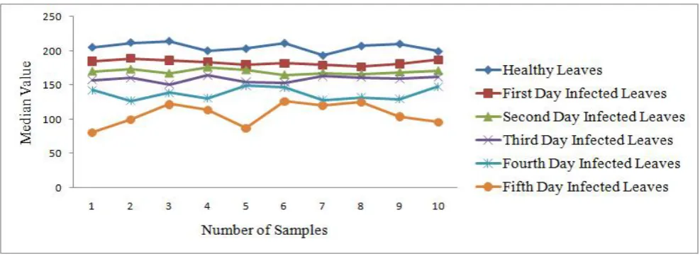
JETIR1602016 Journal of Emerging Technologies and Innovative Research (JETIR) www.jetir.org 77 Figure 5.The blue component median values for front view of healthy betel vine leaves and betel vine leaves
infected by the foot rot disease for first five days
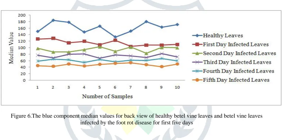
Figure 6.The blue component median values for back view of healthy betel vine leaves and betel vine leaves infected by the foot rot disease for first five days
The median values of the red component in back view ranges are between 151 and 164. For betel vine leaves infected by the foot rot disease on the fourth day, the median values of the red component in front view ranges are between 81 and 95.The median values of the red component in back view ranges are between 127 and 149. For betel vine leaves infected by the foot rot disease on the fifth day, the median values of the red component in front view ranges are between 69 and 80. The median values of the red component in back view ranges are between 81 and 126. The green component median values for front and back view of healthy betel vine leaves and betel vine leaves infected by the foot rot disease for first five days after its identification are shown in Figure 2 and Figure 3. For the healthy betel vine leaves, the median values of the green component in front view ranges are between 171 and 203. The median values of the green component in back view ranges are between 194 and 223. For betel vine leaves infected by the foot rot disease on the first day, the median values of the green component in front view ranges are between 147 and 166. The median values of the green component in back view ranges are between 179 and 191. For betel vine leaves infected by the foot rot disease on the second day, the median values of the green component in front view ranges are between 129 and 146. The median values of the green component in back view ranges are between 163 and 178. For betel vine leaves infected by the foot rot disease on the third day, the median values of the green component in front view ranges are between 108 and 128. The median values of the green component in back view ranges are between 147 and 162. For betel vine leaves infected by the foot rot disease on the fourth day, the median values of the green component in front view ranges are between 82 and 107. The median values of the green component in back view ranges are between 136 and 146. For betel vine leaves infected by the foot rot disease on the fifth day, the median values of the green component in front view ranges are between 54 and 81. The median values of the green component in back view ranges are between 122 and 135.
JETIR1602016 Journal of Emerging Technologies and Innovative Research (JETIR) www.jetir.org 78 front view ranges are between 57 and 68. The median values of the blue component in back view ranges are between 83 and 103. For betel vine leaves infected by the foot rot disease on the third day, the median values of the blue component in front view ranges are between 43 and 56. The median values of the blue component in back view ranges are between 68 and 82. For betel vine leaves infected by the foot rot disease on the fourth day, the median values of the blue component in front view ranges are between 33 and 42. The median values of the blue component in back view ranges are between 55 and 67. For betel vine leaves infected by the foot rot disease on the fifth day, the median values of the blue component in front view ranges are between 21 and 32. The median values of the blue component in back view ranges are between 42 and 54.
V.CONCLUSION
The statistical analysis on RGB color components of healthy and infected betel vine leaves shows that the statistical values for infected leaves varies on the basis of the duration of infection. The median values of all color components have decreased as the day of infection increases for the infected betel vine leaves. This analysis helps to identify the infection of foot rot disease in the early stage for sirugamani variety of betel vine leaves.
Preventive and Corrective Measures for Foot Rot Disease
The most important precautionary measure is the preservation of the seed plant. A 1% mixture of “Phortok” should be drenched monthly once in the plantation soil for the young plants during the period from September to February. The mixing of superphosphate in the soil at a ratio of 600 kilograms for one hector will also reduce the infection. The only corrective measure is that the infected plants along with roots should be carefully removed from the soil and disposed off from the plantation to avoid further spreading of the infection.
VI.REFERENCES
[1] J.Vijayakumar and Dr.S. Arumugam, „Study of betelvine plants diseases and methods of disease identification using digital image processing‟, European Journal of Scientific Research, 240-244,2012.
[2] J.Vijayakumar and Dr.S. Arumugam, „Recognition of powdery mildew disease for betelvine plants using digital image processing‟, International Journal of Distributed and Parallel Systems, 231-241,2012.
[3] J.Vijayakumar and Dr.S. Arumugam, „Foot rot disease identification for the betelvine plants using digital image processing‟, Journal of Computing, 180-183,2011.
[4] J.Vijayakumar and Dr.S. Arumugam, „Early detection of powdery mildew disease for betelvine plants using digital image analysis‟, International Journal of Modern Engineering Research, vol.2, no.4, pp. 2581-2583,2012.
[5] J.Vijayakumar and Dr.S. Arumugam, „Powdery mildew disease identification for vellai kodi variety of betelvine plants using digital image processing‟, European Journal of Scientific Research, 409-415,2012.
[6] J.Vijayakumar and Dr.S. Arumugam, „Foot rot disease identification for vellai kodi variety of betelvine plants using digital image processing‟, ICTACT Journal on Image and Video Processing, 495-501,2012.
[7] J.Vijayakumar and Dr.S. Arumugam, „Disease identification in pachai kodi variety of betelvine plant using digital imaging techniques‟, Archives Des Sciences, 308-312,2013.
[8] J.Vijayakumar and Dr.S. Arumugam, „Foot rot disease identification for karpoori variety of betelvine plants using digital imaging technique‟, Australian Journal of Basic and Applied Sciences, 270-274,2013.
[9] J.Vijayakumar and Dr.S. Arumugam, „Odium piperis fungus identification for piper betel plants using digital image processing,, Journal of Theoretical and Applied Information Technology, 423-427,2013.
[10]J.Vijayakumar and Dr.S. Arumugam, „Powdery mildew disease identification in karpoori variety of betel vine plants using histogram based techniques‟, Advances in Image and Video Processing, 63-75,2103.
[11]J.Vijayakumar and Dr.S. Arumugam, „Disease identification for the prevention of calamity using digital image processing‟, proceedings of the first International Conference on Sensors, Security, Software and Intelligent Systems, pp.32,2009.
[12]J.Vijayakumar and Dr.S. Arumugam, „Certain investigations on foot rot disease for betelvine plants using digital imaging technique‟, proceedings of the fifth International Conference on Emerging Trends in Communication and Computing Applications, pp.79,2013.