© 2015 IJSRSET | Volume 1 | Issue 3 | Print ISSN : 2395-1990 | Online ISSN : 2394-4099 Themed Section: Engineering and Technology
Brain Tumor Detection and Segmentation in MR images Using GLCM and
AdaBoost Classifier
Athira V S, Anand J Dhas, Sreejamole S S
ECE Department, Narayanguru College of Engineering, Anna University, Manjalumoodu, Tamil Nadu, India
ABSTRACT
Brain tumor segmentation is one of the crucial procedures in surgical and treatment planning. Brain tumor segmentation using MRI has been an intense research area. Brain tumors can have various sizes and shapes and may appear at different locations. Varying intensity of tumors in brain magnetic resonance images (MRI) makes the automatic segmentation of tumors extremely challenging. There are various intensity based techniques which have been proposed to segment tumors on magnetic resonance images. Texture is one of most popular feature for image classification and retrieval. The multi fractal texture estimation methods are more time consuming. A texture based image segmentation using GLCM (Gray-Level Co-occurrence Matrix) combined with AdaBoost classifier is proposed here. From the MRI images of brain, the optimal texture features of brain tumor are extracted by utilizing GLCM. Then using these features AdaBoost classifier algorithm classifies the tumor and non-tumor tissues and tumor is segmented. This method provides more efficient brain tumor segmentation compared to the segmentation technique based on mBm and will provide more accurate result.
Keywords: AdaBoost Classifier, Brain tumor, Feature extraction, GLCM, Segmentation
I.
INTRODUCTION
Tumor is the abnormal growth of the tissues. A brain tumor is a mass of unnecessary cells growing in the brain or central spine canal. Brain cancer can be counted among the most deadly and intractable diseases. Today, tools and methods to analyse tumors and their behaviour are becoming more prevalent. Clearly, efforts over the past century have yielded real advances. However, we have also come to realize that gains in survival must be enhanced by better diagnosis tools. Although we have yet to cure brain tumours, clear steps forward have been taken toward reaching this ultimate goal, more and more researchers have incorporated measures into clinical trials each advance injects hope to the team of caregivers and more importantly, to those who live with this diagnosis.
Most of the current conventional diagnosis techniques are based on human experience in interpreting the MRI-scan for judgment; certainly this increases the possibility to false detection and identification of the brain tumor. On the other hand, applying digital image processing ensures the quick and precise detection of the tumor [1]. One of the most effective techniques to extract information from complex medical images that has wide application in medical field is the segmentation process. The main objective of the image segmentation is to partition an image into mutually exclusive and exhausted regions such that each region of interest is spatially contiguous and the pixels within the region are homogenous with respect to a predefined criterion.
The cause of most cases is unknown. Risk factors that may occasionally be involved include: a number of genetic syndrome such as neurofibromatosis as well as exposure to the chemical vinyl chloride, Epstein-Barr virus, and ionizing radiation. The most common types of primary tumors in adults are: meningiomas and astrocytomas such as glioblastomas. In children the most common type is medulloblastomas. Diagnosis is usually by medical examination along with computed tomography or magnetic resonance imaging. This is then often confirmed by biopsy. Based on the finding the tumors are divided into different grades or severity. Treatment may include some combination of surgery, radiation therapy and chemotherapy.
II.
METHODS AND MATERIAL
A.Related Work
Low-level operations such as thresholding, edge detection, and morphological techniques [2], are fast and can be used for brain tumor segmentation. However, the performance of these methods highly depends on the evident difference in the intensities between tumor and non-tumor regions. Watershed segmentation approach is simple and consistently produces complete boundaries [3]. But this method is sensitive to noise and may over segment tumors because of the weak and diffused edges caused by edema.
The asymmetric analysis method [4-5] is also used for tumor segmentation which is based on the principle that tumors which appear in one of the cerebral hemispheres can cause asymmetry between the left and right cerebral
hemispheres. This asymmetry can be detected and tumors can be roughly located in the corresponding cerebral hemisphere. But the difficulty lies in accurately finding the mid-sagittal plane which is a challenging and time-consuming task. Also this method may not be useful when a tumor is located across the mid-sagittal plane [6].
Atlas-based segmentation methods have been extensively used for brain tumor segmentation. Awrfield et al. combined elastic atlas registration with statically classification to mask brain tissue from surrounding structures [7]. Kaus et al. proposed brain tumor segmentation using digital anatomic atlas and MR image intensity [8]. Prastawa et al. use an atlas prior for tumor segmentation requires manual labelling of template MRI. Also because of the intensity variations around the tumor caused by edema and the deformations of healthy tissue morphology caused by the tumor mass effect the deformable registration of the brain atlas to brain images with tumor is an extremely challenging task.
Tao Wang et. Al proposed the contour evolution method which uses a parametric active contour model that facilitates brain tumor detection in MRI. The proposed model makes rather simplistic assumption that there is a single continuous region associated with tumor [9].
B.Proposed Method
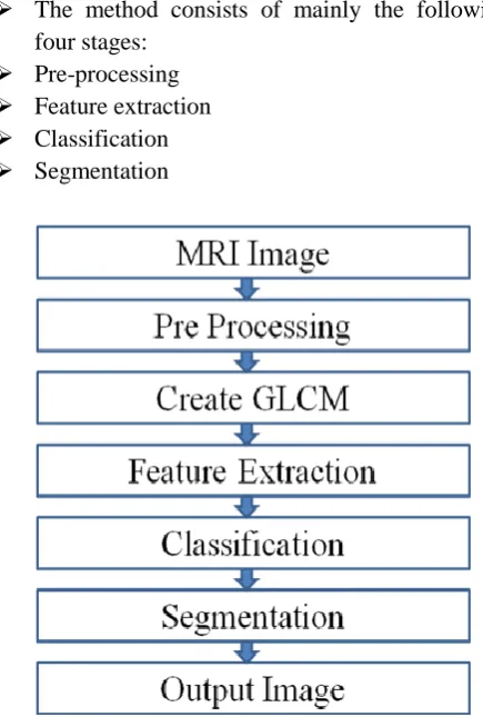
The proposed system introduces a method by which brain tumor can be detected and segmented accurately. The method combines GLCM based feature extraction and AdaBoost classifier for the segmentation of brain tumor in MR images. Analysis with large number of variables requires a large amount of memory and computation power. So feature extraction is done to overcome this problem. Here the GLCM matrix is created for feature extraction and the features thus obtained are used for classifying tumor and non tumor pixels using AdaBoost classifier. Here the texture feature of the MRI image is extracted in which the spatial relationship of the pixel is considered. The different features extracted from the GLCM matrix include Contrast, Correlation, Energy, Entropy, Homogeneity etc.
The method consists of mainly the following four stages:
Pre-processing Feature extraction Classification Segmentation
Figure 1. Flow diagram of the proposed method
i) Pre-Processing:
It is the first step in the proposed method. The purpose of this step is basically to remove the noise and improve the image quality for accurately detecting the tumor.
Here, the input image is passed through a Gaussian filter to remove the noise in order to obtain a better image. Also it improves the image quality.
The Gaussian blur is a type of image-blurring filter that uses a Gaussian function for calculating the transformation to apply to each pixel in the image. Each pixel’s new value is set to a weighted average of that pixel’s neighbourhood. The original pixel’s value receives the heaviest weight (having the highest Gaussian Value) and neighbouring pixels receive smaller weights as their distance to the original pixel increases. This results in a blur that preserves boundaries and edges better than other, more uniform blurring filters; see also scale-space implementation. The input image undergoes smoothing using Gaussian smoothing filter for elimination of noise. Gaussian filter is a linear spatial filter which is used for reducing the high frequency components of an image as a result it smooth’s the edges of the input image.
ii) Feature Extraction
iii) Brain Tumor Segmentation and Classification from Nontumor Tissue:
For tumor/nontumor tissue segmentation and classification, MRI pixels are considered as samples. These samples are represented by a set of feature values extracted from different MRI modalities. Feature from all modalities are fused for tumor segmentation and classification. We follow data driven machine learning approach to fuse different features extracted from different MRI modalities. We let our supervised classifier autonomously exploit multiple features extracted from different modalities in the training dataset.
Due to ineffectiveness of classifying complex tumor texture across various patients, this paper considers an ensemble boosting method. Such boosting method yields a highly accurate classifier by combining many moderately accurate component classifiers. In this method, each component classifier is successively added and trained on a subset of the training data that is “most difficult” given the current set of component classifiers already added to ensemble. Among different variations of boosting methods, adaptive boosting such as AdaBoost [22] is the most common.
Boosting is a machine learning meta-algorithm for reducing bias in supervised learning. A weak learner is defined to be a classifier which is only slightly correlated with the true classification. In contrast, a strong learner is a classifier that is arbitrarily well correlated with the true classification. Boosting problem simply referred to the process of turning a weak learner into a strong learner.
Given a set of training samples, AdaBoost maintains a weight distribution W over these samples. This distribution is initially set uniform. Then AdaBoost calls Component Learn or Weak Learning algorithm repeatedly in a series of cycles. At cycle t, AdaBoost provides training samples with a distribution Wt to weak learner. In response, the weak learner trains a classifier ht. The distribution Wt is updated after each cycle according to the prediction results on the training samples. Easy samples that are correctly classified ht get lower weights and hard samples that are misclassified get higher weights. Thus AdaBoost focuses on the samples with higher weights which seem to be harder
for weak learner. This process continues for T cycles and finally AdaBoost linearly combines all the component classifiers into a single final hypothesis. Greater weights are given to component classifiers with lower training errors. The important theoretical property of AdaBoost is that if the component classifiers consistently have accuracy only slightly better than half, the training error of the final hypothesis drops to zero exponentially fast. This means that the component classifiers need to be only slightly better than random.
III.
RESULTS AND DISCUSSION
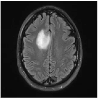
This section provides the experimental results and analysis. Figure 2 shows an example input MRI image.
Figure 2. An example of Brain tumor MRI
Figure 3 shows the testing and the training samples in the AdaBoost classifier. Figure 4 shows the classifier error rate. Here 5 cycles are considered for the classification.
Figure 4. Error rate of the classifier
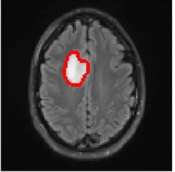
Figure 5 shows an example of tumor segmentation result. The result obtained through this method is compared with the segmentation result obtained through multi-fractal analysis. This proposed method is more efficient than the mBm in terms of computation time and accuracy.
Figure 5. Segmented brain tumor output
IV.
CONCLUSION
In this paper novel GLCM feature extraction and supervised classification techniques for improved brain tumor detection and segmentation are developed. The GLCM feature characterizes intricate tumor tissue texture in brain MRI as a spatially varying process in brain MRI. The AdaBoost algorithm considers wide variability in texture features in MRI slices for improved tumor and nontumor tissue classification. This feature-based brain tumor segmentation does not require deformable image registration with any predefined atlas. The computational complexity of modified AdaBoost
algorithm is linear and increases with number of samples and number of component classifiers.
V.
REFERENCES
[1]. V J Nagalkar and S S Asole, “Brain tumor detection using digital image processing based on soft computing”, Journal of Signal and Image Processing, Vol.3, No:3, 2012
[2]. D. Bhattacharayya and T. H. Kim, “Brain tumor detection using MRI image analysis,” Communications in Computer and Information Science, vol. 151, pp. 307-314, 2011.
[3]. R. Ratan, S. Sharma, and S. K. Sharma, “Brain tumor detection based on multi-parameter MRI image analysis,” International journal on Graphics, Vision and Image Processing, vol.9, no. 3, pp. 9-11,2009.
[4]. B. N. Saha, N. Ray, R. Greiner, A. Murtha, and H. Zhang, “ Quick detection kof brain tumors and edemas: A bounding box method using symmetry,” Computerized Medical Imaging and Graphics, vol. 36, no. 2, pp. 95-107,2012.
[5]. Z. Iscan, Z. Dokur, and T. Olmez, “Tumor detection by using Zernike moments on segmented magnetic resonance brain images,” Expert Systems with Applications, vol. 37, no. 3, pp. 25440-2549,2010. [6]. H. Khotanlou, O. Colliot, J. Atif, and I. Bloch, “3D brain tumor
segmentation in MRI using fuzzy classification, symmetry analysis and spatially constrained deformable models,” Fuzzry Sets and Systems, vol. 160, no. 10, pp. 1457-1473,2009.
[7]. Z. J. Wang, Q. M. Hu, K. F. Loe, A. Aziz, and W. L. Nowinski, “Rapid and automatic detection of brain tumors in MR Images,” in Proc. Of SPIE, 2004.
[8]. M. Prastawa, E. Bullitt, S. Ho, and G. Gerig, “ A brain tumor segmentation framework based on outlier detection,” Med Image Anal, vol. 8, no. 3, pp. 275-83,Sep 2004.
[9]. T. Wang, I. Cheng and A. Basu, “Fluid vector flow and applications in brain tumor segmentation,” IEEE Transactions on Bio-medical Engineering, vol. 56, no. 3, pp. 781-9, Mar 2009.
[10].Y. Wu, W. Yang, J. Jiang, S. Q. Li, Q. J. Feng, and W. F. Chen, “Semi-automatic segmentation of brain tumors using population and individual information,” Journal of Digital Imaging, vol. 26, no. 4, pp. 786-796,2013.