2140
A Comprehensive Review And Analysis On MRI
Based Brain Tumor Segmentation
A.R.Deepa, W.R. Sam Emmanuel
Abstract: Brain tumor is a crucial brain disease that arises because of the group of irregular cells that grows inside and around the brain. Several imaging approaches are used to detect the brain tumor. Among all the imaging technique, a safe and non-invasive imaging technique called the MRI is mainly utilized for brain tumor detection. MRI is considered as a major-medical imaging technique for diagnosing and monitoring brain tumors, and their function for respective treatment. The MRI process of brain tumor identification takes place usually by experienced radiologists. Also for more effective treatment and greater accuracy, clinicians constantly strive in the computer vision based approaches to detect brain tissue from MRI images. In this paper, we presented a comparative study on existing MRI based brain tumor segmentation methods. In addition, we highlight the various algorithms used in different stages of brain tumor segmentation such as pre-processing, feature extraction and classification with its merits and demerits.
Index Terms: Brain tumor, Medical Resonance Imaging, Segmentation, Feature Extraction, Feature Selection, Classification.
————————————————————
1
I
NTRODUCTIONIn general, the tumor is defined as tissue cluster formed due to the aggregation of abnormal cells in the body. Typically, at an appropriate time, the old cells are being replaced by new ones [1]. Due to the advent of a cancerous tumor(s), this cycle is disrupted. The tumor cells grow exponentially and don’t perish, unlike healthy cells. At present imaging technology is must for patient diagnosis [2]. The various medical images like MRI, Ultrasound, CT, X-ray etc play an important role in the field of process of disease, diagnosing and treating [3]. The recent revolution in medical imaging results from techniques such as CT and (MRI) can provide detailed information about disease. and can identify many pathologic conditions giving an accurate diagnosis. Furthermore, the new techniques are helping to advance fundamental biomedical research [4]. Medical imaging is one of the most common techniques used to improving the diagnoses, understanding and treatment of a large variety of diseases [5]. The brain imaging analysis is main objective in the field of medical image analysis [6]. Magnetic resonance (MR) imaging have many benefits over the medical imaging modalities such as a useful non-invasive technique for assisting in clinical diagnoses, the high level of contrast resolution, multispectral characteristics and ability to provide rich information about human soft tissue [7]. MRI provides useful information in the field of surgery, radiotherapy treatment planning, stereotactic neurosurgery [8] Computer Aided Diagnosis system has been developed for Automatic Detection of Brain Tumor through MRI. Improving the ability to identify early stage tumors is an important goal for physicians, because early detection of class of disease is a key factor in producing successful treatments [9]. There is different type of detection techniques which is used to develop the CAD [10].
______________________
A.R.Deepa, Reg.No:12437, Research scholar, Nesamony
memorial Christian college, Marthandam, affiliated ton Manonmaniam sundaranar university, Abishekapatti, Tamil Nadu 627012
W.R. Sam Emmanuel, Associate Professor, Department of
computer science, Nesamony memorial Christian college, Marthandam
To create a CAD system, the integration of various image processing techniques such as segmentation, feature extraction and classification are essential [11] [12].
2
BRAIN TUMORBrain is the center of human central nervous system. The brain is a complex organ as it contains 50-100 billion neurons forming a gigantic network [13]. There are two types of brain tumors namely primary tumor and secondary or metastatic tumor [14]. Usually, the primary brain tumor outsets in the brain and tends to stay during its growth tenure [15]. Whereas, the secondary brain tumor commences elsewhere as cancer in the body and later spreads to the brain region. Further, the primary brain tumor has two sub-division namely, (i) Benign tumor and (ii) Malignant tumor [16]. In the diagnosis of brain tumor, determination of the exact location is an important task, using which helps to find out the shape & size of tumor [17]. In brain tumor detection techniques, image segmentation plays a vital role there are many image segmentation methods are used to extract tumor from magnetic resonance imaging images of brain [18]. Whereas segmentation provides the detailed information about the soft brain tissues such as gray matter (GM), white matter(WM), cerebral spinal fluid (CSF)etc. There are two types of segmentation involves a manual segmentation and automatic segmentation [19]. Manual segmentation technique depends on experience or expert knowledge of human and time consuming technique but reduces the computational efficiency. Whereas automatic segmentation deals with histogram. Which is only based on the intensity of pixels [20].
2.1 Automatic MRI Segmentation
2141
recognition of tumor in brain plays a curial and extreme occupation in the field of medical image processing. This brings the need of introducing automatic MRI segmentation techniques for brain tumor detection [23].
2.2 MRI Modalities
Tissue appearance can be influenced by variable behavior of protons appeared in various tissues. The speed at which versatile hydrogen protons are moving becomes helpful in deciding measure of sign created by explicit tissue [24]. Essentially, MRI images are of three modalities [25]:
T1-Weighted
T2-Weighted
Fluid-Attenuated Inversion Recovery (FLAIR)images
In medical image processing T1-weighted and T2-weighted images are two MRI modalities most often used for visualizations *26+. These MR slice weightings refer to the important signal (whether it be the T1 time or the T2 time) measured to make the contrast in the image. The areas with high fat content have a short T1 time relative to water; T1-weighted images can be taken as visualizing locations of fat *27+. In contrast areas with high water content have a short T2 time related to areas of high fat content, T2-weighted images can be observed as visualizing locations of water *28+. An example T1, T2-weighted and FLAIR images are shown in figure 1. In finding out brain tumors, a second T1-weighted image is collected frequently after the injection of a contrast agent. These contrast agent components use to have elements whose composition makes a decrease in the T1 time of nearby tissue (gadolinium is one example) *29+. The image left: T1-weighted image (light regions visualize locations of fat). The Image Top right: T2-weighted image (light regions visualize locations of water) *30+.
Figure 1: Modalities of MRI Images
To diagnosis a brain tumor, Magnetic Resonance Imaging is commonly used, because of its advantages of high resolution to delicate tissues of brain and it is a non-ionizing, non-radioactive which will not damages human organs *31+. Combined with medical knowledge and clinical experience, the experienced diagnosist can locate the tumor sizes, shapes, anatomical structure and other pathological characteristics of brain tumors which help in suggesting the proper treatment to the patients *32+. Because there are several MRI examinations for every patient in the whole therapeutic treatment, each of which can give data in multiple sequences, it is a large amount of
data to be dealt with for the doctors *33+. Long time of hard work will inevitably lead to mistakes in the diagnosis of the tumor contours for the doctors. Moreover, it is subjective for the doctors to determine the state of the diseases per their medical knowledge and clinical experiences *34+. Therefore, developing an automatic or a semi-automatic computer-aided diagnosis system is meaningful in real medical treatments, which can release the workload of doctors and improve the accuracy by giving objective results *35+.
3
BRAIN TUMOR SEGMENTATIONIdentifying brain tumor is challenging and this process mainly comprises three stages such as pre-processing, feature extraction and classification [36] as shown in figure 2.
Feature Extraction
Pre-processing Segmentation & Classification Input MRI
Figure 2: Stages of Brain Tumor Segmentation
Before feature extraction, the pre-processing phase performs image enhancement functions such as noise reduction, denoising, filtering, and skull removal [37]. The feature extraction refers to the previous function of measuring image segmentation values, such as energy, entropy, correlation, correction, and standard deviation. The second step is segmentation, and the final one is classification. The existing methods in segmentation of tumor imaging and its classification include Artificial Neural Network (ANN), Self-Organizing Mapping (SOM), Learning Vector Quantization (LVQ), segmentation of the hydrographic basin and clustering [38].
3.1 Pre-processing
2142
method has demonstrated by quantitative measurements. Prima et al. [41] offered the popular NL algorithm to propose the alternation of the intensity of the MRI image. In this, each voxel of an image is weighted by the sum of the similar voxel intensities. In some images, there were certain redundancies. Voxel image has generally evaluated in the space quarter of a voxel. In traditional NL, the algorithm and the voxel cell must be processed. Aksam et al. [42] introduced the Enhanced Non-Local Means algorithm for applying magnetic resonance to the brain by providing several extensions of the Improved Adaptive Non Local Means (IANLM). Initially, IANLM is modified to Rician noise in magnetic resonance imaging by applying a Rican diagonal correction procedure. Secondly, a selective medial filtering procedure is used as a post-processing step to overcome the IANLM limits. Several varying parameters of the algorithm is proposed by Enhanced Non-Local Means (ENLM), to optimize the application of brain MRI. Finally, the impact of the ENLM algorithm on segmentation is studied, and improved segmentation results have been achieved compared to other segmentation techniques at the cutting edge. Bauer et al. [43] introduced automatic brain segmentation based on the atlas registration with a Markov Random Field (MRF). This method was based on the Atlas registration and was a combination of biological, medical treatment. Here, the brain tumor has specified as a random field without markers. MRF has performed with basic methods. The advantage of the method is that the image is evaluated qualitatively and compared to the existing method. Also, this method can be used to align the image and adjust the spatial noise for noise estimation. Nie et al. [44] have introduced the MRF space precision field for tumor identification in multi-channel sequences using various techniques such as MRF and Space Precision. This method has several techniques such as Hidden Markov Random Field (HMRF) and spatially weighted spatial accuracy vector based on the precision of the space. The multi-channel segmentation method can be employed to achieve alignment of low-resolution FLAIR, T1, and T2 images. The advantage of this method is that the correct images of inhomogeneity and the implementation of skull removal images are automatically displayed. A series of wavelets is a representation of an integrated square function through an orthonormal series determined by a wavelet which is proposed by Salwe et al. [45]. The wavelet transformation has applied over the grayscale image and all the diagonal, vertical, and horizontal coefficients and stored for later processing. Demirhan et al. [46] established the Wavelet transformation method for the segmentation process. It takes advantage of being adaptable to frequency, time, and discontinuities in the image. Joseph et al. [47] have established the morphology filtering method, which has been applied to detect the size and shape of the structure's image. In this method, the operation has
2143
method was further studied by moving the tumor site and modifying its size in the phantom image. Sikka et al. [53] have designed segmentation of imaging using k-means and optimized c-means. Segmentation performs for all proposed tumor detection methods. Several methods such as hydrographic segmentation for clustering algorithm optimized clustering k -means with a genetic algorithm, and c-optimization means clustering with genetic algorithm has introduced. The comparison was also made regarding the tumor region and search time. From the comparison, the grouping of C-Means after optimization was found to be better than other methods. However, the genetic algorithm improves the convergence, and the calculation time is low. Thus, the issue of segmentation has also been addressed. Park et al. [54] have established the skull stripping technique to address the issues in the segmentation of the brain tumor and the parameter of the MR scanner. The segmentation images obtained are usually in low contrast, and these poor-quality images are very difficult for segmenting precisely. When compared with the clustering methods, the Skull stripping method was difficult to perform. Pereira et al. [55] have established the intensity normalization of image segmentation. The standard deviation and the mean intensity values were computed in each sequence. After that, the sequence will be normalized to unit variance and mean value. The introduced method was used for the MRI image segmentation to increase the image quality, intensities of the image, and image contrast across the acquisition and patients.
3.2 Feature Extraction
The feature extraction method withdraws the features from outlier for detecting the tumor in the image segmentation. Feature extraction was used to detect the tumor and its exact position of the tumor. The feature extract method has classified into three types. Dubey et al. [56] describe the three semi-automated approaches presented for tumor imaging segmentation that exceeds the precision and sensitivity limits of current solutions. In the RoI method, the selected segmentation region has converted to a label matrix, and all the images are extracted to use in the image toolbox. The goal is to examine three techniques: level set, marker-controlled watershed and Modified Gradient Magnitude Region Growing Technique (MGMRGT) and then compared against expert’s manual segmentations. Finally, the reliability of brain tumor area measurements quantitatively compared with manual tracking method and semi-automated segmentation methods. Zulpe et al. [57] introduced GLCM Textural Features for brain tumor identification. The second-order statistic quality feature extraction of image segmentation is known as GLC Matrix. Whereas the GLCM is a co-occurrence matrix, and the images are split into several rows and columns to specify
the grayscale intensity values that are determined between 0 to255. The GLCM textual features of each class have then applied to two layers of Feedforward Neural Network, which offers a 97.5% classification rate. Ghanavati et al. [58] introduced a multimodal frame for automatic tumor detection by combining different MRI modalities, including T1-weighted, T2-weighted, and T1 with gadolinium contrast agent. Initially, the intensity, shape deformation, symmetry, and texture characteristics have extracted from each image where the AdaBoost Classifier was used to select the most discriminating features and to segment the tumor region. MR images with simulated tumors have been used as real truth for training and validating the detection method. Parisot et al. [59] exploited previous knowledge in the form of a limited graph representing the expected spatial positions of the tumor class. Such information was coupled with image-based classification techniques, along with space-smoothing constraints to produce a reliable detection map in a unified graphical formula. Effective linear programming in terms of both performance and the computational load was considered to recover the lowest potential of the objective function.
3.3 Segmentation
2144
that they have two different sets of data in five homogeneous, heterogeneous, different diffusion tumors, and the synthesis of the images. Bhat et al. *63+ introduced an active contour model of the river basin algorithm. Here, the active modeling method was used in the tumor region of the cerebral tumor segmentation extract. This method has been used to reduce the complexity of image computation and to improve image segmentation without noise. The advantage of this method is the high quality of image acquisition interval, clear extraction of the concave tumor region, precise tumor segmentation, and low sensitivity noise. Maiti et al. *64+ introduced the basin type of the tumor detection method. The river basin for the brain color NMR method in HSV color space techniques are used to detect the edges of the image. Using HSV image techniques, images are divided into hue, saturation, and intensity. The advantage of this method is that the segmentation images rounded using the edge detector improve the accuracy. Dhage et al. *65+ introduced river basin segmentation with ICCL coding techniques of connected components to detect tumor segmentation. The paper proposes the potential of hydrographic segmentation, such as image acquisition, image preprocessing, and contrast parameter. The Watershed method divides the abnormal tissue from the usual closure tissue to obtain a real identification of the required area, which helps the surgeon to discriminate the complex area. The advantages are simplicity, consistency, and reduced noise. Moreover, the high-frequency MRI elements were extracted from the MR image and the exact position, shape, and variables like perimeter, eccentricity, entropy, and centroid are also calculated. Vriji *66+ introduced medical image processing using river basin segmentation. The proposed segmentation techniques used Computer Assisted Diagnostic (CAD) segmentation. In the proposed section, there are two phases, such as the pre-processing and improvement phase. Initially, the medical image has been transformed into the standard formatted image. The segmentation method has several advantages since exploring the possible use of MR segmentation imaging accurately improves brain, 2D, and 3D brain tumor shape for surgical planning and gauge cancer. Cuadra et al. *67+ have introduced the method of deformation of the Atlas for complex tumor areas. These explain the growth of the lesion and the condition of the lesion based on three stages. The first step is to bring the bluff record, and the next is to scatter. The last step is for the deformation of the seed, combined with the model of the lesion. The method based on the atlas has exceeded the limitation of the size of the seed per target. The disadvantage is that more time is required to set up the atlas for computational procedures. Gordillo et al. *68+ proposed an automatic and unattended brain tumor method. This method covered the completely new approach explained the operation in two steps: 1. fully
2145
algorithms is FLAIR, T1 and T2 to combine improvement tissues and non-fatal tissues in the tumor area. This method has used several steps, such as recording the T1 and TIE scenes and subtracting by specifying the VIO and seed needed to segment the connectivity. This method can be used for automation and continuous image with accurate high-speed values and can also improve the efficiency of the MRI segmentation system. Solomon et al. [74] established the identification of the brain tumor using a hidden Markov model. Response to treatment responses is classified as complete response, the response in partial, stable disease and progressive disease. The proposed method for paper has several steps, such as 3D spatial segmentation, the hidden model, the trading model. Here they explained the automated approach using probabilistic intelligence both in space and time to segment tumors from RMD space-time RMR data. The advantages of the method are the 4D segmentation method being the improvement over the existing method, the inclusion of positive sentences from the normal intensive tissue was greatly reduced. Bauer et al. *75+ presented an automatic method of segmenting the brain tumor. The approach was based on the atlas method, being the combination of biomedical treatment. Here, the cerebral tumor is specified as a random field marker without mesh, MRF was performed with the basic methods: the tumor would be the first in the displacement pattern, the tumor increased in the three dimensions. The advantage of the method by which the image is evaluated qualitatively and clearly has been compared with the existing method. Bauer et al. *76+have introduced the method based on the atlas using the random field Markov. The proposed techniques have been explained of the mesh-free method applied to compare healthy segmentation of the brain image and abnormal segmentation of brain tissues. The advantages are represented by the 2D image, this method is applied in five different sets of tumor data and visualize the conditions and dimensions of the tumor cell. Mori et al. *77+ have established the Diffusion tensor imaging method to detect tumor status. The proposed methods are based on DTI, color code image, and 3D tract techniques. The proposed techniques utilized to identify the anatomy of white matter. The foremost advantage of the DTI provides new information on the special link between the tumor level and the adjacent white matter sections that may be useful for administrative planning. Zou et al. *78+ presented systematic approaches to validate the accuracy of the results of automatic image segmentation, which led to the probabilistic interpretation of the tumor class by pixel. He then developed an EM algorithm to estimate the latent gold standard. In addition, they modeled the results of probability segmentation using a mix of two beta distributions with different form parameters. Therefore, precision measurements, including the ROC curve, mutual
information and the similarity of the nuts were estimated. Furthermore, an optimal threshold was obtained within each metric. Aslam et al. *79+ presented an improved edge detection algorithm for brain tumor segmentation. It is based on the detection of Sobel edges which Combines the Sobel method with the dependent image threshold method and finds different regions using the boundary cycle algorithm. Eventually, the tumors are extracted from the image using the intensity information in the closed contours. The algorithm is implemented in C and its performance is measured. The results of the simulation show that the algorithm offers superior performance compared to conventional segmentation
3.4 Classification
2146
competitive level. In segmentation, there are more fabrics, which are separated by layers. Here the input data was a competitive layer, used to classify in the self-organizing method so that the output level will compete with the target classes. Using LVQ input data similar to Euclidean factors and distance are determined. Corso et al. [84] present a new method for automatically segmenting heterogeneous image data, which was a step towards overcoming the gap between bottom-up affinity segmentation methods and top-down generalized approaches. The main contribution of the document was a Bayesian formula incorporating the attributions of soft models in computational affinities, which are conventionally non-modular. Then integrate the affinities of the conscious multilevel segmentation model using the weighted aggregation algorithm and apply the technique for detecting and segmenting brain tumor cells and MR multi-channel edema. The efficient calculation method executes orders of magnitude faster than the latest cutting-edge techniques, providing comparable or better results. Patenaude et al. [85] a Bayesian layout model has been proposed that incorporates both form information and intensity from a training set. The idea is similar to the active aspect model, except that it uses a probabilistic structure to estimate the relationship between form and intensity and to a large extent, uses conditional probabilities. Furthermore, the method can be used to perform peak analysis to investigate differences in shape between groups of subjects. It provides a local and direct measurement of geometric changes that are not based on tissue classification or arbitrary extension of leveling. Logeswari and Karnan [86] proposed a cluster Self-Organizing Map (SOM) algorithm for the segmentation of the medical image. This document describes the two-step segmentation method. In the first phase, the image of the MRI brain was obtained from the patient's database. This film eliminates artifact and noise. In the second phase, the image segmentation allows to accurately identify the main tissue structures in these volumes of images. They present a new method of segmentation of unsupervised MR images based on the C-Means fuzzy clustering algorithm for segmentation. Dhanalakshmi and Kanimozhi [87] a K-mean clustering method for identifying MR brain images are mainly based on characteristics and attributes. The number of object groups was called K-means, which is a positive integer. When the Euclidian has minimized, it makes a distance between the cluster's centroid and the data. The K-mean clustering was the unattended method type. In the K-mean method training group, the cluster in random order calculates the values of each cluster and each pixel. Based on the distance value, the cluster center was detected. K indicates that the method was used to position the images in bounds and objects. The main purpose of group
segmentation was the region exhausted in the photo and exclusive. Zhang et al. [88] The Fuzzy C-mean cluster method was introduced and was used to allow two or more data groups to identify brain tumor. This cluster will analyze the data and divide it into the collection of an object in the cluster. The C-mean method has two properties, such as homogeneity data within the cluster, and heterogeneity between cluster data, allowing different data from the different cluster. This method is not suitable for identifying the fuzzy model, but the cluster method will divide the data and create the membership function. Shokouhifar et al. [89] introduced the medium cluster algorithm to identify the brain tumor in the original images with a traditional method of MRI segmentation. The performance of this method has been simple and fast but does not identifies noise and abnormal images. Normally, MRI images present unwanted noises and inaccuracies. But this C-mean method will find the exact shape and size of the target. The neural network region associated with pulses of interest for images is very important for image segmentation, each image segment being a process and analysis for smoothing the image. The primary function of this method is used to identify the front of the image. Pulse-Coupled Neural Networks has managed to solve the image problem that identifies the model, image segmentation, and detects image margins. These define the RoI in segmenting the image. PCNN can find the image pixel neuron. In image segmentation, each neuron divides into three phases: pulse generation, section, and modulation. The output contains a few steps, such as segmenting image, plot, and edge information, which is a dynamic output. Chou et al. [90] presented an extended neural network algorithm coupled to pulses that work in 3-D over the entire image volume. They evaluated performance based on different Signal to Noise Ratio (SNR), resolution, and tested those methods against brain surface extractor algorithm in the brain. The results show that the method goes beyond existing methods and was robust with reduced SNR effects, partial volume at lower resolutions, and facilitate the automatic processing of large-scale studies on rodent brains.
4
P
ERFORMANCEA
NALYSIS&
C
OMPARISON2147
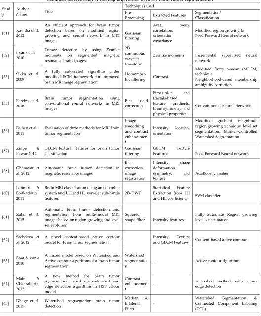
Table 2.1: Comparison of existing algorithms used for brain tumor segmentation
Stud y
Author
Name Title
Techniques used
Pre-Processing Extracted Features
Segmentation/ Classification
[51] Kavitha et al. 2012
An efficient approach for brain tumor detection based on modified region growing and neural network in MRI images
Gaussian filtering
Area, mean, correlation,
orientation, covariance
Modified region growing & Feed Forward Neural network
[52] Iscan et al. 2010
Tumor detection by using Zernike moments on segmented magnetic resonance brain images
2D continuous wavelet transform
Zernike moments Incremental supervised neural network
[53] Sikka et al. 2009
A fully automated algorithm under modified FCM framework for improved brain MR image segmentation
Homomorp
hic filtering Contrast
Modified fuzzy c-mean (MFCM) technique
Neighborhood-based membership ambiguity correction
[55] Pereira et al. 2016
Brain tumor segmentation using convolutional neural networks in MRI images
Bias field correction
First-order and fractals-based texture gradients, brain symmetry, and physical properties
Convolutional Neural Networks
[56] Dubey et al. 2011
Evaluation of three methods for MRI brain tumor segmentation
Image smoothing and contrast enhancemen t
Intensity, location, orientation
Modified gradient magnitude region growing technique, level set segmentation, Marker-Controlled Watershed Segmentation
[57] Zulpe & Pawar 2012
GLCM textural features for brain tumor classification
Gaussian filtering
GLCM Texture
Features Feed Forward Neural network
[58] Ghanavati et al. 2012
Automatic brain tumor detection in magnetic resonance images
Bias correction, image registration
Intensity, shape deformation, symmetry, and texture
AdaBoost classifier
[60]
Lahmiri & Boukadoum 2011
Brain MRI classification using an ensemble system and LH and HL wavelet sub-bands features
2D-DWT
Statistical Feature Extraction from LH
and HL coefficients SVM classifier
[61] Zabir et al. 2015
Automatic brain tumor detection and segmentation from multi-modal MRI images based on region growing and level set evolution
Squared
shape filter Intensity features
Fully automatic Region growing level set estimation
[62] Sachdeva et al. 2012
A novel content-based active contour model for brain tumor segmentation’ -
Intensity, Texture
and GLCM Features Content-based active contour
[63] Bhat & kunte 2010
A mixed model based on Watershed and Active contour algorithms for brain tumor segmentation
Watershed segmentatio n
- Active contour algorithm.
[64]
Maiti & Chakraborty 2012
A new method for brain tumor segmentation based on watershed and edge detection algorithms in HSV colour model
Contrast enhancemen t
- watershed method with canny edge detection
[65] Dhage et al. 2015
Watershed segmentation brain tumor detection
Median & Bilateral Filter
-
2148 [68] Gordillo et
al. 2010
A new fuzzy approach to brain tumor
segmentation Filtering Intensity Fuzzy approach
[71] Zarandi et al . 2007
Systematic image processing for diagnosing brain tumors
Fuzzy based image filtering
Mahalanobis distance, and Kwon validity index
Type-II Approximate Reasoning method
[73] Liu et al. 2012
Classification of MR tumor images based on Gabor wavelet analysis
Wavelet
analysis Texture Features
Gabor wavelet analysis, SVM & LDA labelling
[74] Solomon et al. 2006
Segmentation of brain tumors in 4D MR
images using the hidden Markov model - -
3D-spatial segmentation & Hidden Markov model
[75] Bauer et al. 2011
Fully automatic segmentation of brain tumor images using support vector machine classification in combination with hierarchical conditional random field regularization
Edge-preserving smoothing filter
Intensity Features & mean, variance, skewness, kurtosis, energy, entropy
SVM combined with subsequent hierarchical regularization based on Conditional Random Fields
[79] Aslam et al. 2015
Improved edge detection algorithm for
brain tumor segmentation - Intensity features
Improved Edge Detection algorithm based on Sobel method and thresholding
[80] Kharrat et al. 2010
A hybrid approach for automatic classification of brain MRI using genetic algorithm and support vector machine
Gabor filter Texture features Genetic algorithm (GA) and SVM
In most of the existing works, preprocessing is done before segmenting the original images. Preprocessing is performed in many ways mainly for removing the unwanted noise in the original image or to enhance the quality or intensity of the original image. In addition, some preprocessing techniques are performed for ROI extraction, skull stripping and outline removal of original image. To
remove the unwanted noise from the original image filtering techniques such as Gaussian filter, Median filter, Gabor filter, Bilateral Filter, Smoothing filter, Homomorphic filter, Mean filter, Square shape filter etc are used often. Figure 3 shows the comparison results of different preprocessing techniques.
(f)
Figure 3: Comparison results of different preprocessing techniques (a) original image (b) Gaussian filter (c) Gabor filter (d) Median filter (e) Smoothing Filter (f) Homomorphic Filter
Figure 3 compares the results of different preprocessing techniques such as Gaussian filter [51], Homomorphic filter [53], Median filter [65], Smoothing Filter [75] and Gabor
2149
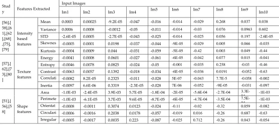
better than the other compared filters. From the literature works we identified that intensity based features, shape based features and texture features are most commonly used for feature extraction. We have randomly selected 10 brain images from the input dataset and the features values extracted from each image which are presented in table 2.2.
The performance of classification algorithms is
evaluated in terms of accuracy, sensitivity and
specificity using the following expressions.
TN
TP
FN
FP
TN
TP
Accuracy
(1)FN
TP
TP
y
Sensitivit
(2)FP
TN
TN
y
Specificit
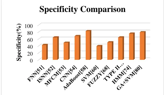
(3)Where TN=True Negative (correctly rejected), TP=True Positive (correctly identified), FN=False Negative (incorrectly rejected), FP=False Positive (incorrectly identified). From figure 4 to 6 we have compared the performance interms of accuracy, sensitivty and specificity of classifiers such as feed forward neural network (FNN) *51+, increamental supervised neural network (ISNN) *52+, Modified fuzzy c-means (MFCM) *53+, convolutional neural network (CNN) *54+, Adaboost *58+, SVM *60+, Fuzzy *68+, Typr II Fuzzy *71+, Hidden Markov Model (HMM) *74+ and Genetic algorithm based SVM *80+.
Figure 4: Accuracy Comparison
From figure 4 it is visible that the ISNN classifier proposed in [52] and the Adaboost classifier proposed in [88] attains a maximum accuracy of 96% with the proposed dataset. Similarly the FNN classifier proposed in [51], CNN classifier proposed in [54] and type II fuzzy classifier proposed in [68] attains the second maximum accuracy of 95%. The other classifiers proposed in [53], [60], [68], [78] and [80] also attains significant accuracies within the range from 80% to 90%.
Table 2.2: Feature Values Extracted for 10 brain images
Stud
y Features Extracted
Input Images
Im1 Im2 Im3 Im4 Im5 Im6 Im7 Im8 Im9 Im10
[56],[ 58],[6 1],[62 ],[68] ,[75], [79]
Intensity based features
Mean 0.0003 0.00025 -9.2E-05 -0.047 -0.016 -0.014 -0.029 0.268 0.037 0.038
Variance 0.0006 0.0008 -0.0012 -0.05 -0.011 -0.014 -0.03 0.076 0.0963 0.602
STD -2.6E-05 0.0005 -2.7E-05 -0.043 -0.025 -0.014 -0.025 0.056 0.197 -2.6E-05 Skewnes
s
-0.0005 0.0001 0.0198 -0.037 -0.044 -9E-05 -0.029 0.005 0.066 -0.035
Kurtosis -0.0004 0.0009 0.044 -0.031 -0.059 -5E-05 -0.42 0.003 0.049 -0.44
[57],[ 62],[7 3],[80 ]
Texture features
Energy -0.0041 0.0008 0.0601 -0.027 -0.061 -6E-05 -0.042 0.077 0.015 -0.041
Entropy -0.0046 0.0078 0.0825 -0.024 -0.05 -0.001 -0.035 0.258 -0.03 -0.46 Contrast -0.0063 0.0057 0.1392 -0.018 -0.034 -6E-05 -0.036 0.0191 -0.052 -0.63 Correlati
on
-0.0082 8.2E-05 0.2325 -0.011 -0.028 5E-07 -0.043 7.7E-5 -0.058 -0.002
Inertia -0.0097 6.6E-06 0.3319 -2.3E-05 -0.028 7E-06 -0.052 -9E-05 -0.031 -0.097
[51],[ 56],[5 8]
Shape features
Area -1.0E-03 -2.4E-05 3.9E-03 5.7E-05 -1.8E-04 -2E-05 -5.6E-04 -2.7E-04
3.3E-04 -1E-03
Perimete r
-1.0E-03 -6.1E-05 3.7E-03 9.6E-05 -8.7E-05 -6E-05 -4.7E-04 -3.5E-04
7.5E-04 -1E-03
Orientat ion
-0.0008 -0.0011 0.3074 0.0123 -0.024 -0.11 -0.02 -0.32 0.059 -0.082 Circulari
ty
-0.0006 -0.0016 0.2038 0.0178 -0.057 -0.019 0.016 -0.26 0.687 -0.63 Irregular
ity
-0.0005 -0.0017 0.0035 0.223 -0.087 -0.025 0.712 -0.26 0.043 -0.054
0 20 40 60 80 100
Acc
ura
cy
(%)
2150 Figure 5: Sensitivity Comparison
From figure 5 it is visible that the sensitivity of CNN [54] and type II fuzzy classifier [68] is maximum than the other classifiers and the other classifiers also shows significant results. From figure 6 it is observed that the specificity of Adaboost classifier [58] is maximum when compared to other classifiers.
Figure 6: Specificity Comparison
5
C
ONCLUSIONA comparative analysis of MRI based brain tumor segmentation and classification techniques are presented in this paper. The goal of segmentation process is to detect brain tumor initially. For this purpose, various automated segmentation techniques are proposed which are highlighted in this paper. In addition, the different stages of brain tumor segmentation techniques such as pre-processing, feature extraction and classification is also explained in this paper. From the comparative analysis, it is observed that each technique has its own advantage and disadvantages and produce significant results in terms of accuracy, sensitivity and specificity. The determination of segmentation techniques is found to be very successful and reliable.
R
EFERENCES[1] Abdulbaqi, H. S, Mat, M. Z, Omar, A. F, Mustafa, I. S. B, & Abood, L. K. 2014, Detecting brain tumor in magnetic resonance images using hidden markov random fields and threshold techniques’. In Research and Development (SCOReD), 2014 IEEE Student Conference on IEEE, pp. 1-5.
[2] Ain, Q, Jaffar, M. A & Choi, T. S. 2014, ‘Fuzzy anisotropic diffusion based segmentation and texture based ensemble classification of brain tumor’. applied soft computing, vol.21, pp. 330-340.
[3] Aksam Iftikhar, M, Jalil, A, Rathore, S, Ali, A, & Hussain, M. 2013, ‘Brain MRI denoizing and segmentation based on improved adaptive nonlocal means’. International journal of imaging systems and technology, vol.23, no.3, pp. 235-248.
[4] Amato, F, López, A, Peña-Méndez, E. M, Vaňhara, P, Hampl, A & Havel, J. 2013, ‘Artificial neural networks in medical diagnosis’.
[5] Bezdek, J. C, Hall, L. O, & Clarke, L. 1993 ‘Review of MR image segmentation techniques using pattern recognition’. Medical physics, vol.20, no.4, pp.1033-1048.
[6] Bezdek, J. C, Hall, L. O, & Clarke, L. 1993, ‘Review of MR image segmentation techniques using pattern recognition’. Medical physics, vol.20, no.4, pp.1033-1048.
[7] Bozkurt S, Gimenez F, Burnside ES, Gulkesen KH & Rubin DL. 2016. ‘Using automatically extracted information from mammography reports for decision-support’. Journal of biomedical informatics. Vol.62, pp. 224-31.
[8] Cabezas, M, Oliver, A, Lladó, X, Freixenet, J & Cuadra, M. B. 2011 ‘A review of atlas-based segmentation for magnetic resonance brain images’. Computer methods and programs in biomedicine, vol.104, no.3, pp. e158-e177.
[9] Cai, H, Verma, R, Ou, Y, Lee, S. K., Melhem, E. R & Davatzikos, C. 2007, ‘Probabilistic segmentation of brain tumors based on multi-modality magnetic resonance images’. In Biomedical Imaging: From Nano to Macro, 2007. ISBI 2007. 4th IEEE International Symposium on IEEE, pp. 600-603.
[10]Calabrese, Christopher, Helen Poppleton, Mehmet Kocak, Twala L. Hogg, Christine Fuller, Blair Hamner, Eun Young Oh et al. "A perivascular niche for brain tumor stem cells." Cancer cell 11, no. 1 (2007): 69-82. [11]Callaghan, M. F, Freund, P., Draganski, B., Anderson,
E., Cappelletti, M., Chowdhury, R & Lutti, A. 2014, ‘Widespread age-related differences in the human brain microstructure revealed by quantitative magnetic resonance imaging’. Neurobiology of aging, vol.35, no.8, pp. 1862-1872.
[12]Castadot P, Lee JA, Parraga A, Geets X, Macq B,
0 20 40 60 80 100
Sens
it
iv
it
y
(%)
Sensitivity Comparison
0 20 40 60 80 100
Sp
ec
if
icit
y
(%)
2151
Grégoire V, 2008. ‘Comparison of 12 deformable registration strategies in adaptive radiation therapy for the treatment of head and neck tumors’. Radiotherapy and oncology. vol.89, no.1, pp.1-2.
[13]Chaddad, A, Zinn, P. O & Colen, R. R. 2014, ‘Brain tumor identification using Gaussian Mixture Model features and Decision Trees classifier’. In Information Sciences and Systems (CISS), 2014 48th Annual Conference on IEEE, pp. 1-4.
[14]Charfi, S, Lahmyed, R & Rangarajan, L. 2014, ‘A novel approach for brain tumor detection using neural network’. International Journal of Research in Engineering and Technology, vol.2, pp. 93-104.
[15]Clarke, L. P, Velthuizen, R. P, Camacho, M. A, Heine, J. J, Vaidyanathan, M., Hall, L. O & Silbiger, M. L. 1995 ‘MRI segmentation: methods and applications’. Magnetic resonance imaging, vol.13, no.3, pp. 343-368.
[16]Coatrieux, G, Huang, H, Shu, H, Luo, L & Roux, C. 2013, ‘A watermarking-based medical image integrity control system and an image moment signature for tampering characterization’. IEEE journal of biomedical and health informatics, vol.17, no.6, pp.1057-1067. [17]Cuadra, M. B, Pollo, C, Bardera, A, Cuisenaire, O,
Villemure, J. G & Thiran, J. P. 2004, ‘Atlas-based segmentation of pathological MR brain images using a model of lesion growth’. IEEE transactions on medical imaging, vol.23, no.10, pp. 1301-1314.
[18]Dahab DA, Ghoniemy SS & Selim GM. 2012. ‘Automated brain tumor detection and identification using image processing and probabilistic neural network techniques’. International journal of image processing and visual communication. Vol.1, no.2, pp. 1-8.
[19]Davnall F, Yip CS, Ljungqvist G, Selmi M, Ng F, Sanghera B, Ganeshan B, Miles KA, Cook GJ & Goh V, 2012, ‘Assessment of tumor heterogeneity: an emerging imaging tool for clinical practice?’. Insights into imaging. Vol.3, no. 6, pp. 573-89.
[20]Daumas-Duport, Catherine, Marie-Louise Tucker, Harry Kolles, Pascale Cervera, Fréderic Beuvon, Pascale Varlet, Naoko Udo, Maria Koziak, and Jean-Paul Chodkiewicz. "Oligodendrogliomas. Part II: A new grading system based on morphological and imaging criteria." Journal of neuro-oncology 34, no. 1 (1997): 61-78.
[21]de Rooij, M, Hamoen, E. H, Witjes, J. A, Barentsz, J. O & Rovers, M. M. 2016, ‘Accuracy of magnetic resonance imaging for local staging of prostate cancer: a diagnostic meta-analysis’. European urology, vol. 70, no.2, pp. 233-245.
[22]Deepa, A. R & Sam Emmanuel, W. R. M. 2016 ‘Identification and classification of brain tumor through mixture model based on magnetic resonance
imaging segmentation and artificial neural network’. Concepts in Magnetic Resonance Part A, vol.45, no.2, pp. e21390.
[23]Deepak, K. S, Gokul, K, Hinduja, R & Rajkumar, S. 2013, ‘An efficient approach to predict tumor in 2D brain image using classification techniques’. In Information Communication and Embedded Systems (ICICES), 2013 International Conference on IEEE.pp. 559-564).
[24]Deepak, K. S, Gokul, K, Hinduja, R & Rajkumar, S. 2013, ‘An efficient approach to predict tumor in 2D brain image using classification techniques’. In Information Communication and Embedded Systems (ICICES), 2013 International Conference on IEEE, pp. 559-564.
[25]El Abbadi NK & Kadhim NE. 2016, ‘Brain Tumor Classification Based on Singular Value Decomposition’. Brain, Vol.5, no.8.
[26]El-Dahshan, E. S. A, Mohsen, H. M, Revett, K & Salem, A. B. M. 2014, ‘Computer-aided diagnosis of human brain tumor through MRI: A survey and a new algorithm’. Expert systems with Applications, vol.41, no.11, pp.5526-5545.
[27]El-Melegy, M. T & Mokhtar, H. M. 2014, ‘Tumor segmentation in brain MRI using a fuzzy approach with class center priors’. EURASIP Journal on Image and Video Processing, vol.2014, no.1, p. 21.
[28]Faisal, A, Parveen, S, Badsha, S & Sarwar, H. 2012, ‘An improved image denoising and segmentation approach for detecting tumor from 2-d mri brain images’. In Advanced Computer Science Applications and Technologies (ACSAT), 2012 International Conference on IEEE.pp. 452-457.
[29]Fletcher-Heath LM, Hall LO, Goldgof DB & Murtagh FR. 2001, ‘Automatic segmentation of non-enhancing brain tumors in magnetic resonance images’. Artificial intelligence in medicine. vol.21, no.1-3, pp.43-63. [30]Gordillo, N, Montseny, E & Sobrevilla, P. 2010, ‘A new
fuzzy approach to brain tumor segmentation’. In Fuzzy Systems (FUZZ), 2010 IEEE International Conference on IEEE, pp. 1-8.
[31]Greenspan, H, Ruf, A & Goldberger, J. 2006, ‘Constrained Gaussian mixture model framework for automatic segmentation of MR brain images’. IEEE transactions on medical imaging, vol.25, no.9, pp.1233-1245.
[32]Halder, A, Giri, C & Halder, A. 2014, ‘Brain tumor detection using segmentation based Object labeling algorithm’. In Electronics, Communication and Instrumentation (ICECI), 2014 International Conference on IEEE, pp. 1-4.
2152
segmenting magnetic resonance images of the brain’. IEEE transactions on neural networks. Vol.3, no. 5, pp.672-82.
[34]Hall LO, Bensaid AM, Clarke LP, Velthuizen RP, Silbiger MS & Bezdek JC. 1992, ‘A comparison of neural network and fuzzy clustering techniques in segmenting magnetic resonance images of the brain’. IEEE transactions on neural networks. Vol.3, no.5, pp. 672-82.
[35]Havaei, M, Davy, A, Warde-Farley, D, Biard, A, Courville, A, Bengio, Y & Larochelle, H. 2017, ‘Brain tumor segmentation with deep neural networks’. Medical image analysis, 35, 18-31.
[36]Hoerr, V & Faber C. 2014, ‘Magnetic resonance imaging characterization of microbial infections’, Journal of pharmaceutical and biomedical analysis. Vol.1, no.93, pp.136-46.
[37]İlhan A. Brain Tumor Detection Using Intensity Adjustment Based Segmentation (Doctoral Dissertation, Near East University).
[38]Işın, A, Direkoğlu, C & Şah, M. 2016, ‘Review of MRI-based brain tumor image segmentation using deep learning methods’. Procedia Computer Science, vol.102, pp.317-324.
[39]Faisal, A, Parveen, S, Badsha, S & Sarwar, H. 2012, ‘An improved image denoising and segmentation approach for detecting tumor from 2-d mri brain images’. In Advanced Computer Science Applications and Technologies (ACSAT), 2012 International Conference on IEEE.pp. 452-457.
[40]Sulaiman, S. N, Ishak, S. M. C, Isa, I. S, & Hamzah, N. 2014, ‘De-noising of noisy MRI brain image using the switching-based clustering algorithm’. In Control System, Computing and Engineering (ICCSCE), 2014 IEEE International Conference on IEEE, pp. 1-6. [41]Prima, S & Commowick, O. 2013, ‘Using bilateral
symmetry to improve non-local means denoising of MR brain images’. In Biomedical Imaging (ISBI), 2013 IEEE 10th International Symposium on IEEE, pp. 1231-1234.
[42]Aksam Iftikhar, M, Jalil, A, Rathore, S, Ali, A, & Hussain, M. 2013, ‘Brain MRI denoizing and segmentation based on improved adaptive nonlocal means’. International journal of imaging systems and technology, vol.23, no.3, pp. 235-248.
[43]Bauer, S, Nolte, L. P & Reyes, M. 2011, ‘Segmentation of brain tumor images based on atlas-registration combined with a Markov-Random-Field lesion growth model’. In Biomedical Imaging: From Nano to Macro, 2011 IEEE International Symposium on IEEE, pp. 2018-2021.
[44]Nie, J, Xue, Z, Liu, T, Young, G. S, Setayesh, K, Guo, L & Wong, S. T. 2009. ‘Automated brain tumor segmentation using spatial accuracy-weighted hidden
Markov Random Field’. Computerized Medical Imaging and Graphics, vol.33, no. 6, pp. 431-441. [45]Salwe, S, Raut, R & Hajare, P. 2016, ‘Brain tumor pixels
detection using adaptive wavelet based histogram thresholding and fine windowing’. In Information Technology (InCITe)-The Next Generation IT Summit on the Theme-Internet of Things: Connect your Worlds, International Conference on IEEE, pp. 256-260). [46]Demirhan, A., Törü, M., & Güler, I. 2015, ‘Segmentation
of tumor and edema along with healthy tissues of brain using wavelets and neural networks’. IEEE journal of biomedical and health informatics, vol.19, no.4, pp. 1451-1458.
[47]Joseph, R. P, Singh, C. S & Manikandan, M. 2014, ‘Brain tumor MRI image segmentation and detection in image processing’. International Journal of Research in Engineering and Technology, vol. 3, no.1, pp. 1-5. [48]Askins, M. A, Ann-Yi, S & Moore, B. D. 2015,
‘Neurocognitive late effects in children treated for cancer: Psychological impact, identification, and prevention and remediation’. In Handbook of long term care of the childhood cancer survivor, pp. 397-409. [49]Ananda, R. S & Thomas, T. 2012, ‘Automatic segmentation framework for primary tumors from brain MRIs using morphological filtering techniques’. In Biomedical Engineering and Informatics (BMEI), 2012 5th International Conference on IEEE, pp. 238-242. [50]Dawngliana, M., Deb, D, Handique, M & Roy, S. 2015,
‘Automatic brain tumor segmentation in MRI: Hybridized multilevel thresholding and level set’. In Advanced Computing and Communication (ISACC), 2015 International Symposium on IEEE, pp. 219-223.
[51]Kavitha, A. R, Chellamuthu, C & Rupa, K. 2012, ‘An efficient approach for brain tumour detection based on modified region growing and neural network in MRI images’. In Computing, Electronics and Electrical Technologies (ICCEET), 2012 International Conference on IEEE, pp. 1087-1095.
[52]Iscan, Z, Dokur, Z & Ölmez, T. 2010, ‘Tumor detection by using Zernike moments on segmented magnetic resonance brain images’. Expert Systems with Applications, vol.37, no.3, pp. 2540-2549.
[53]Sikka, K, Sinha, N, Singh, P. K & Mishra, A. K. 2009, ‘A fully automated algorithm under modified FCM framework for improved brain MR image segmentation’. Magnetic Resonance Imaging, vol.27, no.7, pp. 994-1004.
[54]Park, J. G & Lee, C. 2009, ‘Skull stripping based on region growing for magnetic resonance brain images’. NeuroImage, vol.47, no.4, pp. 1394-1407. [55]Pereira, S, Pinto, A, Alves, V & Silva, C. A. 2016, ‘Brain
2153
imaging, vol.35, no.5, pp. 1240-1251.
[56]Dubey, R. B, Hanmandlu, M & Vasikarla, S. 2011, ‘Evaluation of three methods for MRI brain tumor segmentation’. In Information Technology: New Generations (ITNG), 2011 Eighth International Conference on IEEE, pp. 494-499.
[57]Zulpe, N & Pawar, V. 2012, ‘GLCM textural features for brain tumor classification’. International Journal of Computer Science Issues (IJCSI), vol.9, no.3, p. 354. [58]Ghanavati, S., Li, J, Liu, T., Babyn, P. S., Doda, W &
Lampropoulos, G. 2012, ‘Automatic brain tumor detection in magnetic resonance images’. In Biomedical Imaging (ISBI), 2012 9th IEEE International Symposium on IEEE, pp. 574-577.
[59]Parisot, S, Wells III, W, Chemouny, S, Duffau, H & Paragios, N. 2014, ‘Concurrent tumor segmentation and registration with uncertainty-based sparse non-uniform graphs’. Medical image analysis, vol.18, no.4, pp. 647-659.
[60]Lahmiri, S & Boukadoum, M. 2011, ‘Brain MRI classification using an ensemble system and LH and HL wavelet sub-bands features’. In Computational Intelligence In Medical Imaging (CIMI), 2011, IEEE Third International Workshop On IEEE, pp. 1-7. [61]Zabir, I, Paul, S, Rayhan, M. A, Sarker, T, Fattah, S. A &
Shahnaz, C. 2015, ‘Automatic brain tumor detection and segmentation from multi-modal MRI images based on region growing and level set evolution’. In Electrical and Computer Engineering (WIECON-ECE), 2015 IEEE International WIE Conference on IEEE. pp. 503-506. [62]Sachdeva, J, Kumar, V, Gupta, I, Khandelwal, N &
Ahuja, C. K. 2012, ‘A novel content-based active contour model for brain tumor segmentation’. Magnetic resonance imaging, vol.30, no.5, pp. 694-715.
[63]Bhat, S & Kunte, S. 2010, ‘A mixed model based on Watershed and Active contour algorithms for brain tumor segmentation.’ In Advances in Recent Technologies in Communication and Computing (ARTCom), 2010 International Conference on IEEE, pp. 398-400.
[64]Maiti, I & Chakraborty, M. 2012, ‘A new method for brain tumor segmentation based on watershed and edge detection algorithms in HSV colour model’. In Computing and Communication Systems (NCCCS), 2012 National Conference on IEEE, pp. 1-5).
[65]Dhage, P, Phegade, M. R & Shah, S. K. 2015, ‘Watershed segmentation brain tumor detection’. In Pervasive Computing (ICPC), 2015 International Conference on (pp. 1-5). IEEE.
[66] VRJI, K. A & JAYAKUMARI, J.2011,‘AUTOMATIC DETECTION
OF BRAIN TUMOR BASED ON MAGNETIC RESONANCE
IMAGE USING CAD SYSTEM WITH WATERSHED
SEGMENTATION’. INSIGNAL PROCESSING,
COMMUNICATION, COMPUTING AND NETWORKING
TECHNOLOGIES (ICSCCN), 2011 INTERNATIONAL
CONFERENCE ON IEEE, PP.145-150.
[67]Cuadra, M. B, Pollo, C, Bardera, A, Cuisenaire, O, Villemure, J. G & Thiran, J. P. 2004, ‘Atlas-based segmentation of pathological MR brain images using a model of lesion growth’. IEEE transactions on medical imaging, vol.23, no.10, pp. 1301-1314.
[68]Gordillo, N, Montseny, E & Sobrevilla, P. 2010, ‘A new fuzzy approach to brain tumor segmentation’. In Fuzzy Systems (FUZZ), 2010 IEEE International Conference on IEEE, pp. 1-8.
[69]Khotanlou, H, Colliot, O, Atif, J & Bloch, I. 2009, ‘3D brain tumor segmentation in MRI using fuzzy classification, symmetry analysis and spatially constrained deformable models’. Fuzzy sets and systems, vol.160, no.10.
[70]El-Melegy, M. T & Mokhtar, H. M. 2014, ‘Tumor segmentation in brain MRI using a fuzzy approach with class center priors’. EURASIP Journal on Image and Video Processing, vol.2014, no.1, p. 21.
[71]Zarandi, M. F, Zarinbal, M & Izadi, M. 2011. ‘Systematic image processing for diagnosing brain tumors: A Type-II fuzzy expert system approach’. Applied soft computing, vol.11, no. 1, pp.285-294.
[72]Dou, W, Ruan, S, Chen, Y, Bloyet, D & Constans, J. M. 2007, ‘A framework of fuzzy information fusion for the segmentation of brain tumor tissues on MR images’. Image and vision Computing, vol.25, no. 2, pp. 164-171.
[73]Liu YH, Muftah M, Das T, Bai L, Robson K & Auer D. 2012, ‘Classification of MR tumor images based on Gabor wavelet analysis’. Journal of Medical and Biological Engineering. vol. 32, no.1, pp. 22-8.
[74]Solomon, J, Butman, J. A & Sood, A. 2006 ‘Segmentation of brain tumors in 4D MR images using the hidden Markov model’. Computer methods and programs in biomedicine, vol.84, no.2-3, pp. 76-85. [75]Bauer, S, Nolte, L. P & Reyes, M. 2011, ‘Fully automatic
segmentation of brain tumor images using support vector machine classification in combination with hierarchical conditional random field regularization’. In International Conference on Medical Image Computing and Computer-Assisted Intervention, pp. 354-361.
[76]Bauer S, Wiest R, Nolte LP & Reyes M. 2013, ‘A survey of MRI-based medical image analysis for brain tumor studies. Physics in Medicine & Biology. vol.58, no.13, pp. R97.
2154
neurology, vol.51, no.3, pp.377-380.
[78]Zou, K. H, Wells III, W. M, Kikinis, R & Warfield, S. K. 2004 ‘Three validation metrics for automated probabilistic image segmentation of brain tumours’. Statistics in medicine, vol.23, no.8, pp.1259-1282.
[79]Aslam, A, Khan, E & Beg, M. S. 2015, ‘Improved edge detection algorithm for brain tumor segmentation’. Procedia Computer Science, vol.58, pp. 430-437.
[80]Kharrat, A, Gasmi, K, Messaoud, M. B, Benamrane, N & Abid, M. 2010 ‘A hybrid approach for automatic classification of brain MRI using genetic algorithm and support vector machine’. Leonardo journal of sciences, vol.17, no.1, pp. 71-82.
[81]Cai, H, Verma, R, Ou, Y, Lee, S. K., Melhem, E. R & Davatzikos, C. 2007, ‘Probabilistic segmentation of brain tumors based on multi-modality magnetic resonance images’. In Biomedical Imaging: From Nano to Macro, 2007. ISBI 2007. 4th IEEE International Symposium on IEEE, pp. 600-603.
[82]Menze, B. H, Jakab, A, Bauer, S, Kalpathy-Cramer, J, Farahani, K, Kirby, J & Lanczi, L. 2015, ‘The multimodal brain tumor image segmentation benchmark (BRATS)’. IEEE transactions on medical imaging, vol.34, no.10, pp. 1993.
[83]Tian, D & Fan L. 2007, ‘A brain MR images segmentation method based on SOM neural network’. In: The 1st international conference on bioinformatics
and biomedical engineering, pp 686–689
[84]Corso, J. J, Sharon, E, Dube, S, El-Saden, S, Sinha, U & Yuille, A. 2008, ‘Efficient multilevel brain tumor segmentation with integrated bayesian model classification’. IEEE transactions on medical imaging, vol.27, no. 5, pp. 629-640.
[85]Patenaude, B, Smith, S. M, Kennedy, D. N & Jenkinson, M. 2011, ‘A Bayesian model of shape and appearance for subcortical brain segmentation’. Neuroimage, vol.56, no.3, pp. 907-922. [86]Logeswari T & Karnan M. 2010, ‘An improved
implementation of brain tumor detection using soft computing’. In Communication Software and Networks, 2010. ICCSN'10. Second International Conference on IEEE. pp. 147-151.
[87]Dhanalakshmi P & Kanimozhi T. 2013, ‘Automatic segmentation of brain tumor using K-Means clustering and its area calculation. International Journal of advanced electrical and Electronics Engineering, vol.2, no.2, pp. 130-4.
[88]Zhang, D. Q & Chen, S. C. 2004, ‘A novel kernelized fuzzy c-means algorithm with application in medical image segmentation’. Artificial intelligence in medicine, vol.32, no.1, pp. 37-50.
[89]Shokouhifar, M & Abkenar, G. S. 2011, ‘An artificial bee
colony optimization for mri fuzzy segmentation of brain tissue’. In 2011 International Conference on Management and Artificial Intelligence IPEDR, Vol. 6. [90]Chou, N, Wu, J, Bingren, J. B, Qiu, A & Chuang, K. H.