Cryptococcus neoformans
Connie B. Nichols, Kyla S. Ost, Dayton P. Grogan, Kaila Pianalto, Shirin Hasan, J. Andrew Alspaugh
Departments of Medicine and Molecular Genetics/Microbiology, Duke University School of Medicine, Durham, North Carolina, USA
The localization and specialized function of Ras-like proteins are largely determined by posttranslational processing events. In a highly regulated process, palmitoyl groups may be added to C-terminal cysteine residues, targeting these proteins to specific membranes. In the human fungal pathogenCryptococcus neoformans, Ras1 protein palmitoylation is essential for growth at high temperature but is dispensable for sexual differentiation. Ras1 palmitoylation is also required for localization of this pro-tein on the plasma membrane. Together, these results support a model in which specific Ras functions are mediated from differ-ent subcellular locations. We therefore hypothesize that proteins that activate Ras1 or mediate Ras1 localization to the plasma membrane will be important forC. neoformanspathogenesis. To further characterize the Ras1 signaling cascade mediating high-temperature growth, we have identified a family of proteinS-acyltransferases (PATs), enzymes that mediate palmitoyl-ation, in theC. neoformansgenome database. Deletion strains for each candidate gene were generated by homogenous recombi-nation, and each mutant strain was assessed for Ras1-mediated phenotypes, including high-temperature growth, morphogene-sis, and sexual development. We found that full Ras1 palmitoylation and function required one particular PAT, Pfa4, and deletion of thePFA4gene inC. neoformansresulted in altered Ras1 localization to membranes, impaired growth at 37°C, and reduced virulence.
S
-palmitoylation involves the reversible attachment of palmitic acid moieties to proteins. This process of posttranslational modification is catalyzed by the proteinS-acyltransferase (PAT) enzymes through the thioesterification of a cysteine side chain group. By the addition of this hydrophobic palmitoyl group, pro-teins often become associated with membranes. However, in con-trast to constitutive protein modifications such as prenylation, palmitoylation is often regulated and reversible, allowing for dynamic changes in patterns of subcellular protein localization (1,2).PAT genes were first identified in the budding yeast
Saccharo-myces cerevisiaeas the enzymes responsible for the palmitoylation
of Ras2 (Erf2) and yeast casein kinase (Akr1) (3, 4). The PAT family is characterized by several molecular features, including multiple transmembrane domains, as well as a DHHC motif (Asp-His-His-Cys) within an approximately 50-amino-acid cysteine-rich domain (CRD) (5). Seven PAT family members have now been identified in yeast, while 24 of these proteins have been iden-tified in mammals. Interestingly, nine PATs have been associated with human disease ranging from neuropsychiatric conditions such as Alzheimer’s and Huntington’s disease to developmental conditions (Golz syndrome) and cancer (6).
Unlike other types of lipidation, such as prenylation or N-my-ristoylation, there is no specific consensus sequence for protein palmitoylation. Therefore, simple bioinformatic prediction of palmitoylated proteins has been challenging. However, a compre-hensive analysis of palmitoylated proteins in yeast found that there were conserved features among many of the cysteine resi-dues that are likely to undergo this type of modification: most are located near prenylated/myristoylated residues, most are sur-rounded by basic or hydrophobic amino acids, and many lie in the cytoplasmic regions of membrane-spanning proteins (7).
Lipidation of proteins by palmitoylation mediates membrane localization. The importance of protein localization on function has been extensively explored, especially for Ras-like proteins. In
mammalian cells, palmitoylation of Ras dictates not only localiza-tion to particular membranes, such as the Golgi complex, plasma membrane (PM), and microdomains within the PM, but also downstream signaling from Ras (8,9). In the fission yeast
Schizo-saccharomyces pombe, two distinct Ras protein signaling programs
are largely determined by palmitoylation and localization of Ras1 on different cellular membranes (10).
InC. neoformansthe Ras1 (CnRas1) protein is required for
pathogenesis, morphogenesis, and sexual differentiation. Re-cently, we demonstrated that palmitoylation and subsequent plasma membrane attachment of Ras1 are essential for the main-tenance of normal morphology and pathogenicity. However, pro-tein palmitoylation does not appear to be required for other Ras-related phenotypes such as mating and hyphal differentiation (11).
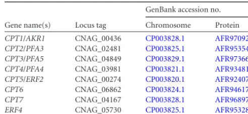
To elucidate the PAT responsible forC. neoformansRas1 pal-mitoylation, we have identified seven genes predicted to encode DHHC motif-containing proteins. Targeted deletion of most of these genes does not affect Ras-dependent cellular functions. However, thepfa4⌬mutant shares some phenotypic similarities with anras1⌬mutant, such as impaired growth at 37°C and atten-uated virulence. Moreover, PM association of a green fluorescent
Received22 January 2015 Accepted5 April 2015
Accepted manuscript posted online10 April 2015
CitationNichols CB, Ost KS, Grogan DP, Pianalto K, Hasan S, Alspaugh JA. 2015.
Impact of protein palmitoylation on the virulence potential ofCryptococcus neoformans. Eukaryot Cell 14:626 –635.doi:10.1128/EC.00010-15.
Address correspondence to J. Andrew Alspaugh, [email protected].
Supplemental material for this article may be found athttp://dx.doi.org/10.1128 /EC.00010-15.
Copyright © 2015, American Society for Microbiology. All Rights Reserved.
doi:10.1128/EC.00010-15
on September 8, 2020 by guest
http://ec.asm.org/
protein (GFP)-Ras1 fusion protein is altered in thepfa4⌬mutant strain compared to that in the wild type, and palmitoylation of Ras1 is reduced in this mutant background. We also found that Pfa4 was required for Ras1-independent phenotypes, indicating that this protein clearly has other targets than Ras proteins. Also, our genetic and biochemical analyses suggest some degree of over-lapping functions among the PATs inC. neoformans, underscor-ing the importance of this process in the growth, differentiation, and virulence of this microbial pathogen.
MATERIALS AND METHODS
Strains and media.C. neoformansstrains used in this study are listed in Table 1. Strains were maintained on YPD (yeast extract, peptone, dex-trose) medium (12). To analyze cell morphology in response to temper-ature stress, strains incubated overnight in liquid YPD cultures with shak-ing were diluted 1:10 in fresh YPD medium and incubated at either 30°C or 37°C with shaking for either 4 h or 24 h prior to imaging. To examine chitin morphology in response to temperature stress, samples were col-lected and resuspended in phosphate-buffered saline (PBS) containing 25 g/ml of calcofluor white and incubated for 10 min. To analyze growth sensitivity in response to temperature and cell wall stress, strains incu-bated overnight in shaking YPD cultures were serially diluted and spotted onto YPD plates prepared with the indicated concentrations of Congo red, calcofluor white, SDS, and caffeine (perFig. 4legend). To examine mating morphology, strains of opposite mating types were mixed together in water and then spotted onto MS mating medium plates (13). Cocul-tures were incubated in the dark prior to imaging.
Molecular biology.C. neoformansgenes identified in this study are listed inTable 2. To generateC. neoformansPAT deletion mutants using a dominant selectable marker, three-piece PCR overlap extension was used to replace theAKR1,PFA3,PFA5, andPFA4open reading frames with the neomycin (neo) resistance cassette to generate strains CBN134 (akr1::
neo), CBN198 (pfa3::neo), CBN124 (pfa5::neo), and CBN201 (pfa4::neo) (14). Thecpt7::neoandcpt6::natconstructs were generated using
three-piece In-Fusion HD cloning (Clontech). Theerf2::neodeletion construct was generated by split-marker double-joint PCR (15). These constructs were used to generate mutant strains DGP1 (erf2::neo), SHC7 (cpt6::nat), and CBN414 (cpt7::neo). All of the PAT mutant strains except for SHC7 were generated in the type H99 background. We were not able to readily generate acpt6::natdeletion mutant strain in the H99 strain, instead re-quiring a strain with a deletion ofCKU80. Ku80 is required for nonho-mologous end joining, and deletion ofCKU80increases homologous re-combination inC. neoformans(16). ThePFA4gene was subcloned into thenatresistance plasmid pCH233 and transformed into thepfa4::neo
mutant strain CBN201 to create aPFA4reconstituted strain (CBN324). Deletion strains and thePFA4reconstituted strain were confirmed by PCR, Southern blot analysis, and reverse transcription-PCR (RT-PCR).
To examine the localization of the Pfa4 protein, thePFA4gene and terminator sequence were amplified by PCR from wild-typeC. neofor-mansgenomic DNA and subcloned into thenatresistance plasmid pCN19 containingGFPunder theHIS3promoter and theneoresistance plasmid pCN52 containingmCHERRYunder theHIS3promoter (11).
Genomic integration of the PAT deletion constructs,PFA4 reconsti-tution construct, and epitope-tagged constructs was performed using the biolistic transformation method as described previously (17,18).
Microscopy and imaging.Differential interference contrast (DIC) microscopy and fluorescent images were captured with a Zeiss Axio Im-ager A1 fluorescence microscope equipped with an AxioCam MRM dig-ital camera. Images were acquired and analyzed by ZEN (black edition) software (Zeiss). High-resolution fluorescent images were captured using a GE DeltaVision Elite deconvolution microscope equipped with a Cool-Snap HQ2 high-resolution charge-coupled-device (CCD) camera. Im-ages were acquired and analyzed by softWoRx software (GE). ImIm-ages were colorized and converted to JPEG format by ImageJ (Fiji) software (19).
Golgi apparatus localization.The green-emitting fluorescent stain C6-NBD-ceramide (6-{N -[(7-nitrobenzo-2-oxa-1,3-diazol-4-yl)amino]-caproylsphingosine}) (Invitrogen) was used to visualize the Golgi appa-ratus. C6-NBD-ceramide has been used to visualize the Golgi complex in yeasts, includingS. cerevisiaeandC. neoformans(20,21). Wild-type cells expressing red fluorescent protein (mCherry)-tagged Ras1 (mCh-Ras1; CBN116) and mCh-Pfa4 (CBN451) were grown overnight in YPD me-dium, diluted 10-fold, and grown for 4 h at 30°C. Cells were fixed in 3.7% formaldehyde for 10 min, washed three times in PBS, and incubated from 4 h to overnight in 5M NBD C6-ceramide.
Palmitoylation assay.We developed a modified version of the acyl-biotinyl switch assay developed by Wan et al. (22) to assess the palmitoyl-ation status ofC. neoformansRas1 (11). Briefly, wild-type andpfa4⌬ mu-tant strains expressingGFP-RAS1were grown overnight in YPD medium to an optical density at 600 nm (OD600) of 1.0. Twenty-milliliter samples were centrifuged and resuspended in 0.5 ml of lysis buffer containing 10 mMN-ethylmaleimide (NEM; Thermo Scientific) and 2⫻protease in-hibitors (Complete Mini, EDTA-free; Roche). After lysis, protein concen-tration was equalized using Precision Red (Cytoskeleton), and the sam-ples were processed as previously described (11) with the exception that GFP-Trap resin (ChromoTek) was used to precipitate GFP-Ras1 in the
TABLE 1C. neoformansstrains
Strain Genotype
Source or reference
H99 MAT␣ 44
KN99a MATa 45
CBN336 MAT␣ras1::neo 11
CBN96 MAT␣pHIS3-GFP-RAS1-nat 11
CBN116 MAT␣pHIS3-mCherry-RAS1-neo 46
CBN167 MAT␣pHIS3-GFP-ras1C203S,C204S-nata 11
CBN134 MAT␣akr1::neo This study
CBN198 MAT␣pfa3::neo This study
CBN124 MAT␣pfa5::neo This study
CBN201 MAT␣pfa4::neo This study
DPG1 MAT␣erf2::neo This study
JLCN437 MAT␣cku80::neo 16
SKC7 MAT␣cku80::neo cpt6::nat This study
CBN414 MAT␣cpt7::neo This study
CBN389 MAT␣pfa3::neo pfa4::neo This study
CBN403 MAT␣pfa3::neo pfa4::neo pHIS3-GFP-RAS1-nat
This study
CBN255 MATapfa4::neo This study
KMP1 MAT␣erf4::neo This study
CBN324 MAT␣pfa4::neo pPFA4-nat This study
CBN241 MAT␣pfa4::neo pHIS3-GFPRAS1-nat This study CBN370 MAT␣pHIS3-mCherry-RAS1-neo
pHIS3-GFP-PFA4-nat
This study
CBN451 MAT␣pHIS3-mCherry-PFA4-neo This study
aras1C203S,C204S, the C203S and C204S substitutions encoded by theras1gene.
TABLE 2C. neoformansgenes
Gene name(s) Locus tag
GenBank accession no.
Chromosome Protein
CPT1/AKR1 CNAG_00436 CP003828.1 AFR97092.1
CPT2/PFA3 CNAG_02481 CP003825.1 AFR95354.1
CPT3/PFA5 CNAG_04849 CP003829.1 AFR97366.2
CPT4/PFA4 CNAG_03981 CP003821.1 AFR93481.1
CPT5/ERF2 CNAG_00274 CP003820.1 AFR92407.2
CPT6 CNAG_06862 CP003824.1 AFR94617.2
CPT7 CNAG_04167 CP003828.1 AFR96897.1
ERF4 CNAG_05730 CP003825.1 AFR95328.2
on September 8, 2020 by guest
http://ec.asm.org/
control samples. Bound proteins were eluted from NeutrAvidin resin (palmitoylated Ras1; ThermoScientific) and GFP-Trap resin (control Ras1) into 30l of 5⫻Laemmli sample buffer and analyzed by Western blotting.
Comparative quantification of Ras1 palmitoylation in thepfa4⌬ mu-tant strain compared to that of the wild type was performed by densito-metric analysis of digitized Western blot signals using ImageJ (Fiji) soft-ware (gel analysis function) (19). Quantified Western blot signals for palmitoylated Ras1 were normalized to total Ras1 signal for each sample and expressed as a percentage of the value for the wild-type strain.
Western blot analysis.Samples were heated to 95°C for 4 min, and half of each sample (15l) was loaded and separated on a NuPAGE 4 to 12% Bis-Tris gel (Invitrogen) using morpholinepropanesulfonic acid (MOPS) running buffer. Samples were electrophoretically transferred to Invitrolon polyvinylidene difluoride (PVDF) membrane (Invitrogen). The membrane blot was blocked with Starting Block T20 (Pierce) for 1 h, incubated with anti-GFP antibody (1/1,000 dilution; Roche) for 1 h, washed three times for 10 min each with Tris-buffered saline plus Tween (TBST), incubated with an anti-mouse peroxidase-conjugated secondary antibody (1/50,000 dilution; Jackson Laboratory), and washed three times for 10 min each with TBST. Reactive proteins were detected by enhanced chemiluminescence (ECL Prime Western blotting detection reagent; GE Healthcare).
Animal studies.Using the murine inhalation model of systemic cryp-tococcosis, we inoculated female A/J (Jackson labs) mice intranasally with 5⫻105C. neoformanscells as previously described (23). Groups of 10 mice were inoculated with one of three strains: the wild type (H99),pfa4⌬
(CBN201) mutant, and reconstitutedpfa4⌬/PFA4(CBN321) strain. Mice were monitored daily for signs of infection and sacrificed at predeter-mined clinical endpoints correlating with an imminently lethal infection. The statistical significance in the difference between the survival curves of the animals inoculated with each strain was evaluated using a log rank test (JMP software; SAS Institute, Cary, NC). All studies were performed in compliance with Duke University institutional guidelines for animal ex-perimentation.
RESULTS
C. neoformans protein S-acyltransferases. We used two ap-proaches to identify an Ras1-specific PAT inC. neoformans. First, we probed theC. neoformansvar.grubiigenome database with a BLAST search for homologs ofS. cerevisiaeErf2, the primary pro-teinS-acyltransferase forS. cerevisiaeRas2 (ScRas2). Second, we queried theC. neoformansgenome databases (Cryptococcus
neo-formansvar. grubiiH99 Sequencing Project, Broad Institute of
Harvard and MIT [24] and Fungi DB [25]) for proteins predicted to contain a DHHC domain (Pfam PF01529). Seven sequences were selected for further analysis and were initially designated
CryptococcusproteinS-acyltransferases (CPTgenes) (Table 2; see
also Fig. S1 in the supplemental material). Two of the sequences, CNAG_04361 (CPT1) and CNAG_03981 (CPT4), have been pre-viously namedAKR1andPFA4based on similarity toS. cerevisiae
sequences. In a phylogenic alignment and tree construction (26) comprised of PAT amino acid sequences fromC. neoformans,S.
cerevisiae, andHomo sapiens, we also found that Cpt1 clustered
with S. cerevisiae Akr1 and Ark2 while Cpt4 clustered with S.
cerevisiaePfa4 and human ZDHC6 (see Fig. S2 in the
supple-mental material). Also, three other C. neoformans PATs (CNAG_02481, CNAG_04849, and CNAG_00274) each clustered with individualS. cerevisiaePATs (Pfa3, Pfa5, and Erf2). Based on these data, we redesignated theC. neoformans CPT1toCPT5genes
AKR1, PFA3,PFA5, PFA4, andERF2, respectively, while CPT6
andCPT7 retain their initial designations. However, our func-tional analysis suggests that, at least in the case of CnPfa4, theC.
neoformansPATs are not true orthologs of theS. cerevisiae
pro-teins and appear to have overlapping substrates.
We also found that CNAG_03981 (PFA4) did not initially ap-pear to encode a protein with an intact DHHC domain as pre-dicted by automated algorithms. However, manual inspection of likely splice junctions revealed errors in automated intron-exon border prediction. Amplification and sequencing of the cDNA associated with this gene confirmed this misannotation, indicat-ing that this sequence actually encoded a DHHC domain-contain-ing protein.
In addition we identified a putative Erf4 homolog inC. neofor-mans(Table 2). InS. cerevisiae, Erf2 palmitoyltransferase activity requires Erf4, a nonenzymatic accessory protein (27). This associ-ation is conserved in mammalian cells, with Gcp16 acting as a cofactor for the Ras PAT DHHC9 (28). InS. cerevisiae, Erf4 acts to stabilize Erf2 against degradation and is also involved in stabiliza-tion of the Erf2-palmitoyl intermediate and transfer of the group to the substrate protein Ras2 (27). Erf4 and Gcp16 share a con-served domain (Erf4 domain; Pfam 10256) that was used to iden-tify CNAG_02504 inC. neoformans.
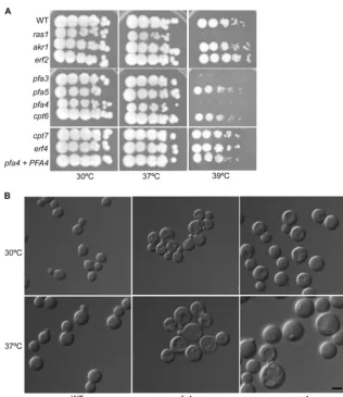
Mutation of Pfa4 and Pfa3 results in various temperature sensitivities.To evaluate the role of these putative protein acyl-transferases in Ras-related cellular processes, we examined strains with targeted mutations in each PAT gene, includingERF4. None of the these genes was essential for growth, and initial phenotypic testing showed that only thepfa4⌬mutant strain was temperature sensitive at 37°C, similar to theras1⌬mutant (Fig. 1A). In addi-tion, thepfa3⌬deletion strain exhibited a subtle temperature sen-sitivity at 39°C (Fig. 1A). These results were confirmed by testing the phenotypes for a subset of these mutants (akr1⌬,pfa3⌬,pfa4⌬, anderf2⌬strains) that were present in an independent collection
ofC. neoformansmutant strains (29).
Strains withras1⌬mutations have pronounced morphology defects in both the yeast and hyphal forms. For the asexual yeast form,ras1cells are larger than those of wild-type strains, resulting from defects in cell polarity and actin cytoskeleton dynamics (30). This cell size difference is noted at 30°C, but it is most apparent at higher growth temperatures. Among the PAT mutant strains, only the pfa4⌬ mutant exhibited a temperature-dependent size in-crease although this was not as dramatic as that of theras1⌬ mu-tant strain (Fig. 1B; also data not shown).
To ensure that any phenotypes in thepfa4⌬mutant strain were appropriately attributed to this specific mutation, an intact copy of thePFA4gene was reintroduced into this mutant strain. The resulting pfa4⌬/PFA4 reconstituted strain demonstrated com-plete restoration of wild-type levels of growth at 37°C, as well as normal morphogenesis at all temperatures (Fig. 1A).
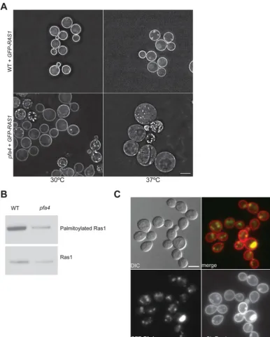
Ras1 localization is impaired in thepfa4⌬mutant strain. Pal-mitoylation of Ras proteins promotes plasma membrane localiza-tion. Previously, we demonstrated that the palmitoylation of amino acid residue C203 or C204 is sufficient for Ras1 function and localization to the plasma membrane (11). To determine if Ras1 localization was impaired in thepfa4⌬mutant strain, we generated apfa4⌬mutant strain constitutively expressing a GFP-Ras1 fusion protein. In wild-type cells, GFP-GFP-Ras1 localizes to the plasma membrane and endomembrane structures resembling punctate dots. In thepfa4⌬mutant there was an overall decrease in GFP-Ras1 localization to the plasma membrane in addition to an increase of punctate dots in some cells (Fig. 2A). The altered PM association of GFP-Ras1 in thepfa4⌬mutant was even more
on September 8, 2020 by guest
http://ec.asm.org/
apparent at 37°C, in contrast to observations in the wild-type strain background that demonstrated equally intense patterns of GFP-Ras1 PM localization at both temperatures (Fig. 2A). We observed no alteration of GFP-Ras1 localization in the other PAT mutant strains or in theerf4⌬mutant strain (data not shown).
InS. cerevisiae, there is a clear overlap of PAT functions. ScRas2
palmitoylation is not completely abrogated in an erf2 mutant strain (31). Additionally, the yeast PATs Akr1, Pfa2, Pfa4, and Pfa3 each exhibitin vitroPAT activity toward Ras2 (32). To begin to explore whether other PAT proteins contribute to Ras1 palmitoyl-ation, we generated apfa3⌬pfa4⌬double mutant. The Pfa3 pro-tein was chosen for specific inclusion, given its modest but mea-surable role in thermotolerance. Thepfa3⌬pfa4⌬double mutant strain displayed no additive phenotypic changes compared with thepfa4⌬mutant in terms of thermotolerance or yeast cell mor-phogenesis. In addition, localization of GFP-Ras1 in thepfa3⌬ pfa4⌬double mutant strain was similar to that in the singlepfa4⌬
mutant strain (data not shown).
Ras1 palmitoylation is decreased in thepfa4⌬mutant strain.
In addition to assessing the localization of Ras1 in thepfa4⌬
mu-tant strain, we directly assessed the palmitoylation status of Ras1 using a modified version of the acyl-biotinyl switch assay (22). In this assay, palmitoyl groups are captured by biotinylation, and relative Ras protein palmitoylation activity can be assessed in dif-ferent strain backgrounds. In two independent experiments, Ras1 palmitoylation was decreased by 50 and 59% in thepfa4⌬mutant compared to the level in the wild type but was not eliminated (Fig. 2B). Similar reductions of Ras1 palmitoylation were observed in thepfa3⌬pfa4⌬double mutant strain (data not shown). These measurements were consistent with the degree of altered GFP-Ras1 PM localization observed in each of these strain backgrounds (Fig. 2A). Together, these results suggest that the Pfa4 protein serves a major role in the palmitoylation of the CnRas1 protein under these growth conditions.
Ras1 and Pfa4 colocalize on endomembrane structures.In current models, palmitoylation promotes the localization of Ras proteins from the Golgi compartment to the PM. If Pfa4 functions as an Ras PAT, we would expect Pfa4 to be localized on endomem-brane structures (Golgi compartment or endoplasmic reticulum [ER]) to catalyze the palmitoylation of Ras1. In fact, other yeast
FIG 1Thepfa4⌬mutant strain exhibitsras1-like defects at high temperature. (A) Overnight cultures of wild-type (WT; H99),akr1⌬(CBN134),pfa3⌬
(CBN198),pfa5⌬(CBN124),pfa4⌬(CBN201),erf2⌬(DPG1),cpt6⌬(SKC7),cpt7⌬(CBN414),ras1⌬(CBN336), anderf4⌬(KMP1) strains and the reconstituted
pfa4⌬/PFA4(CBN324) mutant strain were serially diluted, spotted onto YPD medium, and incubated at 30°C, 37°C, and 39°C for 48 h. (B) Overnight cultures of the wild-type (H99) strain andras1⌬(CBN336) andpfa4⌬(CBN201) mutant strains were diluted in YPD liquid medium and shifted to either 30°C or 37°C for 4 h. Cells were imaged (magnification,⫻100) with DIC optics to assess temperature-dependent alterations in cell size and morphology. Scale bar, 5m.
on September 8, 2020 by guest
http://ec.asm.org/
FIG 2Ras1 palmitoylation is dependent on Pfa4.(A) Ras1 localization to the PM is reduced in thepfa4⌬mutant strain. The wild-type strain andpfa4⌬mutant strain expressingGFP-RAS1under the control of the histone H3 promoter (CBN96 and CBN241) were grown overnight in YPD medium, diluted 10-fold, and grown for 24 h at 30°C or 37°C. Cells were imaged (magnification,⫻63) using DeltaVision microscopy to assess the pattern and intensity of fluorescent fusion protein localization. All images were obtained using identical exposure times. Scale bar, 5m. (B) Ras1 palmitoylation is reduced in thepfa4⌬mutant strain. Cell lysates obtained from the wild-type andpfa4mutant strains expressing GFP-Ras1 (CBN96 and CBN241) were accessed for palmitoylation using a modified version of the acyl-biotinyl switch assay. Lysate samples were split between experimental samples (top panel) and loading control samples (bottom panel) to quantify palmitoylated Ras1 and total Ras1 levels, respectively. Proteins in the experimental samples were labeled with biotin and precipitated using NeutrAvidin beads. In this assay, biotinylation occurs specifically on proteins that contain a free sulfhydryl group generated by hydroxylamine cleavage of a thioester bond (palmitoylated proteins). GFP-Ras1 proteins in the control samples were immunoprecipitated with GFP-Trap resin. Samples were separated by NuPAGE electrophoresis, transferred to PVDF membranes, and immunoblotted with anti-GFP antibodies. Digital images were assessed using densitometry (ImageJ) for signal intensity. Palmitoylated-Ras1 signal was normalized relative to that of total Ras1 signal for each sample. Representative images from multiple independent experiments are demonstrated. (C) Pfa4 and Ras1 colocalize at punctate dots in the cytoplasm. A wild-type strain coexpressing GFP-Pfa4 and mCh-Ras1 (CBN370) was grown overnight in YPD medium, diluted 10-fold, and grown for 4 h at 30°C. Images (magnification,⫻100) were taken using the appropriate filter and merged. Scale bar, 5m.
on September 8, 2020 by guest
http://ec.asm.org/
Ras PATs localize to the Golgi compartment (S. pombe) or ER (S.
cerevisiae) (31,33). To assess the localization of Pfa4, we generated
a GFP-tagged version of this protein. As predicted, we observed that GFP-Pfa4 localized prominently to distinct punctate regions in the cytoplasm as well as in the perinuclear region, consistent with Golgi compartment/ER localization (Fig. 2C).
To probe the association of Pfa4 and Ras1 localization patterns, we examined the localization of GFP-Pfa4 and mCh-Ras1 in a wild-type strain.C. neoformansRas1 localized primarily to the PM but also to punctate dots in the cytoplasm and the vacuole, con-sistent with its predicted progression from endomembranes to the PM (Fig. 2C) (11). However, mCh-Ras1 was not observed in the perinuclear region, nor was Gfp-Pfa4 observed at the PM. Instead, both fluorescent fusion proteins colocalized to the cytoplasmic punctate structures, consistent with previously reported patterns of Golgi compartment localization. We also observed larger re-gions of colocalization in a subset of cells that appeared to be in vacuole-like regions (Fig. 2C).
To determine the precise sites of Ras1 and Pfa4 colocalization, we independently examined the localization of mCh-Ras1 and mCh-Pfa4 in fixed cells incubated with the fluorescent Golgi com-plex stain C6-NBD-ceramide. The C6-NBD-ceramide-stained Golgi complex strongly colocalized with mCh-Ras1 in punctate dots in the cytoplasm (see Fig. S3 in the supplemental material). Additionally, we observed colocalization between mCh-Pfa4 and C6-NBD-ceramide in a subset of cytoplasmic puncta, suggesting that a fraction of Pfa4 also localizes to the Golgi compartment (see Fig. S3 in the supplemental material). Taken together, these data suggest that Pfa4 exists on several endomembrane structures in-cluding the ER (perinuclear) and Golgi complex (cytoplasmic puncta). Additionally, Pfa4 and Ras1 colocalize at the Golgi com-partment to facilitate PM association of the Ras proteins.
Pfa4 is required for pathogenesis.Impaired growth ofC.
neo-formansstrains at elevated temperatures often correlates with
re-duced virulence in animal models of cryptococcosis. We therefore tested the ability of thepfa4⌬mutant strain to survive and cause lethal disease in a murine inhalational model ofC. neoformans
infection. Female C57BL/6 mice were sedated and inoculated by the inhalation of yeast cells. The infected mice were monitored for clinical signs (weight loss or neurological symptoms) consistent with progressive, disseminated cryptococcal infection. Infections with the wild-type orpfa4⌬/PFA4reconstituted strain resulted in a similar progression of lethal disease within approximately 3 weeks, with all infected mice demonstrating neurological symp-toms consistent with meningoencephalitis, the prominent form of this infection. In contrast, none of the mice infected with the
pfa4⌬mutant strain succumbed to the infection; these mice con-tinued to gain weight and appear healthy through the 6-week course of the experiment with no pulmonary or neurological symptoms (Fig. 3). Together, these results indicate that the Pfa4 protein is required for the full virulence potential ofC. neoformans
and that it is likely required for the establishment of the initial infection and/or dissemination to the central nervous system. Therefore, in spite of the presence of multiple genes encoding putative PATs in theC. neoformansgenome, the single Pfa4 pro-tein is required for full Ras1 palmitoylation, high-temperature growth, and virulence in an animal model of cryptococcal infec-tion.
Thepfa4⌬mutant strain exhibitsras1-independent associ-ated phenotypes.Ras1 is unlikely to be the only target of Pfa4 inC.
neoformans. InS. cerevisiae, Erf2 has several confirmed substrates,
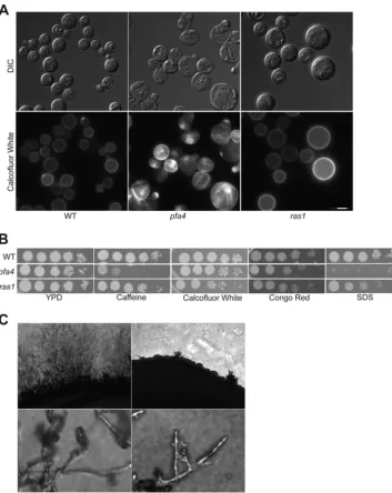
including Ras2, Rho2, and Gpa1 (3,7,31). Consistent with this hypothesis, our analysis of theC. neoformans pfa4⌬mutant strain revealed severalras1-independent phenotypes. During overnight incubations at 37°C,pfa4⌬mutant cells resemble irregular, de-flated balloons, in contrast to the more uniform, large, unbudded phenotype ofras1⌬mutant cells (Fig. 4A). In addition, these cells stain abnormally with the fluorescent chitin stain calcofluor white. In both wild-type andras1⌬mutant cells, calcofluor white uniformly stains the cell wall and septa. In contrast, calcofluor white staining is concentrated in rings and patches over the sur-face ofpfa4⌬mutant cells when they are grown at 37°C (Fig. 4A). Thepfa4⌬mutant strain is also sensitive to reagents that per-turb the cell wall, showing sensitivity to SDS, caffeine, and Congo red. In comparison, in these assays theras1⌬mutant strain exhib-ited only a slight sensitivity to SDS (Fig. 4B). Taken together with the unusual morphology of thepfa4⌬mutant cells, these results suggest that Pfa4 palmitoylates substrates involved in cell wall structure and integrity.
Although Ras1 is required for sexual differentiation inC.
neo-formans, our previous results indicate thatC. neoformansmating is
a palmitoylation-independent process. The Ras1 protein with the C203S and C204S substitutions (Ras1C203S, C204S) is unable to
un-dergo C-terminal palmitoylation, and it is restricted to endomem-branes. However, this palmitoylation-defective mutantras1strain fully supports all stages of sexual differentiation inC. neoformans
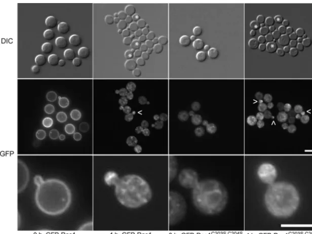
(11). To further characterize the role of palmitoylation and Ras protein localization in the mating process, we assessed the local-ization of the N-terminally tagged Ras1 alleles during a mating reaction and found a dynamic, palmitoylation-independent alter-ation of Ras1 localizalter-ation in cells undergoing fusion. GFP-Ras1 initially primarily localized to the PM in MAT␣wild-type cells.
FIG 3PFA4is required for pathogenesis in a murine model of cryptococcosis. A/Jcr mice were intranasally inoculated with the wild-type H99,pfa4⌬mutant (CBN201), andpfa4⌬/PFA4reconstituted (CBN324) strains. Animal survival was monitored for 40 days.
on September 8, 2020 by guest
http://ec.asm.org/
However, after several hours of incubation with a MATamating partner, the Ras1 localization to the PM was lost, and GFP-Ras1 was instead observed as puncta in the cytoplasm and at the cell bud (Fig. 5). Interestingly, we observed a similar pattern of mating-dependent localization of GFP-Ras1 in cells expressing the mutant form of GFP-Ras1C203S, C204Sthat cannot be
palmitoy-lated, consistent with the intact mating phenotype in this strain (Fig. 5). Previously we found that GFP-Ras1C203S, C204Sis unable to
localize to the PM and is restricted to the endomembranes of
nonmating yeast cells. Taken together, these observations suggest that while the localization of CnRas1 during mating is indepen-dent of palmitoylation, it is very dynamic, moving from the PM to endomembrane structures and polarizing to the emerging bud.
Interestingly, we observed that Pfa4 is required for mating. The
pfa4⌬mutant exhibited a significant mating defect when both partners contained thepfa4 deletion (Fig. 4C, bilateral mating assay). In these assays very few mating foci were observed, mating hyphae were short in comparison to those of the wild type, and few
FIG 4Apfa4strain exhibitsras1-independent phenotypes. (A)pfa4mutant cells exhibit abnormal chitin localization. The wild-type (H99) strain andpfa4⌬
(CBN201) andras1⌬(CBN336) mutant strains were incubated overnight in YPD medium, diluted 10-fold in YPD medium, and then grown for an additional 24 h at 37°C. Cells were stained with calcofluor white prior to imaging (magnification,⫻100) by fluorescence microscopy. Scale bar, 5m. (B) Thepfa4⌬mutant strain is sensitive to cell wall stress. The wild-type (H99) strain andpfa4⌬(CBN201) andras1⌬(CBN336) mutant strains were grown overnight in YPD medium, serially diluted, spotted onto YPD medium containing 1 mg/ml caffeine, 1 mg/ml calcofluor white, 0.5% Congo red, or 0.02% SDS. Plates were incubated at 30°C for 48 h. (C) Pfa4 is required for mating filament formation and differentiation during sexual development in a dose-dependent manner. Bilateral mating mixtures were prepared by mixing overnight cultures of MAT␣and MATawild-type cells (H99 and KN99a) and MAT␣and MATapfa4mutant cells (CBN201 and CBN255). Mixtures were spotted onto MS mating medium and incubated for 5 days in the dark. Images were taken at the periphery of the mating mixtures at magnifications of⫻100 (top panels) and⫻400 (bottom panels).
on September 8, 2020 by guest
http://ec.asm.org/
basidiospores were produced. No defects were observed in unilat-eralpfa4⌬mutant mating assays between apfa4⌬mutant strain and a wild-type strain. In comparison,ras1⌬mutant strains were unable to undergo cell-cell fusion or form filaments in either uni-lateral or biuni-lateral crosses (34,35). These results, together with the prior observation that ras1 palmitoylation mutant strains are completely mating competent (11), suggest that the mating defect of thepfa4⌬mutant strain likely involves the mislocalization of Pfa4 substrates other than Ras1.
DISCUSSION
Protein palmitoylation is a reversible process, allowing a dynamic temporal/spatial movement of targeted proteins to various cellu-lar membranes. In this study, we sought to identify proteins in-volved in the palmitoylation ofC. neoformansRas1. Similar to other organisms,C. neoformanscontains a family of putative pro-teinS-acyltransferases identified by a highly conserved DHHC domain.
Based on sequence analysis and phylogeny,C. neoformansErf2 is the closest homolog toS. cerevisiaeErf2, the primary Ras PAT protein in this species. Additionally,S. cerevisiaeErf2 activity is dependent on Erf4 (3,31,33). However, our experiments suggest that CnPfa4, which is most similar to S. cerevisiaePfa4, is the primary PAT for Ras1.C. neoformans erf2⌬anderf4⌬mutant strains do not exhibit anyras1-like phenotypes. In contrast,pfa4⌬
mutant cells exhibit temperature sensitivity and a temperature-dependent cell size increase, as would be expected with altered CnRas1 function. In addition, the localization of GFP-Ras1 at the PM was perturbed in thepfa4⌬mutant but localized normally in the other PAT mutant strains. Although CnRas1 palmitoylation and PM localization were not completely eliminated in this single-mutant background, the observed level of reduction of Ras
palmi-toylation is similar to that inS. cerevisiaeandS. pombe erf2mutant strains (31,33).
S. cerevisiaePATs and PAT substrates fall into groups. For
ex-ample, Erf2 substrates have additional lipid modifications (preny-lation or myristoy(preny-lation) that occur prior to palmitoy(preny-lation. In contrast, Pfa4 substrates include proteins such as amino acid per-meases that contain transmembrane repeats (7). Our results dem-onstrate that Pfa4 inC. neoformanshas activity on Ras1, offering a different paradigm from that inS. cerevisiae. Whether CnPfa4 has additional activity on transmembrane protein substrates has yet to be determined.
Interestingly,S. cerevisiaePfa4 also has a non-transmembrane protein target, Chs3 (chitin synthase 3), and palmitoylation of Chs3 is required for this protein to exit the ER (36). InC. neofor-mansChs3 is required for cell wall integrity (37,38).chs3⌬and
pfa4⌬ strains exhibit similar sensitivities to cell wall stressors Congo red, caffeine, and SDS, suggesting that CnPfa4 may func-tion as a PAT to Chs3 (37). It is possible that PAT substrate spec-ificity has been altered or expanded inC. neoformansso that Pfa4 serves as the primary PAT for substrates that are targeted by PATs in other species.
Our characterization of the pfa4⌬mutant strain identified
ras1-independent phenotypes, including altered susceptibility to cell wall stress, suggesting additional targets of Pfa4. Both the
ras1⌬andpfa4⌬mutant strains have mating defects, but the na-ture of the altered mating differs between the two strains. The
pfa4⌬mutant strain has a bilateral mating defect in which both mating partners must be mutants for mating to be altered. In contrast, theras1⌬mutant exhibits a unilateral mating defect, and this mating phenotype is independent of palmitoylation (11). In addition to Ras proteins,S. cerevisiaeErf2 substrates include G-al-pha proteins such as Gpa1 and Gpa2 and the G-gamma subunit
FIG 5Ras1 is repolarized in a mating-dependent manner. Mating mixtures were prepared from overnight cultures of MAT␣wild-type cells expressing either GFP-Ras1 (CBN96) or the palmitoylation-defective GFP-Ras1C203S, C204S(CBN167) with wild-type MATacells (KN99a). Mixtures were spotted onto MS
medium plates in 100-l aliquots. At regular intervals, cells were scraped off the plate, resuspended in PBS, and examined for GFP-Ras localization. No fluorescence was detected in wild-type MATacells (representative cells are marked by asterisks in the DIC image). Top panel, DIC images (magnification,⫻100). Middle and bottom panels, fluorescent images (magnification,⫻100). Arrowheads specify cells containing GFP-Ras1 polarized to emerging buds. Bar, 5m.
on September 8, 2020 by guest
http://ec.asm.org/
Ste18 (7). InC. neoformans, G-alpha Gpa1 and G-gamma Gpg1 contain putative palmitoylation sites, and thegpa1andgpg1 mu-tant strains exhibit bilateral mating defects similar to those of the
pfa4⌬mutant strain (39,40).
Previously our lab demonstrated that Ras1 localization to the PM was not required for sexual differentiation. This was surpris-ing since many of the signalsurpris-ing molecules involved in matsurpris-ing re-side on the PM. Here, we observed that Ras1 localization shifts during sexual differentiation from uniform PM localization to cytoplasmic patches and bud tips in a palmitoylation-indepen-dent manner. These results provide an explanation of why palmi-toylation is not required for Ras1 mating competence inC.
neo-formans. Through a mechanism distinct from palmitoylation,
Ras1 is delivered from the endomembranes to specific sites on the PM (bud). This shift of localization is not a result of lipid remod-eling or relocalization of Ras1 within the PM since it occurs even with completely palmitoylation-defective mutant forms of Ras1.
One of the most striking consequences that we observed among theC. neoformans PAT mutations was the dramatically altered virulence of the singlepfa4⌬mutant strain. This virulence attenuation is likely multifactorial, given the observed defects in several phenotypes required for survivalin vivo. Thepfa4⌬ mu-tant is unable to grow at mammalian body temperature, and
sev-eralC. neoformansmutant strains with altered thermotolerance
are hypovirulent (30,34,41,42). Moreover, the prominent cell wall alteration in thepfa4⌬mutant strain also likely contributes to its reduced virulence. Interestingly, expression of the inducible virulence-associated phenotypes of capsule and melanin remains intact with apfa4⌬mutation. Together, these data suggest that Pfa4 is the primary PAT for theC. neoformansRas1 protein and that compensatory palmitoylation by other PATs is not complete in the absence of Pfa4. Also, the Pfa4 protein has substrates in addition to Ras1 that mediate cellular processes involved in mat-ing and cell wall integrity. Therefore, targetmat-ing smat-ingleS -acyltrans-ferases as an antifungal drug target remains a viable strategy de-spite the presence of a large family of potentially functionally related proteins.
ACKNOWLEDGMENTS
This work was supported by PHS grant 1R01-AI050128. Duke Light Mi-croscopy Core Facility DeltaVision instrumentation was funded by grant 1S10RR027528-01.
TheC. neoformansmutant strain collection created by Hiten Madhani (29) was obtained through the Fungal Genetics Stock Center (43). We also acknowledge the Fungal Genomes Initiative at the Broad Institute and Fungi DB (24,25). We thank Sam Johnson at Duke Light Microscopy Core Facility for instruction. We also thank Sandra Breeding for assistance with lab strain curation.
REFERENCES
1.Baekkeskov S, Kanaani J.2009. Palmitoylation cycles and regulation of protein function. Mol Membr Biol26:42–54.http://dx.doi.org/10.1080 /09687680802680108.
2.Salaun C, Greaves J, Chamberlain LH.2010. The intracellular dynamic of protein palmitoylation. J Cell Biol191:1229 –1238.http://dx.doi.org/10 .1083/jcb.201008160.
3.Lobo S, Greentree WK, Linder ME, Deschenes RJ.2002. Identification of a Ras palmitoyltransferase inSaccharomyces cerevisiae. J Biol Chem
277:41268 – 41273.http://dx.doi.org/10.1074/jbc.M206573200. 4.Roth AF, Feng Y, Chen L, Davis NG.2002. The yeast DHHC
cysteine-rich domain protein Akr1p is a palmitoyl transferase. J Cell Biol159:23– 28.http://dx.doi.org/10.1083/jcb.200206120.
5.Mitchell DA, Vasudevan A, Linder ME, Deschenes RJ.2006. Protein
palmitoylation by a family of DHHC proteinS-acyltransferases. J Lipid Res47:1118 –1127.http://dx.doi.org/10.1194/jlr.R600007-JLR200. 6.Hornemann T.2015. Palmitoylation and depalmitoylation defects. J
In-herit Metab Dis38:179 –186.http://dx.doi.org/10.1007/s10545-014-97 53-0.
7.Roth AF, Wan J, Bailey AO, Sun B, Kuchar JA, Green WN, Phinney BS, Yates JR, Davis NG.2006. Global analysis of protein palmitoylation in yeast. Cell125:1003–1013.http://dx.doi.org/10.1016/j.cell.2006.03.042. 8.Prior IA, Hancock JF.2012. Ras trafficking, localization and
compart-mentalized signalling. Semin Cell Dev Biol23:145–153.http://dx.doi.org /10.1016/j.semcdb.2011.09.002.
9.Eisenberg S, Laude AJ, Beckett AJ, Mageean CJ, Aran V, Hernandez-Valladares M, Henis YI, Prior IA.2013. The role of palmitoylation in regulating Ras localization and function. Biochem Soc Trans41:79 – 83. http://dx.doi.org/10.1042/BST20120268.
10. Chang EC, Philips MR.2006. Spatial segregation of Ras signaling: new evidence from fission yeast. Cell Cycle5:1936 –1939.http://dx.doi.org/10 .4161/cc.5.17.3187.
11. Nichols CB, Ferreyra J, Ballou ER, Alspaugh JA. 2009. Subcellular localization directs signaling specificity of theCryptococcus neoformans
Ras1 protein. Eukaryot Cell 8:181–189. http://dx.doi.org/10.1128/EC .00351-08.
12. Sherman F.1991. Getting started with yeast. Methods Enzymol194:3–21. http://dx.doi.org/10.1016/0076-6879(91)94004-V.
13. Xue C, Tada Y, Dong X, Heitman J.2007. The human fungal pathogen
Cryptococcuscan complete its sexual cycle during a pathogenic association with plants. Cell Host Microbe1:263–273.http://dx.doi.org/10.1016/j .chom.2007.05.005.
14. Fraser JA, Subaran RL, Nichols CB, Heitman J.2003. Recapitulation of the sexual cycle of the primary fungal pathogenCryptococcus neoformans
var. gattii: implications for an outbreak on Vancouver Island, Canada. Eukaryot Cell2:1036 –1045.http://dx.doi.org/10.1128/EC.2.5.1036-1045 .2003.
15. Kim MS, Kim SY, Jung KW, Bahn YS.2012. Targeted gene disruption in
Cryptococcus neoformansusing double-joint PCR with split dominant se-lectable markers. Methods Mol Biol845:67– 84.http://dx.doi.org/10.1007 /978-1-61779-539-8_5.
16. Goins CL, Gerik KJ, Lodge JK.2006. Improvements to gene deletion in the fungal pathogenCryptococcus neoformans: absence of Ku proteins in-creases homologous recombination, and co-transformation of indepen-dent DNA molecules allows rapid complementation of deletion pheno-types. Fungal Genet Biol43:531–544.http://dx.doi.org/10.1016/j.fgb.2006 .02.007.
17. Toffaletti DL, Rude TH, Johnston SA, Durack DT, Perfect JR.1993. Gene transfer inCryptococcus neoformansby use of biolistic delivery of DNA. J Bacteriol175:1405–1411.
18. Davidson RC, Cruz MC, Sia RA, Allen B, Alspaugh JA, Heitman J.2000. Gene disruption by biolistic transformation in serotype D strains of Cryp-tococcus neoformans. Fungal Genet Biol29:38 – 48.http://dx.doi.org/10 .1006/fgbi.1999.1180.
19. Schindelin J, Arganda-Carreras I, Frise E, Kaynig V, Longair M, Pietz-sch T, PreibiPietz-sch S, Rueden C, Saalfeld S, Schmid B, Tinevez JY, White DJ, Hartenstein V, Eliceiri K, Tomancak P, Cardona A.2012. Fiji: an open-source platform for biological-image analysis. Nat Methods9:676 – 682.http://dx.doi.org/10.1038/nmeth.2019.
20. Levine TP, Wiggins CA, Munro S.2000. Inositol phosphorylceramide synthase is located in the Golgi apparatus ofSaccharomyces cerevisiae. Mol Biol Cell11:2267–2281.http://dx.doi.org/10.1091/mbc.11.7.2267. 21. Kmetzsch L, Joffe LS, Staats CC, de Oliveira DL, Fonseca FL, Cordero
RJ, Casadevall A, Nimrichter L, Schrank A, Vainstein MH, Rodrigues ML.2011. Role for Golgi reassembly and stacking protein (GRASP) in polysaccharide secretion and fungal virulence. Mol Microbiol81:206 – 218.http://dx.doi.org/10.1111/j.1365-2958.2011.07686.x.
22. Wan J, Roth AF, Bailey AO, Davis NG.2007. Palmitoylated proteins: purification and identification. Nat Protoc2:1573–1584.http://dx.doi.org /10.1038/nprot.2007.225.
23. Cox GM, Mukherjee J, Cole GT, Casadevall A, Perfect JR.2000. Urease as a virulence factor in experimental cryptococcosis. Infect Immun68:
443– 448.http://dx.doi.org/10.1128/IAI.68.2.443-448.2000.
24. Janbon G, Ormerod KL, Paulet D, Byrnes EJ, III, Yadav V, Chat-terjee G, Mullapudi N, Hon CC, Billmyre RB, Brunel F, Bahn YS, Chen W, Chen Y, Chow EW, Coppee JY, Floyd-Averette A, Gaillar-din C, Gerik KJ, Goldberg J, Gonzalez-Hilarion S, Gujja S, Hamlin
on September 8, 2020 by guest
http://ec.asm.org/
JL, Hsueh YP, Ianiri G, Jones S, Kodira CD, Kozubowski L, Lam W, Marra M, Mesner LD, Mieczkowski PA, Moyrand F, Nielsen K, Proux C, Rossignol T, Schein JE, Sun S, Wollschlaeger C, Wood IA, Zeng Q, Neuveglise C, Newlon CS, Perfect JR, Lodge JK, Idnurm A, Stajich JE, Kronstad JW, Sanyal K, Heitman J, Fraser JA, et al.2014. Analysis of the genome and transcriptome ofCryptococcus neoformans
var.grubiireveals complex RNA expression and microevolution leading to virulence attenuation. PLoS Genet 10:e1004261. http://dx.doi.org/10 .1371/journal.pgen.1004261.
25. Stajich JE, Harris T, Brunk BP, Brestelli J, Fischer S, Harb OS, Kissinger JC, Li W, Nayak V, Pinney DF, Stoeckert CJ, Jr, Roos DS. 2012. FungiDB: an integrated functional genomics database for fungi. Nucleic Acids Res40:D675–D681.http://dx.doi.org/10.1093/nar/gkr918. 26. Dereeper A, Guignon V, Blanc G, Audic S, Buffet S, Chevenet F, Dufayard
JF, Guindon S, Lefort V, Lescot M, Claverie JM, Gascuel O.2008. Phylog-eny.fr: robust phylogenetic analysis for the non-specialist. Nucleic Acids Res
36:W465–W469.http://dx.doi.org/10.1093/nar/gkn180.
27. Mitchell DA, Hamel LD, Ishizuka K, Mitchell G, Schaefer LM, De-schenes RJ.2012. The Erf4 subunit of the yeast Ras palmitoyl acyltrans-ferase is required for stability of the acyl-Erf2 intermediate and palmitoyl transfer to a Ras2 substrate. J Biol Chem287:34337–34348.http://dx.doi .org/10.1074/jbc.M112.379297.
28. Swarthout JT, Lobo S, Farh L, Croke MR, Greentree WK, Deschenes RJ, Linder ME.2005. DHHC9 and GCP16 constitute a human protein fatty acyltransferase with specificity for H- and N-Ras. J Biol Chem280:31141– 31148.http://dx.doi.org/10.1074/jbc.M504113200.
29. Liu OW, Chun CD, Chow ED, Chen C, Madhani HD, Noble SM.2008. Systematic genetic analysis of virulence in the human fungal pathogen
Cryptococcus neoformans. Cell 135:174 –188.http://dx.doi.org/10.1016/j .cell.2008.07.046.
30. Nichols CB, Perfect ZH, Alspaugh JA.2007. A Ras1-Cdc24 signal trans-duction pathway mediates thermotolerance in the fungal pathogen Cryp-tococcus neoformans. Mol Microbiol63:1118 –1130.http://dx.doi.org/10 .1111/j.1365-2958.2006.05566.x.
31. Bartels DJ, Mitchell DA, Dong X, Deschenes RJ.1999. Erf2, a novel gene product that affects the localization and palmitoylation of Ras2 in Saccha-romyces cerevisiae. Mol Cell Biol19:6775– 6787.
32. Budde C, Schoenfish MJ, Linder ME, Deschenes RJ.2006. Purification and characterization of recombinant protein acyltransferases. Methods
40:143–150.http://dx.doi.org/10.1016/j.ymeth.2006.07.017.
33. Young E, Zheng Z-Y, Wilkins AD, Jeong H-T, Li M, Lichtarge O, Chang EC.2014. Regulation of Ras localization and cell transformation by evo-lutionarily conserved palmitoyltransferases. Mol Cell Biol34:374 –385. http://dx.doi.org/10.1128/MCB.01248-13.
34. Alspaugh JA, Cavallo LM, Perfect JR, Heitman J.2000. RAS1 regulates filamentation, mating and growth at high temperature ofCryptococcus
neoformans. Mol Microbiol36:352–365.http://dx.doi.org/10.1046/j.1365 -2958.2000.01852.x.
35. Waugh MS, Nichols CB, DeCesare CM, Cox GM, Heitman J, Alspaugh JA.2002. Ras1 and Ras2 contribute shared and unique roles in physiology and virulence ofCryptococcus neoformans. Microbiology148:191–201. 36. Lam KK, Davey M, Sun B, Roth AF, Davis NG, Conibear E. 2006.
Palmitoylation by the DHHC protein Pfa4 regulates the ER exit of Chs3. J Cell Biol174:19 –25.http://dx.doi.org/10.1083/jcb.200602049.
37. Banks IR, Specht CA, Donlin MJ, Gerik KJ, Levitz SM, Lodge JK.2005. A chitin synthase and its regulator protein are critical for chitosan produc-tion and growth of the fungal pathogenCryptococcus neoformans. Eu-karyot Cell 4:1902–1912. http://dx.doi.org/10.1128/EC.4.11.1902-1912 .2005.
38. Baker LG, Specht CA, Donlin MJ, Lodge JK.2007. Chitosan, the deacety-lated form of chitin, is necessary for cell wall integrity inCryptococcus neoformans. Eukaryot Cell 6:855– 867. http://dx.doi.org/10.1128/EC .00399-06.
39. Alspaugh JA, Perfect JR, Heitman J. 1997. Cryptococcus neoformans
mating and virulence are regulated by the G-protein alpha subunit GPA1 and cAMP. Genes Dev11:3206 –3217.http://dx.doi.org/10.1101/gad.11 .23.3206.
40. Li L, Shen G, Zhang ZG, Wang YL, Thompson JK, Wang P. 2007. Canonical heterotrimeric G proteins regulating mating and virulence of
Cryptococcus neoformans. Mol Biol Cell18:4201– 4209.http://dx.doi.org /10.1091/mbc.E07-02-0136.
41. Odom A, Muir S, Lim E, Toffaletti DL, Perfect J, Heitman J.1997. Calcineurin is required for virulence ofCryptococcus neoformans. EMBO J
16:2576 –2589.http://dx.doi.org/10.1093/emboj/16.10.2576.
42. Ballou ER, Nichols CB, Miglia KJ, Kozubowski L, Alspaugh JA.2010. Two CDC42 paralogues modulateCryptococcus neoformans thermotoler-ance and morphogenesis under host physiological conditions. Mol Micro-biol75:763–780.http://dx.doi.org/10.1111/j.1365-2958.2009.07019.x. 43. McCluskey K, Wiest A, Plamann M.2010. The Fungal Genetics Stock
Center: a repository for 50 years of fungal genetics research. J Biosci35:
119 –126.http://dx.doi.org/10.1007/s12038-010-0014-6.
44. Perfect JR, Lang SD, Durack DT.1980. Chronic cryptococcal meningitis: a new experimental model in rabbits. Am J Pathol101:177–194. 45. Nielsen K, Cox GM, Wang P, Toffaletti DL, Perfect JR, Heitman J.2003.
Sexual cycle ofCryptococcus neoformansvar.grubiiand virulence of con-genicaand␣isolates. Infect Immun71:4831– 4841.http://dx.doi.org/10 .1128/IAI.71.9.4831-4841.2003.
46. Selvig K, Ballou ER, Nichols CB, Alspaugh JA.2013. Restricted substrate specificity for the geranylgeranyltransferase-I enzyme inCryptococcus neo-formans: implications for virulence. Eukaryot Cell12:1462–1471.http: //dx.doi.org/10.1128/EC.00193-13.