Tuberculosis of the neuromusculoskeletal
system: a review of two cases presenting as
chiropractic patients
Ismat Kanga,
BSc, DC, FCCS(C)1John A. Taylor,
DC, DACBR2Craig Jacobs,
MSc, DC, FCCS(C)3Geoff Outerbridge,
MSc, DC41 Department of Graduate Studies, Clinical Sciences, Canadian Memorial Chiropractic College, Toronto, Ontario, Canada 2 Professor, Chiropractic Department, D’Youville College, Buffalo, NY
3 Director, Clinical Education and Patient Care, Division of Clinical Education, Canadian Memorial Chiropractic College (CMCC), Toronto,
Ontario, Canada
4 Clinic Director, World Spine Care and Adjunct Faculty, Canadian Memorial Chiropractic College, Toronto, Ontario, Canada
Corresponding author: Ismat Kanga [email protected]
T: (416) 482-2340 F: (416) 482-2560
6100 Leslie Street, Toronto, Ontario, Canada, M2H 3J1
Consent: Written consent was obtained from both patients to use information and images from their files for this case report. ©JCCA 2015
Tuberculosis caused by Mycobacterium tuberculosis is a major public heath problem world-wide, particularly in low-income countries. Increased number of
immunocompromised patients and immigration from countries where tuberculosis is endemic has resulted in increased number of cases in high-income countries. Tuberculosis can affect any organ system, but is of particular interest to chiropractors when it affects the neuromusculoskeletal system. Patients with tuberculosis of the neuromusculoskeletal system can present with mechanical low back pain or with complex neurologic deficits. The aim of this paper is to highlight the importance of considering a diagnosis of tuberculosis in susceptible populations and the devastating
consequences of the disease. The epidemiology, clinical
Introduction
It is estimated that 2.3 billion people worldwide are in-fected with tuberculosis (TB).1 Despite advances in health care, TB continues to be a major public health problem worldwide, particularly in low-income countries.1–3 In-creased numbers of immunocompromised patients and immigration of people from countries where TB is en-demic has resulted in a rise in the number of cases in high-income countries.3,4 Tuberculosis caused by Myco-bacterium tuberculosis, most commonly affects the lungs but can affect any organ system.1–3 TB also affects the neuromusculoskeletal system. The spine is afflicted in 1 to 5% of all patients infected with TB and is the most common site, occurring in approximately 50% of the cases of skeletal TB.2–5 TB of the spine or TB spondylo-discitis is also known as Pott’s disease. It was named after Sir Percival Pott, a British surgeon who first described spinal TB and the surgical treatment of paravertebral ab-scesses in his monograph in 1779.3,6,7 The central nervous system is also involved in approximately 10% of all pa-tients with TB.8
These two cases describe TB in two HIV-positive (Human immunodeficiency virus) patients. The first case chronicles a young male with a history of chronic low back pain, who presented to the chiropractic clinic for treatment. The second patient presented after TB in-volving the central nervous system (CNS) was diagnosed and treated. This patient suffered complex sequelae as a consequence of the infection resulting in paraparesis. The aim of this paper is to emphasize the importance of considering tuberculosis in the differential diagnosis in patients presenting with presumed mechanical back pain and also to remind chiropractors of the devastating conse-quences of the disease. The epidemiology, pathogenesis, imaging features and clinical presentation of TB will be
presented in order to highlight the disease in order to fa-cilitate appropriate management of these patients. Cases:
Case 1
History: A 32-year-old male presented to the World Spine Care Clinic in Mahalapye, Botswana with a complaint of chronic low back pain. He attributed his pain to lowering a 50kg bag from waist height to the ground around 11 months prior. The onset of pain was immediate. He char-acterized the nature of his pain as a dull ache and a cramp down the back of his right thigh, but no neurologic symp-toms were reported. On presentation, he reported constant pain and rated his pain as 10/10 on a scale of 0 to 10 (0 representing no pain at all and 10 being the worst pain ever). Aggravating factors included bending, lifting and prolonged sitting. Lying down helped reduce the intensity of pain somewhat. Prior to his attending the chiropractic clinic, previous treatment for his pain included analgesics (paracetamol), non-steroidal anti-inflammatory (diclo-fenac) injections and tricyclic antidepressant medication (amitriptyline). He reported no constitutional symptoms (fevers, weight loss or chills). Inquiry into his medical history revealed that he was diagnosed with HIV seven years ago. His last CD4 count, assessed a few months pri-or, was 465 cells per microliter (Normal range 500-1000 cells per microliter).
Physical examination: Examination revealed severe-ly restricted and painful lumbar ranges of motion, most notably in rotation and flexion. Kemp’s test was positive bilaterally. Intersegmental joint restrictions and tender-ness were found in the lower lumbar spine from L3 to L5 and bilateral sacroiliac joints. Spinous percussion re-vealed tenderness at the lower lumbar levels. His lumbar features and management of tuberculosis will also be
presented to facilitate early diagnosis, appropriate referral and multidisciplinary care of these patients. (JCCA 2015; 59(1):13-23)
k e y w o r d s: tuberculosis, tuberculous
spondylodiscitis tuberculosis radiculomyelitis, HIV, chiropractic
la lutte contre celle-ci, afin de favoriser un diagnostic précoce, le choix de ressources appropriées et les soins multidisciplinaires donnés à ces patients.
(JCCA 2015; 59(1):13-23)
m o t s c l é s : tuberculose, spondylodiscite
spine, gluteal and posterior thigh musculature were ten-der to direct palpation. Upper and lower limb neurologic examination including reflexes, motor and sensory exam-ination were within normal limits.
Diagnostic Imaging: The patient brought in radio-graphs, which revealed severe disc space narrowing at L4-5 with erosion and partial collapse of the anterior-su-perior and anterior-inferior endplates of the L5 vertebral body, and to a lesser extent the inferior endplate of L4. In addition, prominent sclerosis of the L5 vertebral body was also evident. (Figure 1) Based on radiographic find-ings consistent with infectious spondylodiscitis, the pa-tient was referred to an orthopedic surgeon. The papa-tient was started on immediate anti-tubercular therapy and re-ferred for magnetic resonance imaging (MRI) of the lum-bar spine.
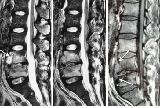
An MRI examination was performed two weeks later. The fluid-sensitive (T2-weighted) images revealed exten-sive high signal intensity with anterior destruction and
collapse of the L4 and L5 vertebral bodies. In addition, the L4-5 disc space was destroyed and a hyperintense inflammatory abscess extended into the anterior soft tis-sues coursing in a subligamentous distribution resulting in bulging of the anterior longitudinal ligament anteriorly. (Figures 2A, B, C) T1-weighted images obtained follow-ing intravenous injection of gadolinium revealed signifi-cant enhancement of the subligamentous abscess. (Figure 2D) The patient history combined with imaging findings confirmed the presence of tuberculous spondylodiscitis of the spine (Pott’s spine).
Case 2
History: A 40-year old male was admitted to a Toron-to area hospital with a fever and postictal (altered con-sciousness) status after having a seizure. Serological testing revealed his serum Venereal Disease Research Laboratory (VDRL) test and Fluorescent Treponemal Antibody-Absorption (FTA-ABS) test were positive, 1A 1B 1C
Figure 1:
Patient 1. Frontal (A), and lateral (B, C) radiographs reveal severe L4-5 disc space narrowing, erosion and partial collapse of the anterior-superior and anterior-inferior L5 vertebral endplates and to a lesser extent the inferior L4
2B 2C 2D 2A
Figure 2:
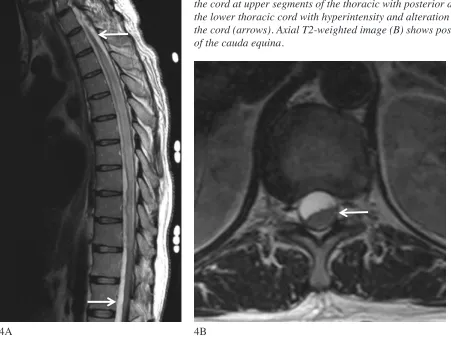
indicating neurosyphilis. At that time he was also diag-nosed with HIV with a CD4 count of 77 cells per micro-liter. Two weeks later, while still at the hospital the pa-tient developed shortness of breath. He was found to have pleural effusion and his pleural fluid tested positive for Acid-Fast Bacilli, confirming pulmonary tuberculosis. He was started on anti-tubercular therapy and antiretroviral medications. During his hospital stay, he started to experi-ence weakness in his lower extremities and subsequently developed a neurogenic bladder. MRI revealed a large plaque located on the dorsal surface of the spinal cord ex-tending from T3 to T11 and increased signal intensity of the cord suggesting edema or ischemia. (Figures 3A, B) Multiple small tubercular granulomas were also
visual-ized on the surface of the brain and cervical spinal cord. The MRI also showed assimilation of the odontoid pro-cess of C2 and the C1 vertebral body. The patient suffered neurologic sequelae from his disseminated TB including TB meningitis, TB arachnoiditis and TB granulomata. Follow-up one year after revealed severe pyramidal weakness bilaterally. Hip flexors were graded as 2/5, hamstrings 4/5 and ankle dorsiflexors 1/5. Other neuro-logic findings included reduced proprioception and vibra-tion sense at the ankles and sensory loss over the surface of the thoracic cage. A repeat MRI revealed anterior placement of the superior thoracic cord and posterior dis-placement of the inferior thoracic cord. The T2-weighted sequences revealed a hyperintense signal with alteration Figure 3:
Patient 2. T2-weighted sagittal image (A) reveals a large linear plaque (arrows) on the dorsal surface of the cord extending from the T3 to T11 segments, resulting in diffuse ventral deviation and cord compression. The T2-weighted axial image (B) shows increased signal intensity in the cord (arrows) due to edema or ischemia.
of the contour of the cord, adhesions and formation of CSF pockets. (Figures 4 A, B)
This patient presented to a Canadian Memorial Chiro-practic College teaching clinic four years after his dis-seminated TB, with complaints of neck pain, low back pain and headaches. His neck pain was also associated with radiation of pain into his left arm and into the third digit. He attributed the onset of his pain to his long hos-pitalization associated with his TB. He reported the pain was constant and rated the intensity of his neck pain as a 10 out of 10. Sitting for prolonged periods aggravated his low back pain, while movement of his neck aggravated his neck pain. Hot showers, acupuncture and manual therapy were reported to relieve his pain. His medical history is significant for Type 2 diabetes, HIV, neurogenic bladder,
obstructive sleep apnea, epistaxis, dyslipidemia, hyper-tension, cognitive impairment and anemia. Past medical history was significant for neurosyphilis with seizures, gastritis and acute respiratory distress syndrome.
Physical Examination: Observation revealed a paretic gait. Active ranges of motion testing in his cervical and lumbar spine were painful and restricted by 25% globally. Upper limb neurologic examination revealed normal sen-sation, motor strength and deep tendon reflexes (DTRs). Hoffman’s test (flicking terminal phalanx of the third or fourth digits) was positive bilaterally. Lower limb DTRs revealed 3+ bilaterally at L4 and 1+ bilaterally at S1. Mo-tor strength was diminished and was rated as 3/5 at L2 and 4/5 in L3-S1 bilaterally. Sensation, plantar reflex and clonus were within normal limits. Tenderness was noted 4A 4B
Figure 4:
bilaterally in his lumbar and cervical paraspinal, trapezius and levator scapulae muscles.
Diagnostic Imaging: Radiographs of his cervical spine revealed mild cervical degenerative disc disease and moderate foraminal stenosis at the C5-7 levels. The atlantoaxial articulation was anomalous with an appar-ent os odontoideum and assimilation of the odontoid process ossicle with the anterior tubercle of C1. (Figure 5) Flexion-extension radiographs revealed marked seg-mental instability with 6 mm of translation of this anom-alous C1 and C2 ossicle in relation to the C2 vertebral body.
Diagnosis: The patient was diagnosed with mechanic-al low back pain, degenerative disc and joint disease and lateral stenosis from C5 to C7, in addition to his persistent Figure 5:
Patient 2. Lateral cervical radiograph reveals assimilation of a C2 os odontoideum with the C1 anterior tubercle, degenerative joint and disc disease and lateral stenosis from C5 through C7.
neurologic deficits as a result of his TB, neurosyphilis and HIV.
Chiropractic Management: The patient was treat-ed with myofascial release of the cervical and lumbar paraspinal muscles, mobilization of the lumbar spine and active rehabilitation including neck and lower limb isometric strengthening and education. The patient re-sponded well to chiropractic care and noticed significant improvement in his strength. Though still requiring a wheelchair for a significant amount of the time, he was able to use a walker for some activities of daily living and accomplish his daily hygiene routine without assistance. Discussion:
Epidemiology
The global prevalence of TB is increasing at a rate of 1.1 % per year.2,9 TB kills 1.4 to 2 million people every year and is second only to HIV as the leading cause of death from infectious disease.1,3 Countries in Africa and South-East Asia have the greatest burden of disease globally.10 Immigration from these regions represents the majority of cases in high-income countries. Risk factors for TB in-clude advanced age, malnutrition, poor sanitation, home-lessness, alcohol and substance abuse, immunocompro-mised states and diabetes.2,3,11 Infection occurs through the respiratory tract, by inhalation of droplet nuclei. In-dividuals with active pulmonary TB spread droplet nuclei when coughing, sneezing or talking. Droplet nuclei are able to penetrate the alveoli, where they multiply before spreading to various organ systems.2
Association with Immunocompromised patients
comprom-ised immune system and are at greater risk of infection with TB.13 It should also be noted that certain patients might be immunocompromised as a result of iatrogenic influences. The immunoinhibitory effects of medications and biologic substances such as corticosteroids and an-ti-tumour necrosis factor (TNF) biologics are well docu-mented.14 Anti-TNF-α agents (infliximab, etanercept and adalimumab), used in the treatment of rheumatoid arth-ritis, ankylosing spondylitis, psoriatic arthritis and juven-ile idiopathic arthritis increase the risk of TB and should be considered in the etiology of tuberculosis infections.15
Tuberculous spondylodiscitis
Pathogenesis: The spread of TB to the vertebral column occurs as a result of haematogenous dissemination via the Batson’s venous plexus or lymphatic drainage via the para-aortic lymph nodes.4,11,16 Once it has reached the ver-tebral column the infection usually arises in the anterior portion of the body.9 The tubercle bacillus begins gradual trabecular destruction and demineralization, progressing to the cortex, adjacent disc and vertebral body. Within the vertebral body a granulomatous lesion develops, com-prised of leukocytes, caseous material, bone debris and tubercle bacilli.4,11,16 The collapse and wedging of mul-tiple adjacent vertebral bodies generate the characteristic gibbus deformity.4,5 Paraspinal abscesses are located in 70% of patients. Psoas abscesses are common and can ex-tend deep to the iliopsoas fascia into the proximal thigh, resulting in a groin mass.11,17 Differential diagnoses for TB spondylodiscitis include pyogenic and fungal infec-tions, brucellosis, compression fractures and metastatic disease.9,11
Clinical Presentation: TB spondylodiscitis has an in-sidious onset of symptoms and slow progression, making early diagnosis of TB fundamentally important in pre-venting deformity and paraplegia. The mean duration be-tween infection and clinical presentation can range from a few days to three years.11,18 The typical presentation of TB spondylodiscitis is low back pain over the affected verte-bral bodies, low-grade fever, chills, weight loss.16 Clinical examination may reveal local tenderness and limitation in spinal motion.11 Neurologic complications such as radicu-lar symptoms, cauda equina and paraplegia can arise from edema, vascular engorgement and retropulsed debris from vertebral body collapse.11
Diagnostic imaging features of tuberculous
spondylo-discitis: TB spondylodiscitis occurs most frequently in the lower thoracic and thoracolumbar spine.9,11,16,18 Con-ventional radiographs are not sensitive in detecting early TB of the spinal column, as vertebral body destruction is not visualized until 50% of the trabeculae have been destroyed, corresponding to approximately six months through the course of the disease.5,11 At later stages in the disease, loss of vertebral height, disc space narrowing, erosions, paravertebral masses and vertebral body col-lapse may be visualized on the radiographs.5,11 Paraverte-bral abscesses are often visualized as areas of soft tissue swelling adjacent to the spine.19 Changes on computed tomography (CT) and MRI are detectable as early as six weeks after infection.4 CT scans are particularly useful in revealing destruction of the vertebral endplates, frag-mentation of the vertebral body, paravertebral calcifica-tions and in facilitating image-guided biopsy to confirm a diagnosis.5,11 Calcification within the paraspinal abscess, seen on CT images are pathognomonic for TB.11 TB spon-dylodiscitis can also present as isolated involvement of the posterior elements, infection limited to one vertebral segment or multiple nonadjacent vertebrae and an ivory vertebrae.11,17 MRI is most sensitive in revealing early changes within the bone and endplate and in demonstrat-ing the extent of spinal cord compromise.5,11 TB lesions appear as low signal intensity on T1-weighted images and heterogeneous increased signal on T2-weighted images.11
Tuberculosis of the Central Nervous System (CNS).
TB of the CNS arises from haematogenous spread sec-ondary to disease in another part of the body. CNS TB is the most dangerous form of systemic TB, responsible for high mortality and severe disability.20 Small tuberculous lesions or Rich’s foci develop in the meninges, surface of the brain or spinal cord. Growth or rupture of these lesions results in the numerous types of CNS tuberculo-sis.8 Tuberculous meningitis (TBM) is the most common presentation of CNS tuberculosis.20 The clinical presenta-tion of TBM includes fever, headache and altered level of consciousness and other meningeal signs such as neck stiffness, photophobia and vomiting.20
in-volving the arachnoid lining or leptomeninges of the spin-al cord.21
Pathogenesis: Three separate mechanisms may be re-sponsible for the development of TBRM. It may result from 1. a primary tuberculosis lesion developed in the central nervous system; 2. direct extension from the ver-tebral body; or 3. descent from intracranial TB meningitis (most common).21,22 The thoracic spine is most frequently involved, and less often the lumbar and cervical spine.22,23 TBRM is characterized by a gross granulomatous reac-tion with associated histiocytic proliferareac-tion, caseareac-tion and tubercle formation that expands and fills the space between the spinal dura mater and the leptomeninges.21 It often extends over several segments, envelops the spinal cord and compresses on it and the nerve roots, but does not penetrate into either.8,20–22 The neurologic symptoms experienced by patients occur as a result of direct com-pression of the cord and ischemia.8 Ischemia often de-velops in the spinal arteries as a result of vasculitis and thrombosis.20,21 Within the parenchyma of the cord syr-ingomyelia and myelomalacia may develop as a result of vacuolization, atrophy, and central necrosis.21,22
Clinical Presentation: TBRM may manifest at any point after infection with TB meningitis. Clinical presentation of TBRM includes paraparesis, paraplegia, radicular pain, paresthesias, muscle atrophy and neurogenic bladder.21,22 Absence of deep tendon reflexes, weakness in the lower limbs and upgoing plantar response are also commonly seen.21 Diagnosis is based on clinical features, presence of tuberculous meningitis, CSF analysis and advanced imaging.24 CSF analysis displays an active inflammatory response with lymphocytosis, decreased glucose content and abnormally high protein levels.21
Diagnostic imaging features of tuberculosis radiculo-myelitis: Loculation and destruction of the subarachnoid space, loss of contour of the spinal cord, myelomalacia and adhesions (thickening, clumping or matting) of the nerve roots particularly in the lumbar spine are common MRI findings of TBRM.21 TBRM on T1-weighted images display increased intensity of the CSF fluid and loss of the contour of the spinal cord, due to increased protein within the CSF. Adhesions of the nerve roots may also be visualized. Fluid-sensitive T2-weighted images display increased signal intensities within the cord as a result of myelitis, edema and ischemia.23 During the chronic phase of the infection, MRI sequences may not exhibit
enhance-ment but continue to show signs of arachnoiditis such as the adhesions of the nerve roots. Development of syringo-myelia and myelomalacia can also be clearly visualized on MRI.21,22
Management
Treatment for TB includes a combination of anti-tuber-cular medications including isoniazid, rifampin, pyraz-inamide, ethambutol, streptomycin, ethionamide and cycloserine, administered over a span of 12 to 13 months.25 Patients with HIV treated with a course of anti-tubercular drugs display the same clinical, radiological and micro-biological response as HIV-negative patients.2 Surgery was previously utilized as a treatment for TB, however it is now used more sparingly and selectively. It is used less for controlling disease and more for reducing or recti-fying spinal deformities and neurologic complications.3 The use of anti-tubercular drugs has resulted in multi-drug resistant strains creating a further complication in the management of TB.26 The first patient presented in the late of stage of infection, after significant vertebral body destruction, but demonstrating only symptoms of mechanical low back pain. He had substantial lower back pain, tenderness and restriction in range of motion but no signs of TB such as weight loss, fever, chills or neurologic symptoms. The diagnosis of TB can easily be missed, especially in patients at early stages of infection with similar signs and symptoms to those of our patient, particularly in the absence of constitutional symptoms or radiographic findings of infection. When patients present to chiropractors with low back pain, infection, both pyo-genic and non-pyopyo-genic should be considered in the dif-ferential diagnosis and should be ruled out during history and physical examination. Red flags suggesting a more serious underlying condition should also be excluded. Any history of constant progressive pain, past history of cancer, trauma, prolonged use of corticosteroids, un-explained weight loss, fever and immunocompromised status warrants further investigation.27
radiculomyelitis as the principal cause of his neurologic deficits. This case is an excellent reminder that TB is a world-wide epidemic and can result in death and disabil-ity. It is a common differential consideration in low-in-come countries, however its recognition is difficult in high-income nations. Because chiropractors encounter it so infrequently, it is often overlooked as a cause of back pain and often mistaken for metastasis or simple compres-sion fracture owing to similar clinical presentation and imaging findings.16 Tuberculous spondylitis presenting as mechanical neck pain has been previously described the in the literature.28 A 21 year-old male presented to a chiropractic office with neck pain, stiffness and difficulty swallowing. Cervical radiographs taken were unremark-able, and the patient was treated with spinal manipulation, trigger point therapy and stretching. Though the patient regained all active ranges of motion, the treatment was unable to alleviate the patients’ persistent tenderness in his suboccipital region. As a result the patient was re-ferred to a physiatrist and subsequently hospitalized with a presumptive diagnosis of tuberculous spondylitis. 28 Despite the low prevalence of TB in North America, these patients – especially immigrants and immunocom-promised patients – may indeed present to our offices. Patients with TB spondylodiscitis present with tender-ness, hypertonicity of the paraspinal muscles and limited mobility. Soft tissue therapy, education and mobilization/ manipulation of unaffected vertebral bodies may be safe-ly applied. Affected vertebral bodies have compromised integrity and are a contra-indication to high-velocity low-amplitude spinal manipulation. Patients who are af-flicted with CNS TB despite early treatment are often left with neurologic deficits including paraparesis and para-plegia. Rehabilitation is important for these patients to maintain and improve strength and to treat soft tissue con-tractures. Patients are treated with anti-tubercular drugs for a period of a year or longer and therefore may even present to our clinics while being on concurrent treat-ment. It would be important for chiropractors to assist in monitoring these patients for disease progression or onset of new symptoms and collaborate with other health care professionals in the care of these patients.
Summary:
It is important to include TB as a differential diagnosis in elderly and immunocompromised patients and
immi-grants from areas where TB is endemic. As it is uncom-mon in high-income countries, its recognition is often dif-ficult and often presents with similar signs and symptoms as mechanical low back pain, metastasis or compression fractures. TB may affect multiple organ systems within the body, but chiropractors should be aware of the clinic-al presentation of neuromusculoskeletclinic-al TB. Presentation of these two cases aims to heighten the awareness of the global burden of disease and remind chiropractors that TB may present in high-income nations as well. Further-more, these cases emphasize the role of chiropractors in the diagnosis and management of the disease.
References
1. CDC Grand Rounds: the TB/HIV syndemic. MMWR Morb Mortal Wkly Rep. 2012;61(26):484–9.
2. Harries AD, Dye C. Tuberculosis. Ann Trop Med Parasitol. 2014;100(5-6):415–31.
3. Tuli SM. Tuberculosis of the spine: a historical review. Clin Orthop Relat Res. 2007;460:29–38.
4. Tuli SM. General principles of osteoarticular tuberculosis. Clin Orthop Relat Res. 2002;(398):11–9.
5. Moore SL, Rafii M. Imaging of musculoskeletal and spinal tuberculosis. Radiol Clin North Am. 2001;39(2):329–42. 6. Dass B, Puet TA, Watanakunakorn C. Tuberculosis of
the spine (Pott’s disease) presenting as “compression fractures”. Spinal Cord. 2002;40(11):604–8.
7. Garg RK, Somvanshi DS. Spinal tuberculosis: a review. J Spinal Cord Med. 2011;34(5):440–54.
8. Garg RK. Tuberculosis of the central nervous system. Postgrad Med J. 1999;75(881):133–40.
9. Burrill J, Williams CJ, Bain G, Conder G, Hine AL, Misra RR. Tuberculosis: a radiologic review. Radiographics. 2007;27(5):1255–73.
10. Zumla A, Raviglione M, Hafner R, von Reyn CF. Tuberculosis. N Engl J Med. 2013;368(8):745–55. 11. De Backer AI, Mortelé KJ, Vanschoubroeck IJ, Deeren D,
Vanhoenacker FM, De Keulenaer BL, et al. Tuberculosis of the spine: CT and MR imaging features. JBR-BTR. 2005;88(2):92–7.
12. Hanekom WA, Lawn SD, Dheda K, Whitelaw A. Tuberculosis research update. Trop Med Int Health. 2010 Aug;15(8):981–9.
13. Jellis JE. Human immunodeficiency virus and osteoarticular tuberculosis. Clin Orthop Relat Res. 2002;(398):27–31.
14. Delogu G, Goletti D. The spectrum of tuberculosis infection: new perspectives in the era of biologics. J Rheumatol Suppl. 2014;91:11–6.
inhibitors: data from clinical trials and national registries. J Rheumatol Suppl. 2014;91:56–64.
16. Dass B, Puet T a, Watanakunakorn C. Tuberculosis of the spine (Pott’s disease) presenting as “compression fractures”. Spinal Cord. 2002;40(11):604–8.
17. Griffith JF, Kumta SM, Leung PC, Cheng JCY, Chow LTC, Metreweli C. Imaging of musculoskeletal tuberculosis: a new look at an old disease. Clin Orthop Relat Res. 2002;(398):32–9.
18. Abou-Raya S, Abou-Raya A. Spinal tuberculosis: overlooked? J Intern Med. 2006;260(2):160–3.
19. Shanley DJ. Tuberculosis of the spine: imaging features. AJR Am J Roentgenol. 1995;164(3):659–64.
20. Bernaerts A, Vanhoenacker FM, Parizel PM, Van Goethem JWM, Van Altena R, Laridon A, et al. Tuberculosis of the central nervous system: overview of neuroradiological findings. Eur Radiol. 2003;13(8):1876– 90.
21. Hernández-Albújar S, Arribas JR, Royo A, González-García JJ, Peña JM, Vázquez JJ. Tuberculous
radiculomyelitis complicating tuberculous meningitis: case report and review. Clin Infect Dis. 2000;30(6):915–21.
22. Chotmongkol V, Kitkuandee A, Limpawattana P. Tuberculous radiculomyelitis (arachnoiditis) associated with tuberculous meningitis. Southeast Asian J Trop Med Public Health [Internet]. 2005;36(3):722–4.
23. Du Plessis J, Andronikou S, Theron S, Wieselthaler N, Hayes M. Unusual forms of spinal tuberculosis. Childs Nerv Syst [Internet]. 2008;24(4):453–7.
24. Sharma A, Goyal M, Mishra NK, Gupta V, Gaikwad SB. MR imaging of tubercular spinal arachnoiditis. AJR Am J Roentgenol [Internet]. 1997;168(3):807–12.
25. Huelskamp L, Anderson S, Bernhardt M. TB of the spine: Pott’s disease. Orthop Nurs. 2000;19(4):31–5.
26. Nathanson E, Nunn P, Uplekar M. MDR tuberculosis – critical steps for prevention and control. NEJM. 2010;363(11):1050-8
27. Van Tulder M, Becker A, Bekkering T, Breen A,
del Real MTG, Hutchinson A, et al. Chapter 3. European guidelines for the management of acute nonspecific low back pain in primary care. Eur Spine J. 2006;15 Suppl 2:S169–91.