SYNTHESIS, INVITRO AND INVIVO ANTICANCER ACTIVITY OF
SUBSTITUTED IMIDAZOLONES
Aravinda Pai*, Rajeev K Singla, Alex Joseph, Tukaram Kedar, Angel Treasa Thomas
Department of Pharmaceutical Chemistry, Manipal College Of Pharmaceutical Sciences,
Manipal-576104, (Karnataka) India
Corresponding Author
Aravinda Pai
Department Of Pharmaceutical Chemistry
Mcops, Manipal University
Email- [email protected]
Summary
Different substitutions of imidazolones were synthesized and screened first for the in vitro anticancer activity at concentrations of 12.5 – 100 µg/ ml on Hela cell lines. Out of these eight synthesized compounds, the most active compound 3C-BRS was selected for in vivo anticancer activity using Ehrlich ascites carcinoma (EAC) , using liquid tumor model. So 3CBRS was found be an effective anticancer agent.
Introduction
If our understanding of the drug action and pathogenesis of different types of neoplasm becomes clearer, more rational approaches to the design of newer drugs which selectively target the tumor with no or reduced side effects may emerge. However, the exact biology of cancer still remains enigmatous at large offering a lot of scope for the research to develop newer compounds to target the malignant cells.
Materials and Methods
Synthesis of substituted 4-Benzylidene-2-Phenyl oxazol-5-one
Place a mixture of 27g,(0.25mol) of redistilled Benzaldehyde,45 g(0.25mol)of Benzoyl glycine ,77 g(71.5ml,0.75mol)of Acetic anhydride and 20.5 g (0.25 mol) of anhydrous sodium acetate in a 500ml conical flask and heat on a electric hot plate with constant shaking. As soon as the mixture has liquefied completely, transfer the flask to a water bath and heat for 2hours.Then add 100ml of ethanol slowly to the contents of the flask and allow the mixture to stand overnight. Filter the crystalline product with suction, wash with two 25 ml portions of ice cold alcohol and then wash with two 25 ml portions of boiling water; dry at 100 c. Recrystallization from benzene. [1]
SYNTHESIS OF SUBSTITUTED HETEROCYCLIC AMINES
Synthesis of 2-amino-5-aryl -1, 3, 4-thiadiazoles
Step 1 Aromatic aldehyde (0.2M)in warm alcohol (300ml)was added to a solution of thiosemicarbazide(0.2M) in hot water(300ml)was mixed slowly with continuous stirring. The product, which separated, was filtered off and recrystallised from 50% aqueous ethanol.
Step 2 Thiosemicarbazone (0.005M) obtained above was suspended in 300 ml distilled water in a 100ml beaker. Ferric chloride (0.15M) in 300 ml distilled water was added to it. The contents were heated and maintained at 80-90°C for an hour. Then it was filtered hot. A mixture of citric acid(0.11M) and sodium citrate(0.05M) was added to the solution and stirred . After cooling the whole solution was taken in a bigger vessel (to account for the increase in volume) and neutralized with 10% aqueous ammonia. The precipitate which separated out was filtered and recrystallised from 25% aqueous ethanol.
Synthesis of 2-amino-5-alkyl -1, 3, 4-thiadiazoles
O
+
NH2CSNHNH2 STEP 1N
NH NH2 H
S
STEP 2 Fe3+
N
S N
NH2 SYNTHESIS OF SUBSTITUTED THIADIAZOLE (METHOD 1)
SUBSTITUTED BENZALDEHYDE THIOSEMICARBAZIDE
SUBSTITUTED THIADIAZOLE THIOSEMICARBAZONE
SYNTHESIS OF SUBSTITUTED THIADIAZOLE(METHOD 2)
NH2NHCSNH2
+
RCOOH H2SO4 NS N
NH2 R
THIOSEMICARBAZIDE SUBS ACID
SUBSTITUTED THIADIAZOLE R
R
R 4 A.2.1)
SYNTHESIS OF SUBSTITUTED IMIDAZOLONES
A solution of 4-benzylidine 2-phenyl oxazol-5-one (0.005M) in 15 ml of dry pyridine,2-amino thiadiazole (0.005M) was added in fraction with constant stirring for 10 min. After that the reaction mixture was refluxed for 9 hours at 1600c. The hot solution was incorporated in a beaker containing 100g of crushed ice and 5ml of conc. HCl. The solid separated was filtered, dried.
N O
O
+
N N
S N H2
R R
N N N
N
S
R O
R
PYRIDINE
130 °C
4.A.3) INSERTION REACTION
SUBSTITUTED OXAZOLONE
SUBSTITUTED AMINE
SUBSTITUTED IMIDAZOLONE
4.3.1)
N O
O R
SUBSTITUTED OXAZOLONE
+
N
S N H2
Br
N N
N
S
Br
O R
SUBSTITUTED IMIDAZOLONE
PYRIDINE
140 °C
In vitro Anticancer Screening
Determination of Mitochondrial Synthesis by Micro culture Tetrazolium (MTT) Assay:
The ability of the cells to survive a toxic insult has been the basis of most cytotoxicity assays. This assay is based on the assumption that dead cells or their products do not reduce tetrazolium. The assay depends both on the number of cells present and on the living cells is clearly a very effective principle on which the assay is based.
The principle involved is the cleavage of tetrazolium salt 3-(4, 5 dimethyl thiazole-2 yl) - 2, 5-diphenyl tetrazolium bromide (MTT) into a blue coloured product (formazan) by mitochondrial enzyme succinate dehydrogenase. The number of live cells was found to be proportional to the extent of formazan production by the cells used. [2]
Cell lines used:, HeLa (Human Cervical cancer cell line).
Standard: Cisplatin (1.25-15 µg/ml).
Procedure:
1. The monolayer cell culture was trypsinized using TPVG and the cell count was adjusted to 1.0x10⁵ cells/ml using medium containing 10% new born calf serum.
2. To each well of the 96 well microtitre plate, 0.1 ml of the diluted cell suspension (approximately 10,000 cells) was added.
3. After 24 hours, when a partial monolayer was formed, the supernatant was flicked off and 100 µl of (1000 to 31.25 µg/ml) test extracts were added to the cells in microtitre plates. The plates were then incubated at 370C for 3 days in 5% CO2 atmosphere, and
microscopic examination was carried out and observations recorded every 24 hours. 4. After 72 hours, the drug solutions in the wells were discarded and 50µl of MTT (MTT:
prepared in Hank’s Balanced Salt Solution without phenol red [(HBSS-PR), 2 mg/ml, Sigma Chemicals] was added to each well.
5. The plates were gently shaken and incubated for 3 hours at 37oC in 5% CO2 atmosphere.
6. The supernatant was removed and 50 µl of 1- propanol was added and the plates were gently shaken to solubilize the formed formazan.
7. The absorbance was measured using a Microplate reader (ELISA Reader, Biotek) at a wavelength of 540nm.
The cytotoxicity effect was expressed as the IC50, which was calculated by bliss.
In Vivo Anticancer Activity against EAC Cell Lines by Liquid Tumour Model
The animal care and handling was carried out in accordance to guidelines issued by the Institutional Animal Ethics Committee, Manipal. The study was also cleared by the committee. (Clearance certificate no. – IAEC/KMC/07/2007-2008).
Animals:
Swiss albino mice were used for the experiments. They were selected from an inbred colony maintained under controlled conditions of light (10:14h, light: dark), temperature (23 ± 3˚C) and humidity (50 ± 5%). Mice were housed in sterile propylene cages containing sterile paddy husk as the bedding material. The animals were fed on mice feed and water. Six to eight weeks old female mice, weighing 25-35 g were used.
Tumor model:
Ehrlich ascites carcinoma (EAC) was used to develop the liquid tumor model. EAC was obtained from Cancer Research Institute, Mumbai and was propagated by serial transplantation in Swiss albino mice in Central animal research facility, Manipal.
Development of EAC:
The ascetic tumor bearing mice (donor) was taken 12 days after tumor transplantation. The ascetic fluid was drawn using an 18 gauge needle into a sterile syringe. A small amount of tumor fluid was tested for microbial contamination. Tumor viability was determined by tryphan blue exclusion test and cells were counted using haemocytometer. The ascetic fluid was suitably diluted with saline to get a concentration of 10 million cells/ml of tumor cell suspension. 250 µl of this fluid was injected in each mouse by i.p. route to obtain ascetic tumor.
Acute toxicity study:
Acute toxicity studies were conducted to determine the safe dose by an up and down staircase method. Drugs were administered by i.p. route to overnight fasted animals. After administration the animals were observed continuously for one hour, frequently for the next four hours and then after 24 hours.
After administration, Irwin's test was conducted, where the animals were observed for gross behavioral changes. For this, the following check list was employed.
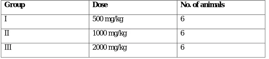
Table -3 Dosage Schedule for Acute toxicity.
Group Dose No. of animals
I 500 mg/kg 6
II 1000 mg/kg 6
III 2000 mg/kg 6
i) Behavioural profile
Awareness: Alertness, Visual placing, stereotypy, passivity.
ii) Neurological profile
Motor activity: Spontaneous activity, reactivity, touches response, pain response, startle response, tremor, gait, grip strength, pinna reflex, and corneal reflex.
iii) Autonomic profile
Writhing, defecation, urination, pile erection, heart rate, respiratory rate.
Selection of doses and dosage schedule:
The doses selected for the drugs were about 1/10th of the maximum tolerated safe dose found from acute toxicity studies. The mice were weighed on the day of tumor inoculation. The drug treatment was started 24 hr after the tumor inoculation. The drug was formulated as suspension in distilled water using 2% acacia and administered by i.p. route daily for 7 days. The mice were weighed every day till 15th day. Cisplatin was given intraperitoneally on only 1st day as standard. Tumor response was assessed on the basis of mean survival time (MST) and % increase in life span (%ILS).
Dosage Schedule and Grouping of EAC induced mice.
Group Compound Dose Route No. of animals
1 2% acacia Equivol i.p. 5
2 Cisplatin 3.5 mg/kg i.p. 6
3 D3+ 50mg/kg i.p 5
4 D3++ 100mg/kg i.p 5
, D3= 3C BRS, D3+= 50mg/kg D3++= 100mg/kg
The dose of standard drug was calculated by computing the minimum human dose to the rats.
RESULTS
Table-6 Anticancer Activity of Synthesized Compounds by MTT Method on HeLa Cell Lines
IC50 value of Cisplatin on HeLa cell lines by MTT method was found to be 6.58±0.81µg/ml.
Table-7. Survival of Mice in Acute Toxicity Study
Compound 500mg/kg 1000mg/kg 2000mg/kg LD50 (mg/kg)
3CBRS 00000 00000 0000X >2000
0-survival, X-death
There was no behavioral, neurological and any additional toxicity observed
Mean Survival Time and % Increase in Life Span
COMPOUND MST(in days) % ILS
CONTROL 17±0.707 -
CISPLATIN 32±3.06b 88.23
D3+ 18.8±1.01 10.5
D3++ 18.2±1.59 7.05
% growth inhibition (µg/ml) Compound code
12.5 25 50 100
IC50
3 C BRS 45.7 70.8 77.9 88.45 14.566
3 CL O 3CL 1.79 6.34 15.52 30.2 >100
IMIDAZOL A 0.36 9.69 17.47 37.6 >100
IMIDAZOL B 5.15 6.2 4.56 18.58 >100
TFC 1 1.8 5.6 6.6 18.6 >100
TFC 2 4.5 8.9 10.9 20.4 >100
TFC 3 1.9 6.9 11.8 15.4 >100
Percentage Change in Body Weight
Percentage change in body weight as compared to day- 0 weight
(MEAN±S.E.M)
Compound code
3rd day 5th day 7th day 9th day 11th day 13th day
Control 11.2±1.02 18.2±2.56 24.5±1.45 28.5±2.27 36.7±5.3b 43.9±4.8b
Cisplatin 0.001±001a 0.53±0.017b 0.55±0.0b 0.56±0.2b 1.66±.01a 2.1±.01a
D3+(50mg/kg) 7.48±2.4b 6.46±1.83a 5.1±1.7b 4.7±2.7b 4.36±4b 10.1±4.8b
D3++(100mg/kg) 2.3±0.96b 0.85±2.4b 7.71±1.5b 2.05±1.4b 4.7±3.8b 2.8±5.4b
All the values are expressed in Mean±S.E.M. a, p<0.05 Vs control; b, p<0.001 Vs control One way ANOVA followed by post hoc Tukey test in SPSS 11.5 in computer package
Discussion
All the synthesized compounds were in conformity with the structure envisaged. The structures were confirmed on the basis of physical and spectral data viz., IR, Mass spectrometry and
1
HNMR.
Synthesis of Imidazolones
The starting compounds substituted oxazolones were prepared by cyclocondensation of various substituted benzaldehydes and hippuric acid in the presence of acetic anhydride. The next step involves synthesis of various heterocyclic amines such as thiadiazolyl amines, thiazolyl amines and 7-amino-2-Quinolones; followed by insertion of these amines in to oxazolone moiety to get substituted Imidazolones. Physiochemical properties of the synthesized compounds are shown in Table – 2.
In vitro anticancer studies.
All the synthesized compounds were screened for cytotoxicity on human cervical cancer cell lines (HeLa) by MTT method. Compound 3CBRS has shown IC 50 value of 14.56 µg/ml which
was comparable with that of standard Cisplatin (6.58 µg/ml). Other compounds showed IC 50
above 100. Based on these results compound 3CBRS was selected for in vivo anticancer studies. Results are shown in table 6. Comparative cytotoxic activity of synthesized compounds were shown in Figure-5. From the figure 6, it can be recognized that the light purple coloured areas show cell growth inhibition. It means that there is less number of living cells in those areas forming less amount of formazan product as compared to control which is having dark purple color.
In vivo anticancer studies
In vivo anticancer studies were done on Ehrlich ascites carcinoma (EAC) cell lines by liquid tumor model using mice. Compound 3CBRS was selected for the study.
Acute toxicity studies. – Acute toxicity studies revealed that maximum tolerated safe dose for all the compounds were at 1000mg/kg .so 1/10 of maximum tolerated safe dose was selected for the study.
EAC Response
Weight Change
Control animals showed a progressive gain in body weights since the tumor inoculation. By day 13, they gained a maximum average weight of 43.9 %. Cisplatin significantly (p< 0.05) reduced tumor induced gain in weight as compared to respective control. On the other hand, Compound 3CBRS reduced body weight significantly till day 13 compared to their respective controls at the both dose levels studied. The results suggest that this compound could able to prevent the growth of tumor as indicated by decrease in progressive gain in body weight when compared to control. The results are shown in Table-8.
Conclusion
Even the latest drug available for the treatment of cancer and infectious diseases is not free from side effects and toxicity. Hence, there is an urgent need for new, effective and inexpensive drugs to be discovered to conquer the world of microbes and cancer. Necessity for new anticancer agent is a ceaseless pursuit to obtain drugs with high potency and least undesirable side effects. The present work has shown that the synthesized compound 3CBRS from imidazolone series was found to be active against human cervical cancer cell lines (HeLa) comparable to that of cisplatin. The activity of 3CBRS might be attributed due to the presence of halogen substituted thiazol ring on Imidazolone moiety. This compound was also found to be effective in preventing the growth of tumor as indicated by decrease in progressive gain in body weight when compared to control. Even though well known mechanisms are there for the anticancer drugs, but in the present work, a study has been done at a cellular level (in vitro & in vivo) and not at the molecular level. So, a well known mechanism of action could not be predicted for the prepared derivatives.
References
[1] Vogel’sText book of practical organic chemistry,2005; Page no – 1153-1156
[2]Eisenbrand G, Pool Zobel, Baker V, Balls M, Blaauboer BJ. Methods of in vitro Toxicology, Food and Chemical Toxicology. 2002; 40:193-236.
[3] Smitha Nair, S P Garg and Pramilla Sah. Synthesis and antimicrobial activity of some new oxazolone derivatives of 4,5 disubstituted-2-amino thiazoles. Journal of the Indian chemical society. 2006;82(2):205-207.